

Key Points
TET2 deficiency leads to clonal expansion of dysfunctional human CFU-E cells.
The clonal expansion is associated with upregulation of c-Kit and tyrosine kinase AXL.

Abstract
Myelodysplastic syndromes (MDSs) are clonal hematopoietic stem cell disorders characterized by ineffective hematopoiesis. Anemia is the defining cytopenia of MDS patients, yet the molecular mechanisms for dyserythropoiesis in MDSs remain to be fully defined. Recent studies have revealed that heterozygous loss-of-function mutation of DNA dioxygenase TET2 is 1 of the most common mutations in MDSs and that TET2 deficiency disturbs erythroid differentiation. However, mechanistic insights into the role of TET2 on disordered erythropoiesis are not fully defined. Here, we show that TET2 deficiency leads initially to stem cell factor (SCF)–dependent hyperproliferation and impaired differentiation of human colony-forming unit–erythroid (CFU-E) cells, which were reversed by a c-Kit inhibitor. We further show that this was due to increased phosphorylation of c-Kit accompanied by decreased expression of phosphatase SHP-1, a negative regulator of c-Kit. At later stages, TET2 deficiency led to an accumulation of a progenitor population, which expressed surface markers characteristic of normal CFU-E cells but were functionally different. In contrast to normal CFU-E cells that require only erythropoietin (EPO) for proliferation, these abnormal progenitors required SCF and EPO and exhibited impaired differentiation. We termed this population of progenitors “marker CFU-E” cells. We further show that AXL expression was increased in marker CFU-E cells and that the increased AXL expression led to increased activation of AKT and ERK. Moreover, the altered proliferation and differentiation of marker CFU-E cells were partially rescued by an AXL inhibitor. Our findings document an important role for TET2 in erythropoiesis and have uncovered previously unknown mechanisms by which deficiency of TET2 contributes to ineffective erythropoiesis.
Introduction
Erythropoiesis is a process during which multipotent hematopoietic stem cells proliferate, differentiate, and eventually form mature erythrocytes. Although this process is a continuum, it can be functionally divided into 2 major stages: early-stage erythropoiesis and terminal erythroid differentiation. During early-stage erythropoiesis, hematopoietic stem cells are first committed to early-stage erythroid progenitor burst-forming unit–erythroid cells, which further differentiate to late-stage erythroid progenitor colony-forming unit–erythroid (CFU-E) cells. During terminal erythroid differentiation, CFU-E cells undergo 3 or 4 mitoses to sequentially generate proerythroblasts, basophilic erythroblasts, polychromatic erythroblasts, and orthochromatic erythroblasts that expel their nuclei to generate enucleated reticulocytes. Disruption of the normal process at any stage of erythroid differentiation can lead to anemia.
Anemia due to disordered or ineffective erythropoiesis is a feature of a large number of human hematological disorders. These include Cooley’s anemia (also known as β-thalassemia),1-3 congenital dyserythropoietic anemia,4-6 Diamond-Blackfan anemia,7,8 malarial anemia,9,10 and various bone marrow failure syndromes, such as myelodysplastic syndromes (MDSs).11-13 Although the causative genes responsible for β-thalassemia,14-16 congenital dyserythropoietic anemia,17-20 and Diamond-Blackfan anemia21,22 have been identified, the molecular basis for the dyserythropoiesis in MDS remains to be fully defined. Recent advances in technologies for the detection of genetic abnormalities have led to the identification of a set of novel recurrent mutations in MDS patients.23-25 Of note, heterozygous loss-of-function mutation of DNA dioxygenase TET2 is one of the most frequently mutated genes in MDS.26 We and other investigators have documented that TET2 deficiency led to disordered erythropoiesis in human and animal models.27-30 However, the mechanisms by which TET2 deficiency leads to altered erythropoiesis are yet to be defined.
In the present study, we used a short hairpin (shRNA)-mediated knockdown approach, including human CD34+ cells and highly purified populations of erythroid cells at distinct stages of differentiation, to study the effects of TET2 deficiency on human erythropoiesis. We show that TET2 knockdown led to hyperproliferation of CFU-E via upregulation of c-Kit, followed by expansion of a dysfunctional population of CFU-E cells via upregulation of AXL. Our findings identified an important role for TET2 in regulating normal human erythropoiesis and uncovered the underlying molecular mechanisms by which deficiency of TET2 contributes to defective erythroid progenitors in MDSs.
Materials and methods
Antibodies and reagents
The antibodies used for western blotting were rabbit anti-human c-Kit, phosphorylated (p-)c-Kit (Y719), ERK, p-ERK, SHP-1, and AXL from Cell Signaling Technology (Beverly, MA), rabbit anti-human AKT and p-AKT from IMAGENTX (Beijing, China), and mouse anti-human glyceraldehyde-3-phosphate dehydrogenase (GAPDH) from PPLYGEN (Beijing, China). Antibodies used for flow cytometry analysis were mouse anti-human CD34-phycoerythrin (PE), CD36-fluorescein isothiocyanate, 7-aminoactinomycin acid, glycophorin A (GPA)–PE, and GPA-allophycocyanin from BD Pharmingen (Franklin Lakes, NJ), annexin V–PE–Cy7 and CD123 (IL-3R)–PE–Cy7 from Thermo Fisher Scientific (Waltham, MA), and α4-integrin–allophycocyanin from Miltenyi Biotec (Bergisch Gladbach, Germany). Band 3–FITC antibody was generated by our laboratory as described previously.30 c-Kit inhibitor (STI571) and AXL inhibitor (R428, BGB324) were obtained from Selleck Chemicals (Houston, TX).
CD34+ cells culture and shRNA-mediated knockdown
The detailed culture medium composition, the culture protocol, preparation of lentivirus, and transduction in CD34+ cells have been described previously.30,31 The plasmids of pLKO.1–luciferase–shRNA, pLKO.1–TET2–shRNA1 (TRCN0000418976), and pLKO.1–TET2–shRNA2 (TRCN0000142853) were from Sigma (Santa Clara, CA).
Flow cytometry analysis and fluorescence-activated cell sorting (FACS) of erythroblasts
The differentiation of erythroid cells was assessed using recently identified surface markers by flow cytometry and the erythroid cells at the distinct developmental stage were sorted using a MoFlo high-speed cell sorter (Beckman-Coulter, Pasadena, CA), as previously described.32,33 Sorted CFU-E cells were cultured in the second-phase media as previously described.33
Single-cell sorting
Luciferase-shRNA and TET2-shRNA1 cells at day 6 of culture were stained for the erythroid progenitors surface markers CD34 and CD36, and single CFU-E cells (CD34− and CD36+) were sorted on a MoFlo high-speed cell sorter into 96-well plates containing 100 μL of culture medium containing erythropoietin (EPO; 1 U/mL) or EPO plus stem cell factor (SCF; 10 ng/mL).
Colony-forming assay
Sorted CFU-E cells were diluted at a density of 200 cells in 1 mL of MethoCult H4330 medium (which contains EPO but not SCF) or H4330 medium, with addition of SCF (10 ng/mL) for colony-forming assay (STEMCELL Technologies), and incubated at 37°C in a humidified atmosphere with 5% CO2. The CFU-E colonies were defined according to previously described criteria.32
Cytospin assay
The detailed protocols of cell collection, staining, and image acquisition have been described previously.33
qRT‐PCR
The primer sequences are listed in supplemental Table 1 (available on the Blood Web site), and the detailed protocols of RNA extraction, reverse transcription, primer design, and quantitative real-time polymerase chain reaction (qRT-PCR) have been described previously.30
Western blotting
Whole-cell lysates of erythroid cells were prepared with radioimmunoprecipitation buffer (PPLYGEN) in the presence of protease inhibitor and PhosSTOP cocktails (Roche, Basel, Switzerland). Protein concentration was measured using a BCA protein assay kit (PPLYGEN). All of the procedures were performed as described previously.33
RNA sequencing and bioinformatics analysis
RNA was extracted from FACS-sorted cells, and cDNA libraries were prepared using an Illumina TruSeq kit and sequenced on an Illumina HiSeq 2500 System (Epigenomics Core, Weill Cornell Medical College, New York, NY). The data were analyzed as previously described.30-32
Global DNA methylation quantitation
Genomic DNA was extracted from 1 × 10⁶ cells using a PureLink Genomic DNA Mini Kit (Thermo Fisher Scientific). DNA was denatured, and twofold serial dilutions were spotted on the activated polyvinylidene difluoride membrane with methyl alcohol. The blotted membrane was air-dried, UV-cross-link, blocked with 5% nonfat milk, and incubated with rabbit polyclonal anti-5hmC (1:10 000) and anti-5mC (1:300) antibodies, as well as horseradish peroxidase–conjugated anti-rabbit immunoglobulin G secondary antibody (1:3 000). Signal density was quantified using an Azure c600 imaging system (Azure Biosystems).
Site-specific quantitative analysis of 5mC and 5hmC
The 5mC and 5hmC levels at the promoter region of AXL were determined using an EpiJET 5hmC and 5mC Analysis Kit (K1501; Thermo Fisher Scientific). The detailed protocol was described previously.30 The primers used for the analysis, Fwd/Rev-AXL-CCGG (−414) and Fwd/Rev-AXL-CCGG (+320), are listed in supplemental Table 1.
Statistical analysis
Statistical evaluations of different experimental groups were performed using GraphPad Prism 7 (unpaired t test), and P < .05 was considered to indicate statistical significance.
Results
TET2 knockdown led to SCF-dependent hyperproliferation of human CFU-E cells
Li et al have previously shown that Tet2−/− mice exhibited mild anemia accompanied by erythroblast proliferation in bone marrow, spleen, and liver.27 We recently documented that TET2 knockdown in human CD34+ cells led to hyperproliferation of erythroid cells.30 These findings strongly suggest an important regulatory role for TET2 in erythropoiesis. However, the function of TET2 in normal erythropoiesis and the mechanisms by which TET2 deficiency contributes to defective erythropoiesis have yet to be fully defined. Because erythropoiesis involves multiple developmental stages, we sought to examine whether TET2 deficiency causes stage-specific defects. To this end, we used an shRNA-mediated knockdown approach in human CD34+ cells in conjunction with an in vitro erythroid-differentiation system. We sorted erythroid cells at all distinct developmental stages using the experimental strategy that we described previously.32,33 RT-PCR analysis revealed that, with the exception of the burst-forming unit–erythroid cell stage, efficient knockdown was achieved at all other developmental stages (Figure 1A). We then compared the colony-forming ability of luciferase control and TET2-knockdown CFU-E cells. In the presence of EPO only, the size of luciferase and TET2-knockdown colonies is similar (Figure 1B, left panel). Interestingly, in the presence of both EPO and SCF, the size of the luciferase CFU-E colonies increased compared with their size in the presence of EPO only, indicating that SCF enhances proliferation of CFU-E cells. Notably, the size of ∼20% of TET2-knockdown CFU-E colonies was significantly larger than that of luciferase CFU-E colonies (Figure 1B, right panel; Figure 1C). We further examined the cell growth using liquid culture in the presence of both EPO and SCF. As shown in Figure 1D, proliferation of TET2-knockdown cells was higher than that of luciferase CFU-E cells. Moreover, although the growth of control cells plateaued starting at day 10 of culture, TET2-knockdown cells continued to grow until day 30. In contrast, growth of sorted proerythroblasts, basophilic erythroblasts, polychromatic erythroblasts, and orthochromatic erythroblasts was not affected by TET2 knockdown (Figure 1E). These findings demonstrate that TET2 knockdown led to SCF-dependent hyperproliferation of a subpopulation of human CFU-E cells.
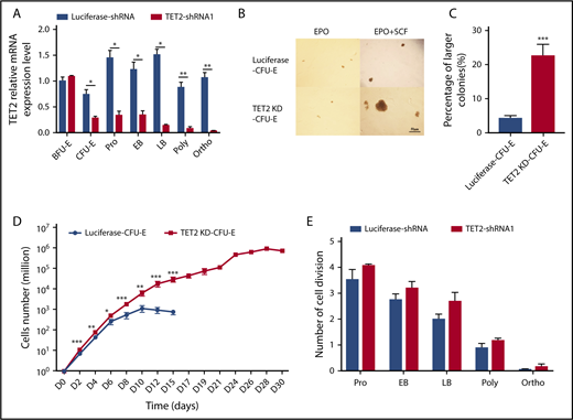
Hyperproliferation of TET2-knockdown CFU-E cells in the presence of SCF. (A) qRT‐PCR analyses of TET2 mRNA levels (normalized to actin) of sorted luciferase and TET2-knockdown erythroid cells. (B) Colony-forming ability of sorted luciferase and TET2-knockdown CFU-E cells in the presence of EPO only (left panel) or in the presence of EPO plus SCF (right panel). Scale bar, 50 μm. (C) Percentage of larger-size colonies in panel B. (D) Growth curves of sorted luciferase and TET2-knockdown CFU-E cells cultured in the presence of EPO plus SCF. (E) Numbers of cell divisions of sorted erythroblasts at the indicated stages in culture. All results are from 3 independent experiments. *P < .05, **P < .01, ***P < .001.
Hyperproliferation of TET2-knockdown CFU-E cells in the presence of SCF. (A) qRT‐PCR analyses of TET2 mRNA levels (normalized to actin) of sorted luciferase and TET2-knockdown erythroid cells. (B) Colony-forming ability of sorted luciferase and TET2-knockdown CFU-E cells in the presence of EPO only (left panel) or in the presence of EPO plus SCF (right panel). Scale bar, 50 μm. (C) Percentage of larger-size colonies in panel B. (D) Growth curves of sorted luciferase and TET2-knockdown CFU-E cells cultured in the presence of EPO plus SCF. (E) Numbers of cell divisions of sorted erythroblasts at the indicated stages in culture. All results are from 3 independent experiments. *P < .05, **P < .01, ***P < .001.
TET2 knockdown significantly impaired human terminal erythroid differentiation
Terminal erythroid differentiation refers to the process by which CFU-E cells differentiate into morphologically recognizable proerythroblasts that undergo 3 or 4 mitoses to sequentially generate basophilic erythroblasts, polychromatic erythroblasts, and orthochromatic erythroblasts. Because the differentiation of CFU-E cells to proerythroblasts is characterized by expression of the erythroid lineage–specific marker GPA, we monitored the changes in GPA expression. Figure 2A shows the representative flow cytometry analysis of GPA expression of sorted CFU-E cells cultured for different periods of time. It shows that, on day 0, all cells were GPA−. On day 5, although only ∼17% of control cells were GPA−, ∼35% of TET2-knockdown cells were still GPA−. Furthermore, on day 15, although almost all control cells were GPA+, ∼30% of TET2-knockdown cells continued to be GPA−. Quantitative analysis revealed that, although almost all control cells progressively became GPA+, a fraction of TET2-knockdown cells remained GPA−, even on day 28 of culture (Figure 2B). We further examined the terminal erythroid differentiation of GPA+ cells to late-stage erythroblasts by monitoring surface expression of band 3 and α4 integrin.33 Figure 2C shows the representative plots of band 3 vs α4 integrin of GPA+ cells at days 5 and 15 of culture, which reveal that, although control cells progressively became band3hiα4 integrinlow, the majority of TET2-knockdown cells still express high levels of α4 integrin, demonstrating impaired differentiation of proerythroblasts to late-stage erythroid cells. Quantitative analyses clearly showed that there were more proerythroblasts and basophilic erythroblasts in the TET2-knockdown group than in the control group (Figure 2D). Consistent with flow cytometric analysis, cytospin analyses show that, although luciferase CFU-E cells progressively differentiate to orthochromatic erythroblasts, TET2-knockdown cells are still at the early stages of differentiation, even on day 15 of culture (Figure 2E). These findings demonstrate that TET2 knockdown significantly impaired terminal erythroid differentiation.
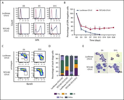
TET2 knockdown impaired human terminal erythroid differentiation. (A) Representative flow cytometry analysis of GPA expression of sorted luciferase and TET2-knockdown CFU-E cells cultured in the presence of EPO plus SCF for different days, as indicated. (B) Quantitative analysis of changes in GPA− population from 3 independent experiments. (C) Terminal erythroid differentiation was monitored on the indicated days by flow cytometric analysis based on the expression of band 3 and α4 integrin. Representative plots of α4-integrin vs band 3 of GPA+ cells are shown, and the erythroblasts are separated into 5 populations: proerythroblasts (Pro; α4-integrinhiband 3neg), early basophilic erythroblasts (EB; α4-integrinhiband 3low), late basophilic erythroblasts (LB; α4-integrinhiband 3med), polychromatic erythroblasts (Poly; α4-integrinmedband 3med), and orthochromatic erythroblasts (Ortho; α4-integrinlowband 3hi). (D) Quantitative analyses of results shown in panel C from 3 independent experiments. (E) Representative cytospin images of erythroblasts cultured for different days as indicated. *P < .05, **P < .01.
TET2 knockdown impaired human terminal erythroid differentiation. (A) Representative flow cytometry analysis of GPA expression of sorted luciferase and TET2-knockdown CFU-E cells cultured in the presence of EPO plus SCF for different days, as indicated. (B) Quantitative analysis of changes in GPA− population from 3 independent experiments. (C) Terminal erythroid differentiation was monitored on the indicated days by flow cytometric analysis based on the expression of band 3 and α4 integrin. Representative plots of α4-integrin vs band 3 of GPA+ cells are shown, and the erythroblasts are separated into 5 populations: proerythroblasts (Pro; α4-integrinhiband 3neg), early basophilic erythroblasts (EB; α4-integrinhiband 3low), late basophilic erythroblasts (LB; α4-integrinhiband 3med), polychromatic erythroblasts (Poly; α4-integrinmedband 3med), and orthochromatic erythroblasts (Ortho; α4-integrinlowband 3hi). (D) Quantitative analyses of results shown in panel C from 3 independent experiments. (E) Representative cytospin images of erythroblasts cultured for different days as indicated. *P < .05, **P < .01.
TET2 knockdown led to upregulation and activation of c-Kit
The findings that SCF promoted proliferation and impaired differentiation of TET2-knockdown CFU-E cells strongly suggest a role for the c-Kit signaling pathway in the enhanced cell proliferation of TET2-knockdown CFU-E cells. To test this thesis, we compared the expression of c-Kit in sorted luciferase CFU-E cells and TET2-knockdown CFU-E cells by qRT-PCR and western blot analysis. Figure 3A shows that there was a modest, but significantly higher, messenger RNA (mRNA) level (∼1.4-fold) of c-Kit in TET2-knockdown CFU-E cells. Consistent with the increased mRNA level, the protein level of c-Kit was also increased (∼1.3-fold) (Figure 3B). Moreover, the phosphorylation levels of c-Kit were also increased (approximately two-fold). Because the increase in c-Kit phosphorylation is higher than the noted increase in c-Kit expression, we reasoned that additional factors contribute to the activation of c-Kit. In this context, we measured the expression level of phosphatase SHP-1, which has been shown to interact with p–c-Kit and dephosphorylate the receptor.34,35 As shown in Figure 3C, the protein level of SHP-1 was significantly decreased in TET2-knockdown cells compared with control cells. To demonstrate that the increased activation of c-Kit is responsible for the enhanced proliferation and impaired differentiation, we examined the effects of c-Kit inhibitor STI571 on cell growth and differentiation. As shown in Figure 3D, STI571 dramatically inhibited the growth of TET2-knockdown cells (∼150-fold), with far less inhibitory effect on the growth of control cells (∼10-fold). Furthermore, the impaired differentiation of TET2-knockdown cells, as assessed by surface expression of GPA (Figure 3E) and α4 integrin/band 3 (Figure 3F-G), as well as cytospin analysis (Figure 3H), was also rescued by STI571 treatment. Thus, the hyperproliferation and impaired differentiation of TET2-knockdown cells are associated with upregulation and activation of c-Kit.

TET2 knockdown led to upregulation and activation of c-Kit. (A) mRNA levels (normalized to actin) of c-Kit, as assessed by qRT-PCR. (B) Representative western blot analysis of total c-Kit, p–c-Kit, and SHP-1. GAPDH was used as loading control. (C) Quantitative analysis of c-Kit, p–c-Kit, and SHP-1 from 3 independent experiments. (D) Effects of c-Kit inhibitor STI571 (0.5 μM) on proliferation of sorted luciferase and TET2-knockdown CFU-E cells. Dimethyl sulfoxide (DMSO) was used as control. (E-H) Effects of c-Kit inhibitor STI571 (0.5 μM) on differentiation of luciferase and TET2-knockdown CFU-E cells. Sorted luciferase and TET2-knockdown CFU-E cells were cultured for 13 days in the presence of DMSO or STI571. Expression of GPA (E) or band 3/α4 integrin (F) was examined by flow cytometry analysis. (G) Quantitative analysis of results shown in panel F from 3 independent experiments. (H) Representative cytospin images of erythroblasts. Note that the TET2-knockdown–induced impairment in differentiation was reversed by STI571 treatment. *P < .05, **P < .01, ***P < .001.
TET2 knockdown led to upregulation and activation of c-Kit. (A) mRNA levels (normalized to actin) of c-Kit, as assessed by qRT-PCR. (B) Representative western blot analysis of total c-Kit, p–c-Kit, and SHP-1. GAPDH was used as loading control. (C) Quantitative analysis of c-Kit, p–c-Kit, and SHP-1 from 3 independent experiments. (D) Effects of c-Kit inhibitor STI571 (0.5 μM) on proliferation of sorted luciferase and TET2-knockdown CFU-E cells. Dimethyl sulfoxide (DMSO) was used as control. (E-H) Effects of c-Kit inhibitor STI571 (0.5 μM) on differentiation of luciferase and TET2-knockdown CFU-E cells. Sorted luciferase and TET2-knockdown CFU-E cells were cultured for 13 days in the presence of DMSO or STI571. Expression of GPA (E) or band 3/α4 integrin (F) was examined by flow cytometry analysis. (G) Quantitative analysis of results shown in panel F from 3 independent experiments. (H) Representative cytospin images of erythroblasts. Note that the TET2-knockdown–induced impairment in differentiation was reversed by STI571 treatment. *P < .05, **P < .01, ***P < .001.
TET2 knockdown led to generation of “marker CFU-E” cells
As described above, TET2 knockdown led to the persistent presence of a GPA− cell population. To further characterize this cell population, we FACS sorted TET2-knockdown GPA− cells on day 13 of culture (Figure 4A). Cytospin analysis shows that, morphologically, TET2-knockdown GPA− cells are indistinguishable from luciferase CFU-E cells isolated from the first phase of culture at day 6 (Figure 4B). Consistent with cytospin analysis, flow cytometry analysis reveals that, similar to luciferase CFU-E cells (characterized as IL-3R−GPA−CD36+CD34−CD71high), TET2-knockdown GPA− cells are immunophenotypically the same as CFU-E cells (Figure 4C). However, as shown in Figure 4D, in contrast to luciferase CFU-E cells, which require only EPO for proliferation, TET2-knockdown GPA− cells required EPO and SCF for proliferation. Furthermore, differentiation of the TET2-knockdown GPA− cells, as assessed by surface expression of GPA (Figure 4E) and α4 integrin/band 3 (Figure 4F-G), as well as cell morphology (Figure 4H), was also significantly impaired. Given the fact that TET2-knockdown GPA− cells are immunophenotypically similar to CFU-E cells, but are functionally different, we termed them “marker CFU-E” cells. Intriguingly, in contrast to the increased phosphorylation of c-Kit and decreased SHP-1 in TET2-knockdown CFU-E cells isolated at day 6 of culture (Figure 3C), the phosphorylation level of c-Kit in marker CFU-E cells isolated at day 13 of culture was decreased (Figure 4I-J), with no change in SHP-1 protein level. Together, our findings demonstrate that TET2 knockdown led to SCF-dependent expansion of dysfunctional CFU-E cells with time.

Knockdown of TET2 led to the generation of marker CFU-E cells. (A) TET2-knockdown CFU-E cells were cultured for 13 days, and the GPA− population was sorted. (B) Representative cytospin images of sorted luciferase CFU-E and TET2-knockdown GPA− cells. (C) Flow cytometry analysis of luciferase CFU-E and TET2-knockdown GPA− cells using the surface markers IL-3R, GPA, CD34, CD36, and CD71. (D) Proliferation of luciferase CFU-E and TET2-knockdown GPA− cells in the presence of EPO only or EPO plus SCF. (E-F) Differentiation of luciferase CFU-E and TET2-knockdown GPA− cells. Sorted luciferase and TET2-knockdown GPA− cells were cultured for 5 or 15 days. Expression of GPA (E) or band 3/α4 integrin (F) was examined by flow cytometry analysis. (G) Quantitative analysis of results shown in panel F from 3 independent experiments. (H) Representative cytospin images of erythroblasts. (I) Western blot analysis of c-Kit, p–c-Kit, AKT, p-AKT, ERK, p-ERK, and SHP-1. GAPDH was used as loading control. (J) Quantitative analysis of results shown in panel H from 3 independent experiments. *P < .05, **P < .01, ***P < .001.
Knockdown of TET2 led to the generation of marker CFU-E cells. (A) TET2-knockdown CFU-E cells were cultured for 13 days, and the GPA− population was sorted. (B) Representative cytospin images of sorted luciferase CFU-E and TET2-knockdown GPA− cells. (C) Flow cytometry analysis of luciferase CFU-E and TET2-knockdown GPA− cells using the surface markers IL-3R, GPA, CD34, CD36, and CD71. (D) Proliferation of luciferase CFU-E and TET2-knockdown GPA− cells in the presence of EPO only or EPO plus SCF. (E-F) Differentiation of luciferase CFU-E and TET2-knockdown GPA− cells. Sorted luciferase and TET2-knockdown GPA− cells were cultured for 5 or 15 days. Expression of GPA (E) or band 3/α4 integrin (F) was examined by flow cytometry analysis. (G) Quantitative analysis of results shown in panel F from 3 independent experiments. (H) Representative cytospin images of erythroblasts. (I) Western blot analysis of c-Kit, p–c-Kit, AKT, p-AKT, ERK, p-ERK, and SHP-1. GAPDH was used as loading control. (J) Quantitative analysis of results shown in panel H from 3 independent experiments. *P < .05, **P < .01, ***P < .001.
TET2 knockdown led to changes in gene expression in marker CFU-E cells
To explore the molecular basis for the generation of marker CFU-E cells after TET2 knockdown, we performed RNA sequencing (RNA-seq) analysis on luciferase CFU-E cells and TET2-knockdown CFU-E cells isolated at day 6 of culture and TET2-knockdown marker CFU-E cells isolated at day 13 of culture. Bioinformatics analysis revealed that only 51 genes were differentially expressed between luciferase CFU-E cells and TET2-knockdown CFU-E cells. In contrast, 400 genes were differentially expressed between luciferase CFU-E cells and TET2-knockdown marker CFU-E cells, whereas 305 genes were differentially expressed between TET2-knockdown CFU-E cells and TET2-knockdown marker CFU-E cells (Figure 5A). The list of differentially expressed genes is shown in supplemental Table 2. Gene-set enrichment analysis of the differentially expressed genes in TET2-knockdown marker CFU-E cells compared with luciferase CFU-E cells or TET2-knockdown CFU-E cells revealed alterations in several pathways, including upregulation of genes associated with an “organ-regeneration” pathway (Figure 5B). Notably, the expression of AXL, a gene associated with hematological malignancy and transformation,36-39 is significantly upregulated in TET2-knockdown marker CFU-E cells (Figure 5C). The increased AXL mRNA level was confirmed by qRT-PCR analysis (Figure 5D). Consistent with the increased mRNA level, western blotting analysis revealed that AXL is abundantly expressed in TET2-knockdown marker CFU-E cells but is not detectable in luciferase CFU-E cells or TET2-knockdown CFU-E cells (Figure 5E-F). Because activation of AXL triggers the AKT and ERK pathways,39 we examined the phosphorylation of AKT and ERK in marker CFU-E cells, and, as expected, phosphorylation of AKT and ERK was increased in marker CFU-E cells (Figure 4I-J). To further document that the upregulation of AXL contributes to expansion of marker CFU-E cells, we examined the effect of R428, an AXL inhibitor, on cell growth in liquid culture with EPO and SCF. Similar to c-Kit inhibitor, AXL inhibitor R428 significantly inhibited the growth of TET2-knockdown cells (∼135-fold), with a much smaller effect on luciferase cells (∼20-fold) (Figure 5G). Moreover, the delayed differentiation of TET2-knockdown CFU-E cells was partially rescued by the inhibitor, as assessed by surface expression of GPA (Figure 5H) and α4 integrin/band 3 (Figure 5I-J) and cytospin evaluation of erythroid cells (Figure 5K). These findings suggest that upregulation of the AXL pathway contributes to impaired erythropoiesis caused by TET2 knockdown.
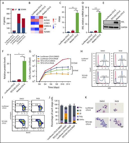
Upregulation of AXL in marker CFU-E cells. (A) Number of differentially expressed genes among luciferase CFU-E, TET2-knockdown CFU-E, and TET2-knockdown marker CFU-E cells, as revealed by RNA-seq analysis. Fold change > 2. (B) Heat map of the 6 genes involved in the “organ regeneration” pathway. (C) mRNA levels of AXL, as revealed by RNA-seq. (D) mRNA levels of AXL, as assessed by qRT-PCR. (E) Western blot analysis of AXL. (F) Quantitative analysis of AXL protein levels from 3 independent experiments. (G) Growth curves of sorted luciferase and TET2-knockdown CFU-E cells in the presence of DMSO or 0.2 μM R428. (H) Flow cytometry analysis showing GPA expression of luciferase and TET2-knockdown CFU-E cells cultured for 13 days in the presence of DMSO or 0.2 μM R428. (I) Flow cytometry analysis showing band 3/α4 integrin expression of luciferase and TET2-knockdown CFU-E cells cultured for 13 days in the presence of DMSO or 0.2 μM R428. (J) Quantitative analysis of results shown in panel I. (K) Representative cytospin images of erythroblasts. *P < .05, **P < .01, ***P < .001.
Upregulation of AXL in marker CFU-E cells. (A) Number of differentially expressed genes among luciferase CFU-E, TET2-knockdown CFU-E, and TET2-knockdown marker CFU-E cells, as revealed by RNA-seq analysis. Fold change > 2. (B) Heat map of the 6 genes involved in the “organ regeneration” pathway. (C) mRNA levels of AXL, as revealed by RNA-seq. (D) mRNA levels of AXL, as assessed by qRT-PCR. (E) Western blot analysis of AXL. (F) Quantitative analysis of AXL protein levels from 3 independent experiments. (G) Growth curves of sorted luciferase and TET2-knockdown CFU-E cells in the presence of DMSO or 0.2 μM R428. (H) Flow cytometry analysis showing GPA expression of luciferase and TET2-knockdown CFU-E cells cultured for 13 days in the presence of DMSO or 0.2 μM R428. (I) Flow cytometry analysis showing band 3/α4 integrin expression of luciferase and TET2-knockdown CFU-E cells cultured for 13 days in the presence of DMSO or 0.2 μM R428. (J) Quantitative analysis of results shown in panel I. (K) Representative cytospin images of erythroblasts. *P < .05, **P < .01, ***P < .001.
Global DNA methylation changes in TET2-knockdown marker CFU-E cells
TET2 modulates gene expression by regulating DNA methylation. Specifically, it has been documented that TET family members promote active DNA demethylation by converting 5mC to 5hmC.40,41 Unexpectedly, we recently showed that TET2 or TET3 knockdown did not cause significant changes in global 5mC or 5hmC levels in erythroid cells.30 Our current finding that only 51 genes are differentially expressed between luciferase CFU-E cells and TET2-knockdown CFU-E cells, but ∼400 genes are differentially expressed between luciferase CFU-E cells and TET2-knockdown marker CFU-E cells, suggests that TET2 knockdown might cause changes in DNA methylation in TET2-knockdown marker CFU-E cells. To test this thesis, we examined 5mC and 5hmC levels by dot blot analysis. As shown in Figure 6A-C, the 5mC level increased and the 5hmC level decreased in TET2-knockdown marker CFU-E cells compared with luciferase CFU-E cells, implying that changes in global DNA methylation occur at later stages of cell differentiation following TET2 knockdown.
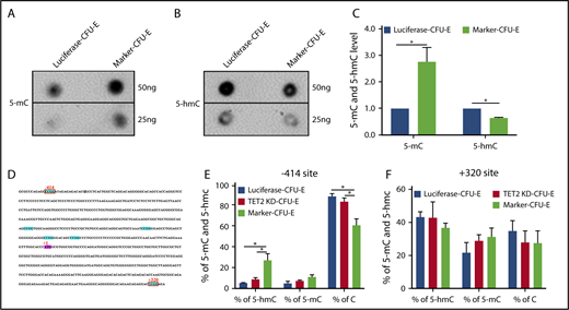
Effects of TET2 knockdown on DNA methylation. (A) DNA dot blots for 5mC. (B) DNA dot blots for 5hmC. (C) Quantitative analysis of 5mC and 5hmC levels from 3 independent experiments. (D) AXL promoter region containing 6 CCGG sites (blue). (E-F) Methylation-sensitive restriction enzyme analysis of 5mC and 5hmC changes at −414, +320 CCGG sites of AXL promoter. *P < .05.
Effects of TET2 knockdown on DNA methylation. (A) DNA dot blots for 5mC. (B) DNA dot blots for 5hmC. (C) Quantitative analysis of 5mC and 5hmC levels from 3 independent experiments. (D) AXL promoter region containing 6 CCGG sites (blue). (E-F) Methylation-sensitive restriction enzyme analysis of 5mC and 5hmC changes at −414, +320 CCGG sites of AXL promoter. *P < .05.
To understand how TET2 knockdown led to upregulation of AXL, we examined the methylation status of the AXL promoter region using methylation-sensitive restriction enzyme analysis, followed by quantitative polymerase chain reaction. Among the 6 CCGG sites (−414, −146, −96, −69, −58, +320) in the AXL promoter region (Figure 6D), 2 CCGG sites (−414, +320) could be examined by methylation-sensitive restriction enzyme analysis. As shown in Figure 6E, the 5hmC level at site −414 in the AXL promoter was increased in TET2-knockdown marker CFU-E cells, whereas no changes in 5mC and 5hmC levels were observed at site +320 (Figure 6F). These findings suggest that AXL could be a direct target of TET2.
TET2 knockdown led to SCF-dependent clonal expansion of CFU-E cells
MDS is characterized by the clonal expansion of hematopoietic stem and progenitor cells.42-45 The persistent presence of GPA− erythroid cells following TET2 knockdown strongly suggests clonal expansion of erythroid progenitors. To test this thesis, we performed a single-cell proliferation assay of FACS-sorted luciferase CFU-E cells and TET2-knockdown CFU-E cells. As shown in Figure 7A, in the presence of EPO alone, no significant difference in cell growth was noted between individual luciferase CFU-E cells and TET2-knockdown CFU-E cells. In the presence of EPO and SCF, the growth of ∼20% of individual TET2-knockdown CFU-E cells was significantly more than that of individual luciferase CFU-E cells (Figure 7B). Cytospin analyses revealed that, although luciferase and low-proliferative (LP) TET2-knockdown CFU-E cells differentiated into late-stage erythroblasts, there was persistence of early-stage erythroblasts in highly proliferating (HP) TET2-knockdown CFU-E cells, even after 15 days in culture (Figure 7C). Interestingly, AXL expression is significantly higher in HP cells than that in LP cells (Figure 7D-F). Taken together, our findings demonstrate that TET2 knockdown led to SCF-dependent clonal expansion of erythroid progenitors, at least in part by upregulation of c-Kit and AXL.
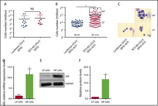
TET2 knockdown led to the SCF-dependent clonal expansion of CFU-E cells. (A) Growth of sorted single luciferase or TET2-knockdown CFU-E cells cultured in the presence of EPO for 7 days. (B) Growth of sorted single luciferase or TET2-knockdown CFU-E cells cultured in the presence of EPO plus SCF for 15 days. Note that cells can be divided into HP and LP populations. (C) Representative cytospin images of erythroblasts from panel B. (D) mRNA levels of AXL in the LP cells (pooled) and HP cells (pooled). (E) Western blot analysis of AXL of pooled LP and HP cells. (F) Quantitative analysis of AXL protein levels from 3 independent experiments. *P < .05. NS, not statistically significant.
TET2 knockdown led to the SCF-dependent clonal expansion of CFU-E cells. (A) Growth of sorted single luciferase or TET2-knockdown CFU-E cells cultured in the presence of EPO for 7 days. (B) Growth of sorted single luciferase or TET2-knockdown CFU-E cells cultured in the presence of EPO plus SCF for 15 days. Note that cells can be divided into HP and LP populations. (C) Representative cytospin images of erythroblasts from panel B. (D) mRNA levels of AXL in the LP cells (pooled) and HP cells (pooled). (E) Western blot analysis of AXL of pooled LP and HP cells. (F) Quantitative analysis of AXL protein levels from 3 independent experiments. *P < .05. NS, not statistically significant.
Discussion
Ineffective erythropoiesis and the resultant anemia are common features of MDS. However, our understanding of the molecular and mechanistic basis for ineffective erythropoiesis in MDS remains to be fully defined. Recent studies revealed that the gene encoding DNA dioxygenase TET2 is one of the most frequently mutated genes in MDS. Studies by us and other investigators have demonstrated that TET2 deficiency impairs erythroid differentiation.20-22 Here, we document that TET2 knockdown enhanced proliferation and impaired differentiation of human erythroid progenitor CFU-E cells, and, with time, there is a clonal expansion of dysfunctional CFU-E cells. We further uncovered that this results from sequential upregulation of c-Kit and the tyrosine kinase AXL.
Erythropoiesis is a complex multiple-step process. Using methods that we developed to highly enrich erythroid cells at each distinct developmental stage,32,33 we were able to identify the effect of TET2 deficiency in a stage-specific manner. The hematopoiesis of MDS is characterized by the hyperproliferation and impaired differentiation of progenitor cells and apoptosis of late-stage cells.23,24,46 Our finding that TET2 deficiency leads to clonal expansion of erythroid progenitor CFU-E cells is in line with the pathologic changes noted in MDS patients. In addition to TET2, TET3 is expressed in erythroid cells; interestingly, in contrast to the effects of TET2 deficiency on erythroid progenitors, TET3 knockdown impaired human terminal erythroid differentiation with no effect on erythroid progenitors.30 Overlapping and/or redundant roles for TET family members have been documented in other cells.27,47-49 Our findings demonstrate that TET family members play distinct and stage-specific roles in erythropoiesis.
It has been well documented that EPO is sufficient for the proliferation and survival of CFU-E cells.50,51 Interestingly, when we perform CFU-E colony assays and cell-proliferation assays in the presence of EPO and in the presence of EPO and SCF, we find that the colony size is larger when EPO and SCF are present compared with EPO alone. Thus, it appears that, although SCF is not essential for CFU-E cell proliferation, it does enhance their proliferative capacity. Notably, SCF further enhanced the proliferation of TET2-knockdown CFU-E cells significantly. Our finding is consistent with the previous report that c-Kit expression was upregulated in Tet2 shRNA-expressing mouse bone marrow progenitor cells.52 Moreover, Tet2 restoration in Tet2-knockout mice decreased the expression of c-Kit.53 Additionally, the extent of the increase in c-Kit phosphorylation is greater than the noted increased expression level of c-Kit protein, suggesting additional mechanism(s) contribute to the increased c-Kit phosphorylation. Supporting this hypothesis, we found that phosphatase SHP-1, a negative regulator of c-Kit,35 is significantly decreased in TET2-knockdown CFU-E cells. Interestingly, decreased expression of SHP-1 protein has been reported in MDS patients.54 In future studies, it will be interesting to examine the relationship between TET2 mutation and expression of SHP-1 in these diseases. Together, our findings suggest that 1 mechanism by which TET2 deficiency contributes to the increased proliferation and impaired differentiation of myeloid progenitors is through upregulation and activation of c-Kit. Importantly, our findings that c-Kit inhibitor reversed the proliferation and differentiation of TET2-knockdown CFU-E cells suggest that c-Kit inhibitor could be a potential therapeutic approach for patients with TET2 mutations.
During normal in vitro erythropoiesis, GPA− cells progressively decrease and eventually disappear on day 15. Our finding that TET2 knockdown led to the persistent presence of the GPA− cell population suggests clonal selection and expansion. Although these cells are characterized by the expression of normal CFU-E cell surface markers, they are functionally different from normal CFU-E cells in that SCF is required for their continued survival. We termed this cell population marker CFU-E cells. Intriguingly, in contrast to the increased activation of the c-Kit pathway in TET2-knockdown CFU-E cells, the phosphorylation of c-Kit was significantly decreased in these clonally expanded TET2-knockdown marker CFU-E cells, explaining the requirement for SCF for their survival. More surprisingly, in spite of the decreased phosphorylation of c-Kit, phosphorylation levels of AKT and ERK are increased. In exploring the mechanisms for the clonal expansion of the marker CFU-E cells, we found that a tyrosine kinase AXL is significantly increased. Similar to c-Kit inhibitor, AXL inhibitor reversed the proliferation and differentiation of marker CFU-E cells, suggesting that AXL inhibitor could also be a potential therapeutic approach for patients with TET2 mutations.
Regarding the molecular mechanisms for transformation or hyperproliferation due to Tet2 deficiency, 1 previous study showed that Tet2 loss and Nras mutation cooperatively drove myeloid transformation via downregulation of Spry2 and activation of mitogen-activated protein kinase signaling.55 Another 1 showed that Tet2 loss decreased Pten expression, leading to hyperproliferation of bone marrow–derived mast cells in a phosphatidylinositol 3-kinase–dependent manner.56 In the present study, we show that sequential upregulation of c-Kit and AXL is responsible for the hyperproliferation and clonal expansion of human erythroid progenitors. These findings suggest that Tet2 deficiency leads to transformation or hyperproliferation via different pathways in a cell-type–specific manner.
In further defining the mechanism for the increased AXL expression, we found that the 5hmC level at −414 in the AXL promoter is increased. This finding seems to be contradictory to the predominant role of TET2 in converting 5mC to 5hmC. However, it should be noted that TET family members play dual roles in regulating 5hmC levels and in regulating gene expression. For example, it has been reported that TET2 deletion in mouse embryonic stem cells results in an increase in 5hmC levels at some promoter/transcription start site regions.57 Moreover, in addition to catalyzing the conversion of 5mC to 5hmC, TET family members can further oxidize 5hmC to 5fC and 5caC.58,59 Alternatively, the increased 5hmC level could be due to impaired oxidation of 5hmC to 5fC and 5caC. Thus, the effect could be more complex and worth future in-depth analyses.
In summary, our findings document that TET2 deficiency leads to clonal selection of human erythroid progenitor CFU-E cells. Initial hyperproliferation of CFU-E cells occurs via upregulation of c-Kit, followed by clonal expansion of dysfunctional CFU-E cells as the result of upregulation of AXL. These findings provide new insights into the mechanisms for the ineffective erythropoiesis associated with TET2 mutation. Moreover, because TET2 mutations frequently occur in many hematologic malignancies,26 it is likely that our findings may also provide novel insights into these diseases.
The online version of this article contains a data supplement.
The publication costs of this article were defrayed in part by page charge payment. Therefore, and solely to indicate this fact, this article is hereby marked “advertisement” in accordance with 18 USC section 1734.
Acknowledgments
This work was supported in part by grants 81530005, 81570099, 81770112, and 81700102 from the Natural Science Foundation of China; by grants DK100810 and DK32094 from the National Institute of Diabetes and Digestive and Kidney Diseases, National Institutes of Health; and by the Hugoton Foundation. This study is also supported by the Outstanding Young Talent Research Fund of Zhengzhou University (F0001059) and the National Key Research and Development Program of China (2018YFA0107800).
Authorship
Contribution: X.Q., S.Z., S.W., Y.W., W.L., Y.H., X.W., C.A., X.G., and J. Li performed research and analyzed data; J.H. and H.Z. performed bioinformatics analyses; C.D.H., N.M., J. Liu, K.Y., and F.V. analyzed data and edited the manuscript; L.C. and Q.K. designed experiments and analyzed data; and X.A. designed experiments, analyzed data, and wrote the manuscript.
Conflict-of-interest disclosure: The authors declare no competing financial interests.
Correspondence: Xiuli An, Laboratory of Membrane Biology, New York Blood Center, 310 E 67th St, New York, NY 10065; e-mail: xan@nybc.org; Qiaozhen Kang, Department of Protein Function and Immunomodulatory Laboratory, School of Life Sciences, Zhengzhou University, #100 Kexue Rd, Zhengzhou 450001, People's Republic of China; e-mail: qzkang@zzu.edu.cn; and Lixiang Chen, Erythrocyte Biology Laboratory, School of Life Sciences, Zhengzhou University, #100 Kexue Rd, Zhengzhou 450001, People's Republic of China; e-mail: lxchen@zzu.edu.cn.
