Key Points
CLL lymphocytes show high intracellular and extracellular NAMPT levels, further increased upon activation.
eNAMPT prompts differentiation of CLL monocytes into M2 macrophages that sustain CLL survival and reduce T-cell proliferation.
Abstract
Nicotinamide phosphoribosyltransferase (NAMPT) is the rate-limiting enzyme in nicotinamide adenine dinucleotide biosynthesis. In the extracellular compartment, it exhibits cytokine-/adipokinelike properties, suggesting that it stands at the crossroad between metabolism and inflammation. Here we show that both intracellular and extracellular NAMPT levels are increased in cells and plasma of chronic lymphocytic leukemia (CLL) patients. The extracellular form (eNAMPT) is produced by CLL lymphocytes upon B-cell receptor, Toll-like receptor, and nuclear factor κB (NF-κB) signaling pathway activation. eNAMPT is important for differentiation of resting monocytes, polarizing them toward tumor-supporting M2 macrophages. These cells express high levels of CD163, CD206, and indoleamine 2,3-dioxygenase and secrete immunosuppressive (interleukin [IL] 10, CC chemokine ligand 18) and tumor-promoting (IL-6, IL-8) cytokines. NAMPT-primed M2 macrophages activate extracellular-regulated kinase 1/2, signal transducer and activator of transcription 3, and NF-κB signaling; promote leukemic cell survival; and reduce T-cell responses. These effects are independent of the enzymatic activity of NAMPT, as inferred from the use of an enzymatically inactive mutant. Overall, these results reveal that eNAMPT is a critical element in the induction of an immunosuppressive and tumor-promoting microenvironment of CLL.
Introduction
Besides being the first line of defense against pathogens, macrophages orchestrate tissue plasticity and homeostasis. They are classified into classically activated (M1) or alternatively activated (M2) macrophages, reflecting a different functional role.1 In cancer tissues, macrophages tend to be of the M2 phenotype, acquired and maintained through multiple interactions with tumor cells.2 Evidence indicates that these macrophages enhance tumor progression, mainly through the secretion of chemokines/cytokines that sustain neoplastic the cell proliferation and suppress immune responses.3,4
Chronic lymphocytic leukemia (CLL) is a disease of mature B cells, which rely on the host environment for progression.5-7 Tumor-host interactions occur predominantly in protected niches in the lymph nodes (LNs) and in the bone marrow, known as proliferation centers.8,9 Within these areas, CLL cells are in contact with a population of CD68+ elements, resembling tumor-associated macrophages.10-13 They may be also differentiated in vitro by coculturing peripheral blood monocytes with CLL cells. These so-called nurselike cells (NLCs) protect leukemic cells from apoptosis through multiple interactions regulated by soluble or cell-surface-anchored molecules.14,15 Leukemic cells play an essential role in driving NLC differentiation, as inferred from the lack of differentiation when monocytes from CLL patients are cultured with normal B lymphocytes.16 However, the signals and factors that regulate NLC differentiation are incompletely understood.
The enzyme nicotinamide phosphoribosyltransferase (NAMPT) was first identified in the supernatants of activated lymphocytes during the search for novel cytokinelike proteins.17 A few years later, NAMPT, dubbed visfatin, was recognized as a novel adipokine exerting insulinlike properties.18 The unexpected finding that the same protein possessed significant homology with a bacterial enzyme, termed NadV, turned NAMPT into a unique enzyme-cytokine molecule.19,20 NAMPT utilizes 5-phosphoribosyl-1-pyrophosphate (PRPP) and nicotinamide to generate nicotinamide mononucleotide (NMN), which is subsequently adenylated to nicotinamide adenine dinucleotide (NAD) by the enzyme NMN adenylyltransferase. The NAMPT-catalyzed reaction is considered the rate-limiting step in NAD biosynthesis from nicotinamide.20,21 The cytokinelike functions of NAMPT prevail in the extracellular environment, where the enzymatic activity seems to be dispensable.22,23 Extracellular NAMPT levels are increased in various metabolic and inflammatory diseases,24,25 as well as in tumors,26-29 rendering this pleiotropic molecule a novel player in tumor/host cross-talk.
This work shows that NAMPT levels are increased in CLL cells and that extracellular NAMPT (eNAMPT) production is induced upon activation of the leukemic cell. In the tumor microenvironment, eNAMPT is an important element in inducing monocyte polarization to M2 macrophages secreting tumor-promoting cytokines and inhibiting T-cell responses. Lastly, drugs that block CLL cell activation or that restore immune competence decrease eNAMPT production.
Methods
Patient and healthy donor (HD) samples
The study was approved by the Human Genetics Foundation Ethical Committee. Blood samples were obtained in accordance with Institutional Guidelines and the Declaration of Helsinki. Patient characteristics are reported in supplemental Table 1 (available on the Blood Web site). Blood samples of HDs were obtained through the local blood bank.
Purified B lymphocytes were prepared as described30 and cultured as detailed in supplemental Methods.
Antibodies and reagents
The full list of antibodies and reagents is provided in supplemental Methods.
Purification of monocytes and NLC generation
Circulating monocytes were isolated by cell sorting using a fluorescence-activated cell sorter (FACS) ARIA III sorter (BD Biosciences, Milan, Italy). NLCs were generated as described.31 When indicated, recombinant NAMPT (rNAMPT; 200 ng/mL, Adipogen, San Diego, CA), blocking anti-NAMPT (50 μg/mL) polyclonal antibody (pAb),32,33 or lenalidomide (0.5 μM) were added to the cultures.
NAMPT activity in lysates or plasma
NAMPT activity was determined by a novel, multicoupled fluorometric assay, developed to measure NAMPT activity in cell crude extracts and biological fluids.34 Full details are provided in supplemental Methods.
NAD and NMN determination
eNAMPT quantification by enzyme-linked immunosorbent assay (ELISA)
eNAMPT concentrations were determined using human NAMPT ELISA kit (Adipogen).
Immunocytochemistry
Cell morphology and numbers were studied by Giemsa staining.37 For immunocytochemistry, coverslips were stained as detailed in supplemental Methods.
Immunohistochemistry
Formalin-fixed, paraffin-embedded sections of CLL (n = 9) or reactive (n = 3) LNs were stained as described38 and analyzed by light microscopy as detailed in supplemental Methods.
Confocal microscopy
Slides were analyzed using a TCS SP5 laser scanning confocal microscope; images were acquired with LAS AF software (both from Leica Microsystems, Milan, Italy). Pixel intensity was calculated using the ImageJ software (http://rsbweb.nih.gov/ij/).
Phagocytosis assay
NLCs were incubated (15 minutes, 37°C) with fluorescein isothiocyanate (FITC)-dextran (1 mg/mL in phosphate-buffered saline [PBS] + 5% fetal calf serum [FCS]) to allow internalization. Where indicated, NLCs were pretreated with neutralizing monoclonal antibody to CD206 (60 minutes, 37°C).39 Coverslips were stained40 and analyzed by confocal microscopy.
FACS analyses
Data were acquired using a FACSCanto II cytofluorometer (BD Biosciences) and processed with DIVA-v7 (BD Biosciences) and FlowJo-v9.01 softwares (TreeStar, Ashland, OR).
Immunoprecipitation and western blot
Supernatants and CLL plasma samples were concentrated using Microcon 30k (Merck-Millipore, Vimodrone, Italy). Albumin was removed using Albumin Depletion Kit (Pierce-Thermo-Scientific, Rockford, IL). Anti-NAMPT monoclonal antibody (Adipogen) was employed for immunoprecipitation (Protein-G-Mag Sepharose; GE Healthcare, Milan, Italy).
Whole cell lysates41 were resolved by sodium dodecyl sulfate– polyacrylamide gel electrophoresis and transferred to nitrocellulose membranes (Bio-Rad, Hercules, CA). rNAMPT (200 ng, Adipogen) was used as reference control. The shift in molecular weight between endogenous and rNAMPT is because of the FLAG (DYKDDDDK sequence peptide) tag.
Images were acquired using the ImageQuant Las4000 gel imager (GE Healthcare).
RNA extraction and quantitative real-time polymerase chain reaction (qRT-PCR)
RNA was extracted using RNeasy Plus Mini kit (Qiagen, Milan, Italy) and converted to complementary DNA using the High Capacity cDNA Reverse Transcription kit (Life Technologies, Milan, Italy). qRT-PCR was performed using the 7900 HT Fast Real Time PCR system (SDS2.3 software) using the TaqMan assays (Life Technologies) listed in supplemental Methods. Relative gene expression was calculated as described.42
Cell viability, proliferation, and chemotaxis assays
Cell viability was measured using the Annexin-V Apoptosis Kit (Valter Occhiena, Turin, Italy), and proliferation using carboxyfluorescein diacetate succinimidyl ester (CFSE; Life Technologies). Chemotaxis experiments were performed using classical Boyden chamber assays.30
Cytokine/chemokine measurement
Interleukin (IL) 6, IL-8, IL-10, and CC chemokine ligand (CCL) 3 concentrations were determined using the Bio-Plex/Luminex technology (http://www.bioclarma.com). CCL18 was determined using a specific ELISA assay (R&D Systems, Milan, Italy).
T-cell proliferation
Coculture of NLCs with autologous peripheral blood mononuclear cells (PBMCs) was performed after CFSE labeling of preactivated PBMCs with anti-CD3 (2 μg/mL) and IL-2 (15 IU/mL) for 3 to 5 days with or without rNAMPT.43 T-cell proliferation was analyzed by flow cytometry, after gating on CD3+ lymphocytes.
Statistical analysis of data
Statistical analyses were performed with GraphPad version 6.0 (GraphPad Software Inc., La Jolla, CA). Continuous variables were compared by Mann-Whitney U (unpaired data) or Wilcoxon signed rank (paired data) tests. Correlation between continuous variables was assessed using Pearson’s coefficient.
Results
Intracellular NAMPT (iNAMPT) is overexpressed in CLL cells and is enzymatically active
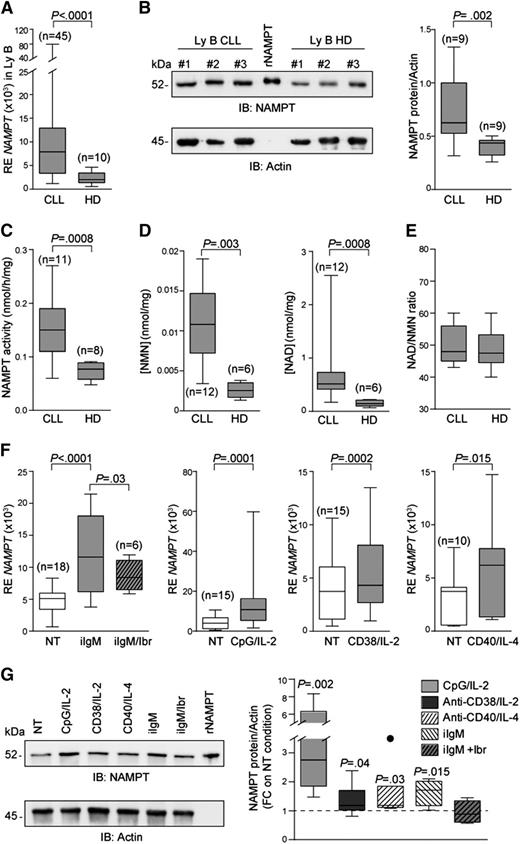
NAMPT messenger RNA (mRNA) and protein expression in lysates of CLL and normal B lymphocytes were comparatively investigated by qRT-PCR and western blot. NAMPT mRNA levels in purified CLL cells were significantly higher than those of normal B lymphocytes obtained from the peripheral blood of age- and sex-matched donors (Figure 1A). Analysis of protein levels in whole cell lysates confirmed higher levels of iNAMPT in purified CLL compared with normal circulating B cells (Figure 1B). Accordingly, the activity of the enzyme was higher in CLL than in normal B lymphocytes (Figure 1C). In keeping with the NAMPT activity profile, CLL cells also contained higher NMN and NAD levels than normal B lymphocytes (Figure 1D). The NAD/NMN ratio was constant and similar in all cells tested (Figure 1E), confirming that the enzyme converting NMN to NAD (ie, NMN adenylyltransferase) is not the rate-limiting enzyme in the nicotinamide-NAD pathway.44
An enzymatically active NAMPT is overexpressed by CLL cells. (A) Box plot showing expression levels of NAMPT mRNA in B lymphocytes from CLL patients (n = 45) or HDs (n = 10). (B) Western blot analysis of NAMPT protein expression in CLL (n = 9) or HD (n = 9) B lymphocytes. A FLAG-tagged rNAMPT was used as internal control. (C-E) Box plots representing NAMPT activity expressed as nmol/hour/mg of protein (C), NMN and NAD intracellular concentration (nmol/mg) (D), and NAD/NMN ratio (E) in B lymphocytes from CLL patients and HDs. (F) qRT-PCR analysis showing expression of NAMPT mRNA in purified CLL lymphocytes cultured with the indicated stimuli (24 hours). When indicated, cells were pretreated with ibrutinib (10 µM, 30 minutes). (G) Purified CLL cells activated as indicated in panel F were lysed, and iNAMPT expression levels determined by western blot. Cumulative results (n = 8) are shown in the box plot.
An enzymatically active NAMPT is overexpressed by CLL cells. (A) Box plot showing expression levels of NAMPT mRNA in B lymphocytes from CLL patients (n = 45) or HDs (n = 10). (B) Western blot analysis of NAMPT protein expression in CLL (n = 9) or HD (n = 9) B lymphocytes. A FLAG-tagged rNAMPT was used as internal control. (C-E) Box plots representing NAMPT activity expressed as nmol/hour/mg of protein (C), NMN and NAD intracellular concentration (nmol/mg) (D), and NAD/NMN ratio (E) in B lymphocytes from CLL patients and HDs. (F) qRT-PCR analysis showing expression of NAMPT mRNA in purified CLL lymphocytes cultured with the indicated stimuli (24 hours). When indicated, cells were pretreated with ibrutinib (10 µM, 30 minutes). (G) Purified CLL cells activated as indicated in panel F were lysed, and iNAMPT expression levels determined by western blot. Cumulative results (n = 8) are shown in the box plot.
When CLL signaling pathways were activated, NAMPT mRNA was significantly upregulated. The increase in NAMPT mRNA could be highlighted after engagement of B-cell receptor (BCR), Toll-like receptor 9 (TLR9), CD38, and CD40 (24 hours, Figure 1F). These signals are known to induce CLL cell activation and CCL3 gene transcription,45,46 as confirmed here (supplemental Figure 1A). Higher levels of NAMPT mRNA were paralleled by increased intracellular protein (Figure 1G). Treatment of activated CLL cells with ibrutinib, a selective bruton tyrosine kinase (BTK) inhibitor, abrogated BCR-, TLR9-, and CD38-induced NAMPT mRNA and protein upregulation (Figure 1F-G and supplemental Figure 1B-C), confirming that BTK is a critical intermediate in these signaling pathways47,48 and linking this kinase with NAMPT transcription. In the conditions used49 (10 μM dose, 24 hours), we detected complete inhibition of BTK phosphorylation, with limited cytotoxicity (supplemental Figure 1D-E).
NAMPT is present in plasma and can be secreted by activated CLL cells
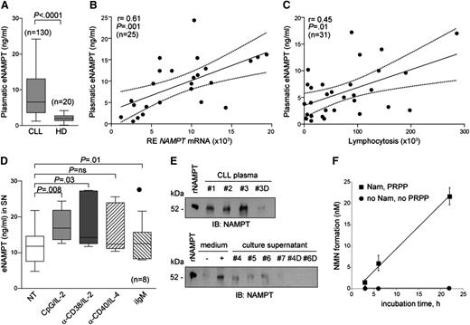
We then asked whether NAMPT protein could also be found in the extracellular environment (eNAMPT). By using an ELISA assay, eNAMPT was detected in the plasma of CLL patients (n = 130) at significantly higher concentrations than in HDs of a comparable age (Figure 2A). The finding of a positive correlation between NAMPT mRNA expression in purified CLL cells and eNAMPT levels in plasma (Figure 2B) suggested that leukemic lymphocytes may contribute to the production of eNAMPT. Consistently, a positive correlation between plasmatic NAMPT and absolute lymphocytosis was observed in CLL patients with comparable percentages of circulating monocytes, known contributors to NAMPT secretion (Figure 2C).50
eNAMPT is present in CLL plasma and can be produced by activated CLL cells. (A) Box plot showing eNAMPT concentrations measured with a quantitative ELISA assay performed on plasma samples from CLL patients (n = 130) or HDs of a comparable age (n = 20). (B) Regression line showing a positive correlation between NAMPT mRNA levels (x-axis) and plasmatic eNAMPT (y-axis) in 25 CLL patients. Pearson coefficient (r) and the corresponding P value are noted. (C) Regression line showing a positive correlation between lymphocytosis (x-axis) and plasmatic eNAMPT (y-axis) in 31 CLL patients. Pearson coefficient (r) and the corresponding P value are noted. (D) ELISA assay showing eNAMPT concentrations in supernatants (SN) from cultures of purified CLL cells cultured with the indicated stimuli (n = 8, 24 hours). (E) The presence of eNAMPT was confirmed by western blot performed on concentrated (×10) albumin-depleted CLL plasma samples (#1-#3) and culture supernatants (#4-#7) from different CLL patients with high (>20 ng/mL, #4-#6) or low (<5 ng/mL, #7) eNAMPT levels, as detected by ELISA assay. The condition (–) and (+) medium indicates concentrated (×10) albumin-depleted RPMI +10% FCS alone (–) or with 20 ng of rNAMPT (+). #3D, #4D, #6D indicate NAMPT-deprived fractions by immunoprecipitation from #3 CLL plasma, or from supernatants #4 and #6. rNAMPT was loaded as control. (F) Time course of eNAMPT activity determined in the plasma of a prototype CLL patient, as described in supplemental Methods.
eNAMPT is present in CLL plasma and can be produced by activated CLL cells. (A) Box plot showing eNAMPT concentrations measured with a quantitative ELISA assay performed on plasma samples from CLL patients (n = 130) or HDs of a comparable age (n = 20). (B) Regression line showing a positive correlation between NAMPT mRNA levels (x-axis) and plasmatic eNAMPT (y-axis) in 25 CLL patients. Pearson coefficient (r) and the corresponding P value are noted. (C) Regression line showing a positive correlation between lymphocytosis (x-axis) and plasmatic eNAMPT (y-axis) in 31 CLL patients. Pearson coefficient (r) and the corresponding P value are noted. (D) ELISA assay showing eNAMPT concentrations in supernatants (SN) from cultures of purified CLL cells cultured with the indicated stimuli (n = 8, 24 hours). (E) The presence of eNAMPT was confirmed by western blot performed on concentrated (×10) albumin-depleted CLL plasma samples (#1-#3) and culture supernatants (#4-#7) from different CLL patients with high (>20 ng/mL, #4-#6) or low (<5 ng/mL, #7) eNAMPT levels, as detected by ELISA assay. The condition (–) and (+) medium indicates concentrated (×10) albumin-depleted RPMI +10% FCS alone (–) or with 20 ng of rNAMPT (+). #3D, #4D, #6D indicate NAMPT-deprived fractions by immunoprecipitation from #3 CLL plasma, or from supernatants #4 and #6. rNAMPT was loaded as control. (F) Time course of eNAMPT activity determined in the plasma of a prototype CLL patient, as described in supplemental Methods.
Culture of CLL cells for 24 hours was followed by the appearance of eNAMPT in spent media (Figure 2D), independently of the number of cells undergoing apoptosis or proliferation, evincing an active rather than a passive mechanism (not shown). Furthermore, after activation of purified CLL lymphocytes through the BCR, TLR9, CD38, or CD40 for 24 hours, eNAMPT levels were invariably increased (Figure 2D). No significant modification in the total number of cells was apparent at this time point. The nature of the protein present in CLL plasma or culture supernatants (Figure 2E and supplemental Figure 2A) was confirmed by western blot, showing a protein compatible with NAMPT selectively in eNAMPT-rich samples. Furthermore, deprivation of eNAMPT by immunoprecipitation was followed by the disappearance of the western blot band (Figure 2E).
These results suggest that eNAMPT could derive, at least partly, from leukemic cells. The generation of eNAMPT from CLL cells is apparent under resting conditions and increases upon activation, arguing in favor of a role for this cytokine/enzyme in the CLL microenvironment.
The critical issue in the field is whether eNAMPT is enzymatically active and whether this activity is relevant in the extracellular compartment.22,51,52 Using an ad hoc devised assay,34 we detected NMN production when CLL plasma was incubated with nicotinamide and PRPP (Figure 2F), with an activity of 1.7 ± 0.1 pmol NMN/hour per mL plasma. However, in the absence of added nicotinamide and PRPP, NAMPT activity was undetectable, in line with previous studies.53 Accordingly, NMN, the product of the reaction, was undetectable in both plasma and media (supplemental Figure 2B).
Treatment of CLL monocytes with NAMPT induces M2 macrophage differentiation
Next we analyzed the functional role of eNAMPT in the CLL microenvironment. Exposure of purified CLL cells to rNAMPT did not result in increased proliferation or chemotaxis, but it significantly enhanced survival of the leukemic cells (supplemental Figure 3A-C). The protective effect may be explained by de novo transcription of TNF-α (supplemental Figure 3D), which is involved in antiapoptotic effects through the activation of B cell activating factor and a proliferation inducing ligand.54,55 Conversely, the increased transcription of TGF-β and of CCL3 (supplemental Figure 3E-F) in response to rNAMPT argued in favor of a “microenvironmental effect” of eNAMPT. This later hypothesis is in agreement with previous data implicating the molecule in the differentiation and activation of monocytes.56-58
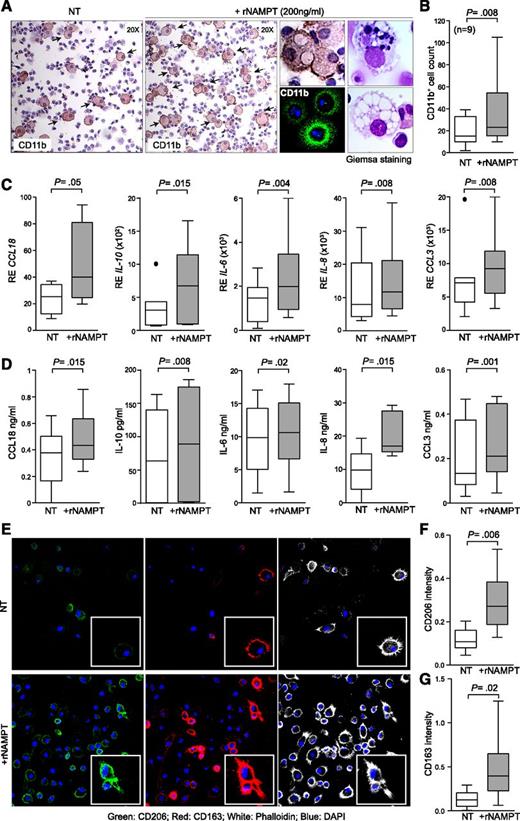
For this reason, we further investigated whether eNAMPT produced by activated leukemic cells modifies the myeloid component. After 5 days of exposure to rNAMPT, PBMCs from normal donors showed a significant increase in CD11b+ macrophages (supplemental Figure 4A-C). This finding was confirmed using PBMCs of CLL patients. Macrophage increases were substantiated by Giemsa staining (highlighting typical intracellular vacuoles and granules) and by CD11b and CD68 staining (Figure 3A-B and supplemental Figure 5A-D).
rNAMPT induces monocytes to differentiate into macrophages with M2 features. (A) PBMC preparations from CLL patients were plated in complete medium with or without rNAMPT (200 ng/mL). After 5 days, cells were fixed and stained for CD11b. Original magnification ×20. Middle panel represents a zoomed area of the same sample. CD11b staining was also confirmed by immunofluorescence. The panels on the right show Giemsa staining to evaluate cellular morphology. (B) Cumulative data showing the number of CD11b+ cells in at least 4 ×20 fields of 9 different samples. (C-D) Box plots reporting the mRNA (C) or the protein (D) expression levels of CCL18, IL-10, IL-6, IL-8 and CCL3, in monocytes sorted from leukemic PBMCs (n = 9) cultured with or without rNAMPT (24 hours). (E) Confocal microscopy analysis of CD163 (red) and CD206 (green) expression in sorted CLL monocytes differentiated for 5 days with or without rNAMPT. Nuclei were counterstained with 4,6-diamidino-2-phenylindole (DAPI; blue); actin filaments were visualized using phalloidin (white). Original magnification ×63. (F-G) Cumulative analysis of CD206 (F) and CD163 (G) pixel intensity (arbitrary units, a.u.), scoring at least 10 different cells for 3 different samples.
rNAMPT induces monocytes to differentiate into macrophages with M2 features. (A) PBMC preparations from CLL patients were plated in complete medium with or without rNAMPT (200 ng/mL). After 5 days, cells were fixed and stained for CD11b. Original magnification ×20. Middle panel represents a zoomed area of the same sample. CD11b staining was also confirmed by immunofluorescence. The panels on the right show Giemsa staining to evaluate cellular morphology. (B) Cumulative data showing the number of CD11b+ cells in at least 4 ×20 fields of 9 different samples. (C-D) Box plots reporting the mRNA (C) or the protein (D) expression levels of CCL18, IL-10, IL-6, IL-8 and CCL3, in monocytes sorted from leukemic PBMCs (n = 9) cultured with or without rNAMPT (24 hours). (E) Confocal microscopy analysis of CD163 (red) and CD206 (green) expression in sorted CLL monocytes differentiated for 5 days with or without rNAMPT. Nuclei were counterstained with 4,6-diamidino-2-phenylindole (DAPI; blue); actin filaments were visualized using phalloidin (white). Original magnification ×63. (F-G) Cumulative analysis of CD206 (F) and CD163 (G) pixel intensity (arbitrary units, a.u.), scoring at least 10 different cells for 3 different samples.
These results suggest the existence of a paracrine circuit, where activated CLL cells produce eNAMPT, that in turn recruits monocytes through CCL3 secretion59 and induces macrophage differentiation. The use of purified monocyte preparations confirmed that eNAMPT acts directly on this cell population (supplemental Figure 5E).
The monocyte subset of CLL patients showed constitutive M2 skewing, based on high interferon regulatory factor 4 (IRF4) transcription factor and low IL-12 levels, considered M2 and M1 markers, respectively (supplemental Figure 6A). Surface expression of the scavenger receptor CD163 and of the mannose receptor CD206, considered M2 markers, strengthened this finding (supplemental Figure 6B).60,61 Treatment of purified CLL monocytes with rNAMPT further enhanced M2 features, as inferred on the basis of (1) a transcriptional profile, which showed induction of CCL18, IL-10, IL-6, IL-8, and CCL3 genes (Figure 3C); (2) evidence of increased concentrations of the same cytokines/chemokines in the supernatants of rNAMPT-treated monocytes (Figure 3D); and (3) increased expression of CD163 and CD206 (Figure 3E-G). Similar effects following rNAMPT treatment were observed when using monocytes from HDs (supplemental Figure 7A-B), suggesting that both HD and CLL monocytes can respond to rNAMPT.
NLCs express high levels of NAMPT
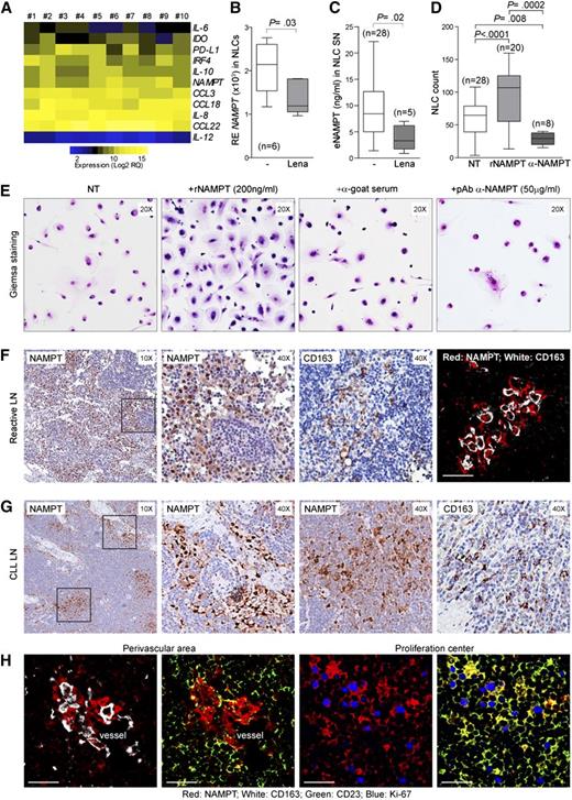
Monocytes from CLL patients spontaneously differentiate in vitro into NLCs, large, multinucleated, adherent cells that protect the leukemic clone from apoptosis.14,16 NLCs are also present in lymphoid tissues where they presumably deliver prosurvival signals to CLL cells.62 In agreement with evidence suggesting that NLCs are closely related to tumor-associated macrophages,11,12,43 they expressed high levels of IRF4 and of the tryptophan-metabolizing enzyme indoleamine 2,3-dioxygenase (IDO; Figure 4A). Their chemokine/cytokine profile was typical of M2 macrophages, including IL-10, IL-6, IL-8, CCL18, CCL22, and CCL3. PD-L1, the ligand for the inhibitory programmed cell death-1 (PD-1) receptor, was also highly expressed. Conversely, IL-12 mRNA, which distinguishes M1 macrophages, was undetectable (Figure 4A), as was expression of the costimulatory molecule CD80 (supplemental Figure 8A).
NAMPT is expressed by NLCs and by myeloid elements in CLL LNs. (A) Heat map showing gene expression profiling performed on conventionally differentiated NLCs (n = 10) highlighted elevated levels of genes classically associated with M2 macrophages, including NAMPT. Expression values are represented as Log2 of relative quantification (RQ) calculated as relative expression on ACTB housekeeping gene. (B-C) Box plot showing NAMPT mRNA expression levels (B) and eNAMPT soluble levels (C) in NLCs from CLL patients obtained using conventional culture conditions (n = 28) or differentiated in the presence of lenalidomide (n = 5). (D-E) NLC numbers (D) and morphology assessed by Giemsa staining (E) were determined in CLL samples differentiated with or without addition of rNAMPT. A blocking pAb anti-NAMPT or control preimmune goat serum were added to conventional NLC cultures to inhibit constitutive eNAMPT. Original magnification ×20 in panel E. (F-G) Immunohistochemical analysis of NAMPT expression in reactive (F) or CLL (G) LN. CD163 was used to detect macrophages in both normal tissues and CLL LN. Images at ×4 and ×20 original magnifications. The immunofluorescence image in panel F shows complete overlap between NAMPT (red) and CD163 (white) staining in reactive LN (magnification ×63). (H) Immunofluorescence images showing partial overlap between NAMPT staining (red) and CD163 (white) in CLL LN samples. Within the proliferation center, NAMPT shows partial colocalization with CD23+/Ki-67+ CLL lymphocytes. Original magnification ×63.
NAMPT is expressed by NLCs and by myeloid elements in CLL LNs. (A) Heat map showing gene expression profiling performed on conventionally differentiated NLCs (n = 10) highlighted elevated levels of genes classically associated with M2 macrophages, including NAMPT. Expression values are represented as Log2 of relative quantification (RQ) calculated as relative expression on ACTB housekeeping gene. (B-C) Box plot showing NAMPT mRNA expression levels (B) and eNAMPT soluble levels (C) in NLCs from CLL patients obtained using conventional culture conditions (n = 28) or differentiated in the presence of lenalidomide (n = 5). (D-E) NLC numbers (D) and morphology assessed by Giemsa staining (E) were determined in CLL samples differentiated with or without addition of rNAMPT. A blocking pAb anti-NAMPT or control preimmune goat serum were added to conventional NLC cultures to inhibit constitutive eNAMPT. Original magnification ×20 in panel E. (F-G) Immunohistochemical analysis of NAMPT expression in reactive (F) or CLL (G) LN. CD163 was used to detect macrophages in both normal tissues and CLL LN. Images at ×4 and ×20 original magnifications. The immunofluorescence image in panel F shows complete overlap between NAMPT (red) and CD163 (white) staining in reactive LN (magnification ×63). (H) Immunofluorescence images showing partial overlap between NAMPT staining (red) and CD163 (white) in CLL LN samples. Within the proliferation center, NAMPT shows partial colocalization with CD23+/Ki-67+ CLL lymphocytes. Original magnification ×63.
In NLC samples, NAMPT mRNA was invariably elevated (Figure 4A-B). Moreover, conditioned media from NLC cultures contained high amounts of eNAMPT (Figure 4C). Differentiation of NLCs from CLL PBMC preparations was significantly increased when rNAMPT was added to the cultures during the differentiation process (Figure 4D). This conclusion was based on (1) morphologic parameters (Figure 4E) and (2) expression of lineage-specific markers such as CD11b and CD68 (supplemental Figure 8B-D), suggesting that NAMPT is important for full NLC differentiation. Confirmation was obtained by deriving NLCs in the presence of a blocking anti-NAMPT goat pAb,33 which significantly decreased the total number of differentiated cells (Figure 4D-E). Furthermore, treatment with the immunomodulatory drug lenalidomide, which partially corrects the M2 phenotype,63 robustly decreased NAMPT mRNA levels and in eNAMPT concentrations in spent media (Figure 4B-C), indicating that NAMPT is part of the M2 signature. As expected, IRF4 expression was significantly downregulated (supplemental Figure 8E).64
NAMPT is expressed in the proliferation centers of CLL LN
In reactive LN samples NAMPT was expressed by residential macrophages (CD11b+/CD68+/CD163+; Figure 4F and supplemental Figure 9A), as inferred both from morphologic parameters and from colocalization between NAMPT and CD163 fluorescent signals (Figure 4F). Accordingly, by qRT-PCR myeloid cells in the peripheral blood express the highest NAMPT levels, both in HD and CLL patients (supplemental Figure 8F). Residential CD68+/CD163+ macrophages clearly stained positive for NAMPT also in CLL LN samples (Figure 4G and supplemental Figure 9B). However, NAMPT+ elements were also present in the paler areas classically associated with proliferation centers (Figure 4G). NAMPT+ cells were a mixture of CD163+ and CD23+ elements, confirming that the leukemic clone can express NAMPT at high levels, as observed in vitro (Figure 4H and supplemental Figure 9C-D). In general, NAMPT+ cells overlapped or were in contact with Ki-67+ cells (Figure 4H and supplemental Figure 9E-F), consistent with our data indicating upregulation of NAMPT in proliferating CLL cells in culture.
Treatment of NLCs with rNAMPT induces chemokine secretion through the activation of NF-κB and STAT3
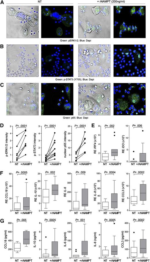
The final phase of the study examined the effects induced by rNAMPT on NLCs. Exposure of conventionally differentiated NLCs to rNAMPT induced a rapid and prominent activation of intracellular signaling. Phosphorylation of extracellular regulated kinase 1/2 (ERK1/2) started a few minutes after rNAMPT exposure, peaked at 20 to 30 minutes, and slowly decreased (Figure 5A,D). Nuclear translocation of phospho–signal transducer and activator of transcription 3 (p-STAT3) and nuclear factor κB (NF-κB) component p65 was also observed with comparable kinetics (Figure 5B-D), consistent with what was reported for murine macrophages22 and a human monocytic cell line.65 Pretreatment with a blocking anti-NAMPT pAb completely inhibited signal transduction (supplemental Figure 10A-D). Furthermore, exposure of NLCs to rNAMPT for 24 hours was followed by enhanced transcription of IRF4, IDO, CCL18, IL-10, IL-6, IL-8, and CCL3 (Figure 5E-F), the same transcripts that were previously found in rNAMPT-treated monocytes (Figure 3C-D). The corresponding cytokines and chemokines were present in media of rNAMPT-treated NLC preparations (Figure 5G). rNAMPT treatment in the presence of specific inhibitors of both NF-κB and STAT3 failed to modulate cytokine transcription, clearly indicating that activation of both pathways is required to drive gene expression in response to NAMPT (supplemental Figure 10E-I).
Exposure of NLCs to rNAMPT activates signal transduction and expression of a panel of genes specifying the M2 phenotype. NLCs were exposed to rNAMPT (30 minutes, 37°C) before fixing, permeabilizing, and staining for phospho-ERK1/2 (A), p-STAT3 (B), and the p65 subunit of the NF-κB complex (C). DAPI (blue) was used to counterstain. Original magnification ×63. (D) Graphs show cumulative analyses plotting the mean values of all the fluorescence measurements for each independent experiment. At least 4 randomly chosen fields from different samples were analyzed after defining a region of interest. (E-F) Conventionally differentiated NLCs were treated with rNAMPT for 24 hours before RNA extraction and qRT-PCR analysis of the expression of IRF4 and IDO (E) or for a panel of chemokines/cytokines defining the M2 phenotype (F). (G) Increased expression of immunosuppressive chemokines/cytokines in cultured supernatants (n = 14) was confirmed at the protein level.
Exposure of NLCs to rNAMPT activates signal transduction and expression of a panel of genes specifying the M2 phenotype. NLCs were exposed to rNAMPT (30 minutes, 37°C) before fixing, permeabilizing, and staining for phospho-ERK1/2 (A), p-STAT3 (B), and the p65 subunit of the NF-κB complex (C). DAPI (blue) was used to counterstain. Original magnification ×63. (D) Graphs show cumulative analyses plotting the mean values of all the fluorescence measurements for each independent experiment. At least 4 randomly chosen fields from different samples were analyzed after defining a region of interest. (E-F) Conventionally differentiated NLCs were treated with rNAMPT for 24 hours before RNA extraction and qRT-PCR analysis of the expression of IRF4 and IDO (E) or for a panel of chemokines/cytokines defining the M2 phenotype (F). (G) Increased expression of immunosuppressive chemokines/cytokines in cultured supernatants (n = 14) was confirmed at the protein level.
These results suggest that eNAMPT enhances M2 features in fully differentiated NLCs as well.
rNAMPT enhances the immunosuppressive features of NLCs
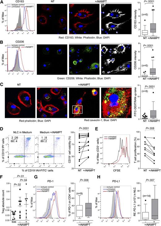
rNAMPT-treated NLCs were characterized by increased expression of the M2 macrophage markers CD163 (Figure 6A) and CD206 (Figure 6B). They were also characterized by increased phagocytosis of FITC-conjugated dextran particles (Figure 6C), which was however independent of CD206, as inferred by using a neutralizing anti-CD206 antibody (supplemental Figure 11).39
Exposure of NLCs to rNAMPT enhances their immunosuppressive function. NLCs (n = 6) were generated in the presence of rNAMPT before assessing expression of CD163 (A) and CD206 (B) by flow cytometry and confocal microscopy. Box plots show cumulative analyses of pixel intensity (a.u.) in at least 4 fields for the different samples. (C) Conventionally obtained NLCs (n = 8) were incubated (15 minutes, 37°C) with FITC-dextran (1 mg/mL in PBS + 5% FCS) with or without rNAMPT. Phagocytosis was confirmed by costaining with caveolin-1 (in red, last 2 panels) and confocal microscopy analysis. Original magnification ×63. (D) Dot plots showing Annexin-FITC (AV) and propidium iodide (PI) staining of CD19+ cells cultured with NLC derived with or without NAMPT for 14 days. The graph represents cumulative data (n = 14). (E) CFSE-labeled, preactivated (anti-CD3/IL-2, 24 hours) autologous PBMCs were cocultured with predifferentiated NLCs (3-5 days with or without rNAMPT). Graph shows cumulative data (n = 8). (F) FACS analysis of basal Treg (CD4+/CD25high/CD127low) expression and after 5 days of coculture (with or without rNAMPT) with autologous NLCs. (G) FACS analysis of PD-1 expression on CD4+ T cells after 5 days of coculture (with or without rNAMPT) with autologous NLCs. The gray histogram represents the isotype control. Box plot represent the cumulative data (n = 9) showing the percentage of PD-1 expression. (H) PD-L1 expression on NLCs treated with rNAMPT for 24 hours was checked by flow cytometry and confirmed by quantitative PCR (n = 14).
Exposure of NLCs to rNAMPT enhances their immunosuppressive function. NLCs (n = 6) were generated in the presence of rNAMPT before assessing expression of CD163 (A) and CD206 (B) by flow cytometry and confocal microscopy. Box plots show cumulative analyses of pixel intensity (a.u.) in at least 4 fields for the different samples. (C) Conventionally obtained NLCs (n = 8) were incubated (15 minutes, 37°C) with FITC-dextran (1 mg/mL in PBS + 5% FCS) with or without rNAMPT. Phagocytosis was confirmed by costaining with caveolin-1 (in red, last 2 panels) and confocal microscopy analysis. Original magnification ×63. (D) Dot plots showing Annexin-FITC (AV) and propidium iodide (PI) staining of CD19+ cells cultured with NLC derived with or without NAMPT for 14 days. The graph represents cumulative data (n = 14). (E) CFSE-labeled, preactivated (anti-CD3/IL-2, 24 hours) autologous PBMCs were cocultured with predifferentiated NLCs (3-5 days with or without rNAMPT). Graph shows cumulative data (n = 8). (F) FACS analysis of basal Treg (CD4+/CD25high/CD127low) expression and after 5 days of coculture (with or without rNAMPT) with autologous NLCs. (G) FACS analysis of PD-1 expression on CD4+ T cells after 5 days of coculture (with or without rNAMPT) with autologous NLCs. The gray histogram represents the isotype control. Box plot represent the cumulative data (n = 9) showing the percentage of PD-1 expression. (H) PD-L1 expression on NLCs treated with rNAMPT for 24 hours was checked by flow cytometry and confirmed by quantitative PCR (n = 14).
NLC generated in the presence of rNAMPT had a greater ability to sustain leukemic cells (Figure 6D). Conversely, when autologous T lymphocytes preactivated using anti-CD3 antibody and IL-2 were cultured with NLCs in the presence of rNAMPT, proliferation was markedly suppressed (Figure 6E). These culture conditions also increased the number of regulatory T cells (Treg’s), as determined on the basis of a CD4+/CD25high/CD127low phenotype (Figure 6F). Lastly, culture of T cells in the presence of rNAMPT-treated NLCs led to the upmodulation of the PD-1/PD-L1 axis (Figure 6G-H).66 Upon total PBMCs coculturing with autologous NLCs in the presence of rNAMPT, CD4+ T lymphocytes showed higher PD-1 expression than when the same cells were cultured with NLCs without rNAMPT (Figure 6G). Consistently, in the presence of rNAMPT, NLCs expressed higher levels of PD-L1 (Figure 6H).
Together, these findings suggest that eNAMPT is among the soluble factors produced by leukemic cells to turn nonneoplastic bystander cells into supporters of tumor growth and survival.
eNAMPT effects are independent of the enzymatic functions of the molecule
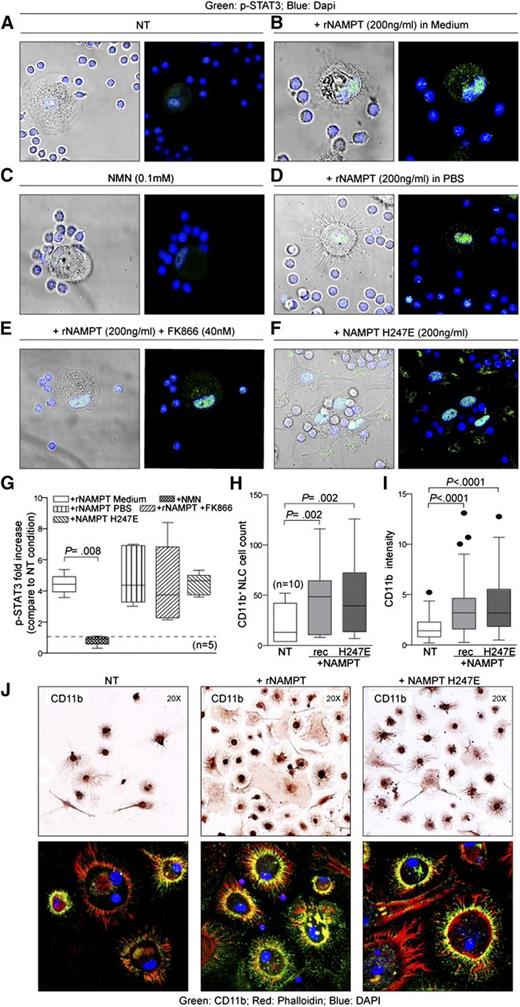
To explore the role of enzymatic activity in modulating eNAMPT effects, we selected STAT3 activation and NLC differentiation as prototypes of short- and long-term rNAMPT effects. Four lines of evidence argue in favor of a nonenzymatic function of eNAMPT in this model. First, exposure of NLCs to extracellular NMN failed to induce STAT3 phosphorylation, suggesting that the product of the reaction is inactive (Figure 7A-C). Second, treatment of NLCs with rNAMPT diluted in PBS, without medium that contains high levels of nicotinamide, the substrate of the reaction, induced prominent activation of STAT3 (Figure 7D). Third, treatment with the NAMPT inhibitor FK866 did not block the ability of rNAMPT to stimulate the phosphorylation of STAT3 (Figure 7E). Fourth, the enzymatically inactive NAMPT H247E mutant67 retained the ability to activate STAT3 phosphorylation (Figure 7F-G).22 Considered together, these results indicate that the enzymatic activity of eNAMPT is dispensable when triggering NLC activation. Similarly, the H247E mutant enhanced NLC differentiation, as determined by morphologic parameters and protein expression (CD11b and CD68; Figure 7H-J and supplemental Figure 12A-B).
eNAMPT activities are independent of the enzymatic activities. (A-F) NLCs were treated (30 minutes) in different conditions and then stained for the presence of p-STAT3 (green fluorescence). DAPI (blue) was used to visualize nuclei. (G) Cumulative data (n = 5) are shown. (H) Box plots representing the quantification of CD11b+ NLCs count (n = 10) and the corresponding fluorescence pixel intensity (I) in the indicated conditions. (J) CD11b staining in NLC preparation by immunocytochemistry (upper panels, original magnification ×20) and by immunofluorescence (green, bottom panels, original magnification ×63).
eNAMPT activities are independent of the enzymatic activities. (A-F) NLCs were treated (30 minutes) in different conditions and then stained for the presence of p-STAT3 (green fluorescence). DAPI (blue) was used to visualize nuclei. (G) Cumulative data (n = 5) are shown. (H) Box plots representing the quantification of CD11b+ NLCs count (n = 10) and the corresponding fluorescence pixel intensity (I) in the indicated conditions. (J) CD11b staining in NLC preparation by immunocytochemistry (upper panels, original magnification ×20) and by immunofluorescence (green, bottom panels, original magnification ×63).
Overall these results indicate that the enzymatic activity of eNAMPT is not essential for activating signaling or inducing long-term effects such as NLC differentiation.
Discussion
This study demonstrates that NAMPT possesses 2 distinct functions in the CLL microenvironment. Inside the cell, it regulates production of NMN from nicotinamide, the limiting step in the generation of NAD. CLL lymphocytes expressed higher levels of iNAMPT and contained higher amounts of NMN and of NAD than circulating normal B lymphocytes from donors of similar ages, a finding consistent with increased NAD needs to allow for continued glycolytic flux.68 Consistently, iNAMPT levels were further upregulated when CLL cells were activated through distinct receptors, suggesting also that NAMPT transcription may be controlled by signaling intermediates common to these pathways, including BTK. The significant difference in NAD levels between CLL cells and normal B lymphocytes was also noted in a previous study using a different method.69 These data are also in line with reports indicating that CLL lymphocytes are particularly sensitive to the NAMPT inhibitor FK866, which induces rapid NAD deprivation.70,70-72
The second main finding of this work is that elevated amounts of eNAMPT are present in the plasma of CLL patients. In the CLL samples examined, eNAMPT levels were significantly higher than those scored by controls, which were in line with the literature.58 After correcting for the number of circulating monocytes, we observed a direct correlation between eNAMPT levels and lymphocytosis, indirectly suggesting that the leukemic cells contribute to eNAMPT production. In agreement with this hypothesis, activation of purified CLL lymphocytes increased the concentrations of eNAMPT, and within LN proliferating CLL cells were clearly NAMPT+.
eNAMPT produced by CLL cells enhanced polarization of circulating monocytes into macrophages with an M2 phenotype. This conclusion was reached after showing (1) expression of nuclear factors and surface markers involved in M2 differentiation, (2) secretion of immunomodulatory cytokines/chemokines, and (3) production of mediators of immune suppression. The use of a blocking anti-NAMPT antibody significantly compromised NLC differentiation and phenotype, arguing for a direct role of the molecule in the acquisition of M2 properties.
NAMPT is also part of the M2 signature, as inferred from the finding that both monocytes and NLCs contained high levels of iNAMPT and could produce eNAMPT. Consistently, the use of lenalidomide to correct M2 polarization of NLCs was followed by a marked decrease of iNAMPT and eNAMPT.
When NLCs were exposed to NAMPT ERK1/2, STAT3 and NF-κB were activated, arguing for a direct effect of this protein on the target cells. The blocking anti-NAMPT antibody completely abrogated pathway activation, in line with a receptor-mediated effect. Culture with rNAMPT also enhanced the immunosuppressive phenotype of NLCs, as well as their ability to sustain CLL survival and suppress autologous T-cell proliferation and Treg expansion. This could be achieved through the production of IL-6 and IL-8 as tumor-promoting cytokines,73,74 while IL-10, CCL18, and IDO could act as immunosuppressors.75
One of the intriguing questions in the field concerns the role of the enzymatic activity in the functions exerted by eNAMPT. Our data indicated that eNAMPT is catalytically active, as expected,76,77 even if NMN, the reaction product, was undetectable both in plasma and in media, confirming previous data.78 This finding, together with the observation that eNAMPT might be unable to catalyze NMN formation in the extracellular space because of the lack of suitable substrate concentrations (particularly PRPP),53 suggests that the functional responses elicited by eNAMPT are independent of its catalytic activity. Four independent lines of evidence, including the use of inactive eNAMPT mutant (H247E),67 are provided to support the conclusion that NAMPT activity is dispensable to activate STAT3 and NF-κB pathways in NLCs and long-term NLC differentiation. This finding leaves open the issue of how eNAMPT may exert its functional activities. Future studies will tell whether there is a NAMPT receptor or whether alternative mechanisms, such as internalization of the enzyme, are in place.
From the translational point of view, an important observation is that drugs interfering with CLL signaling, such as ibrutinib, can suppress NAMPT transcription, reducing both iNAMPT and eNAMPT. Furthermore, NLCs differentiated in the presence of lenalidomide express lower levels of NAMPT, suggesting that drugs that restore immune functions interfere with the production of eNAMPT.
To conclude, we propose that a vicious circle based on CLL cell activation through antigen and accessory signals increases eNAMPT and CCL3 production. CCL3 serves as an attractant for circulating monocytes, which, in the presence of high levels of eNAMPT, differentiate into NLCs, with an enhanced M2 phenotype and functional characteristics, contributing to CLL survival, activation, and proliferation and inhibition of T-cell responses.
The online version of this article contains a data supplement.
The publication costs of this article were defrayed in part by page charge payment. Therefore, and solely to indicate this fact, this article is hereby marked “advertisement” in accordance with 18 USC section 1734.
Acknowledgments
The authors thank M. Lamusta and K. Gizzi for excellent technical support.
This work was supported by grants from the Italian Ministry of Education, University and Research (Futuro in Ricerca 2012 RBFR12D1CB); the Italian Ministry of Health (Bando Giovani Ricercatori 2008 GR-2008-1138053, GR-2010-2317594, and GR-2011-02349282); the Associazione Italiana per la Ricerca sul Cancro AIRC (IG 12754 and Special Program Molecular Clinical Oncology 5 x 1000 No. 10007); and the Fondazione Cariplo (grant 2012-0689). V.A. is supported by an FIRC/AIRC triennial fellowship (#15047).
Authorship
Contribution: V.A. planned and performed research, analyzed data, and helped write the manuscript; S.S., D.B., F.M., F.A., T.V., and R.M. performed research; M.C., D.R., G.G., and G.I. provided blood or tissue samples; T.W., C.W., and J.G.N.G. contributed vital reagents; D.R., G.G., and M.R. analyzed and discussed data; N.R. planned research, analyzed data, and contributed to writing the manuscript; and S.D. designed the study and wrote the manuscript.
Conflict-of-interest disclosure: J.G.N.G. is the founder and president of Aqualung Therapeutics Corp., a nonpublicly traded startup company, and is currently on the Scientific Advisory Board of Lovelace Research Institute. The remaining authors declare no competing financial interests.
Correspondence: Silvia Deaglio, Department of Medical Sciences, University of Turin School of Medicine & Human Genetics Foundation (HuGeF), via Nizza, 52, 10126 Torino, Italy; e-mail: silvia.deaglio@unito.it.

