Abstract
Background: HLA-A allele-lacking leukocytes (HLA-LLs) derived from hematopoietic stem cells (HSCs) with copy-number neutral loss of heterozygosity (LOH) in chromosome 6p (6pLOH) are detected in 13% of patients with acquired aplastic anemia (AA). These defective leukocytes account for more than 90% of granulocytes in some AA patients in remission after immunosuppressive therapy (IST). However, it remains unclear how a few HSC clones with genetic abnormalities sustain hematopoiesis for a long time in AA patients. The fate of 6pLOH(+) HSCs can be easily monitored by revealing HLA-LLs using monoclonal antibodies (mAbs) specific for HLA-A alleles. Phenotypic analyses of hematopoietic progenitor cells (HPCs) in bone marrow (BM) and gene expression profiling of HLA-A allele-lacking HPCs may help to clarify the mechanisms underlying clonal hematopoiesis by 6pLOH(+) HSCs in patients with AA.
Methods: The HLA-A allele expression of BM CD34+ cell subsets, including common myeloid progenitors (CMPs), megakaryocyte/erythroid progenitors (MEPs) and granulocytic/monocyte progenitors (GMPs) in three patients possessing HLA-LLs was determined using mAbs specific for CD34, CD38, CD45RA, CD123 and three different HLA-A alleles. The HLA-A allele-lacking and wild-type CMPs were sorted, and the gene expression levels were compared between the two subsets using an Agilent Whole Human Genome Oligo Microarray (4x44K). The CXCR4 expression by CMPs was compared among different types of BM failure patients.
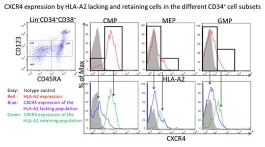
Results: The percentages of HLA-A allele-lacking cells in peripheral blood granulocytes, GMPs, MEPs and CMPs of the three patients were 54.1%/67.3%/63%/1.7%, 98.1%//97.2%/98.8%/4.5% and 97.2%/97.8%/96.9%/12.9%, indicating that the hematopoiesis of these patients was being supported by HLA-A-lacking cells at the CMP level, which accounted for only a small percentage of the total CMPs. To characterize the CMPs capable of supporting hematopoiesis, the gene expression profiles were compared between HLA-A-lacking CMPs that substantially contributed to hematopoiesis and HLA-A-retaining (wild type) CMPs that did not contribute to hematopoiesis. One of the striking differences in the gene expression profile between the two groups was a lower expression of CXCL12 in the HLA-lacking CMPs compared to the wild type CMPs (1 vs. 27.6). Because CD34+ cells are known to negatively regulate themselves by secreting CXCL12, the expression of its receptor, CXCR4, on CMPs was examined using flow cytometry. Surprisingly, all HLA-A-lacking cells were negative for CXCR4, while most of the wild-type cells expressed the CXCL12 receptor (Figure). Examination of the CD34+ cell subsets from 10 healthy individuals showed that the percentages of CXCR4-negative cells were 25.9±8.7% (mean±SD) in CMPs, 98.7±6.4 in MEPs, 98.7±2.1 in GMPs and 77.1±11.5 in CD34+ cells. When the CXCR4 expression levels of CMPs were compared among different types of BM failure, the mean fluorescence intensity (MFI) of CXCR4 in the CMPs from four patients with AA (550±76, mean±SD) was significantly lower than that in three patients with MDS (901±332) and 10 healthy individuals (1440±229).
Conclusions: Hematopoiesis in patients with AA is supported by a limited number of CXCR4(-) cells at the CMP level. Decreased CXCR4 expression by CMPs of AA patients may represent immune pressure applied to HSCs that elicits the participation of dormant CMPs in hematopoiesis.
No relevant conflicts of interest to declare.
Author notes
Asterisk with author names denotes non-ASH members.


This feature is available to Subscribers Only
Sign In or Create an Account Close Modal