Abstract
Autoimmune lymphoproliferative syndrome (ALPS) is classically defined as a disease with defective FAS-mediated apoptosis (type I-III). Germline NRAS mutation was recently identified in type IV ALPS. We report 2 cases with ALPS-like disease with somatic KRAS mutation. Both cases were characterized by prominent autoimmune cytopenia and lymphoadenopathy/splenomegaly. These patients did not satisfy the diagnostic criteria for ALPS or juvenile myelomonocytic leukemia and are probably defined as a new disease entity of RAS-associated ALPS-like disease (RALD).
Introduction
Autoimmune lymphoproliferative syndrome (ALPS) is a disease characterized by dysfunction of the FAS-mediated apoptotic pathway,1,2 currently categorized as: type Ia, germline TNFRSF6/FAS mutation; type Ib, germline FAS ligand mutation; type Is, somatic TNFRSF6/FAS mutation; and type II, germline Caspase 10 mutation. Patients exhibit lymphadenopathy, hepatosplenomegaly, and autoimmune diseases, such as immune cytopenia and hyper-γ-globulinemia. An additional subclassification has been proposed that includes types III and IV, whereby type III has been defined as that with no known mutation but with a defect in FAS-mediated apoptosis and type IV as one showing germline NRAS mutation.3 Type IV is considered exceptional because the FAS-dependent apoptosis pathway is not involved in the pathogenesis, and this subclass is characterized by a resistance to interleukin-2 (IL-2) depletion-dependent apoptosis. Recent updated criteria and classification of ALPS suggested type IV ALPS as a RAS-associated leukoproliferative disease.4
Juvenile myelomonocytic leukemia (JMML) is a chronic leukemia in children. Patients show lymphadenopathy, hepatosplenomegaly, leukocytosis associated with monocytosis, anemia, thrombocytopenia, and occasional autoimmune phenotypes. Approximately 80% of patients with JMML have been shown to have a genetic abnormality in their leukemia cells, including mutations of NF1, RAS family,5 CBL, or PTPN11. The hallmarks of the laboratory findings of JMML include spontaneous colony formation in bone marrow (BM) or peripheral blood mononuclear cells (MNCs) and hypersensitivity to granulocyte-macrophage colony-stimulating factor (GM-CSF) of CD34+ BM-MNCs.6
Germline RAS pathway mutations cause Costello (HRAS), Noonan (PTPN11, KRAS, and SOS1), and cardio-facio-cutaneous syndromes (KRAS, BRAF, MEK1, and MEK2). Patients with Costello and Noonan syndromes have an increased propensity to develop solid and hematopoietic tumors, respectively7 ; among these tumors, the incidence of JMML in patients with germline mutation of NF1 or PTPN11 is well known.
We present 2 cases with autoimmune cytopenia and remarkable lymphadenopathy and hepatosplenomegaly, both of which were identified as having a somatic KRAS G13D mutation without any clinical features of germline RAS mutation, such as cardio-facio-cutaneous or Noonan syndrome.
Methods
All studies were approved by the ethical board of Tokyo Medical and Dental University.
Case 1
A 9-month-old boy had enormous bilateral cervical lymphadenopathy and hepatosplenomegaly (supplemental Figure 1A-B, available on the Blood Web site; see the Supplemental Materials link at the top of the online article). Blood test revealed the presence of hemolytic anemia and autoimmune thrombocytopenia. Hyper-γ-globulinemia with various autoantibodies was also noted. ALPS and JMML were nominated as the diseases to be differentially diagnosed. Detailed clinical history and laboratory data are provided as Supplemental data. The patient did not satisfy the criteria for the diagnosis of ALPS or JMML as discussed in “Results and discussion.”
Case 2
A 5-month-old girl had a fever and massive hepatosplenomegaly (supplemental Figure 1D). She was initially diagnosed with Evans syndrome based on the presence of hemolytic anemia and autoimmune thrombocytopenia with hyper-γ-globulinemia and autoantibodies. Spontaneous colony formation assay and GM-CSF hypersensitivity of BM-MNCs showed positivity. Then, tentative diagnosis of JMML was given, even though she showed no massive monocytosis or increased fetal hemoglobin. Detailed clinical history and laboratory data are provided in supplemental data.
Detailed methods for experiments are described in supplemental data.
Results and discussion
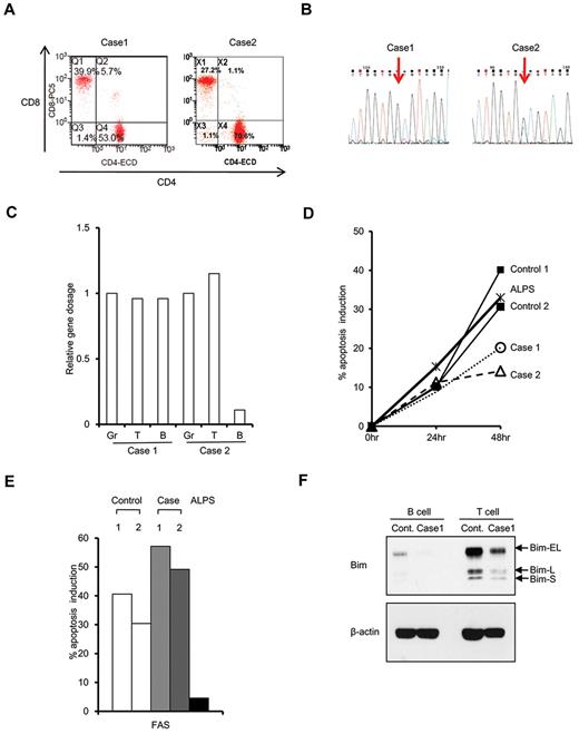
Case 1 showed a high likelihood of being a case of ALPS according to the symptoms and clinical data presented (supplemental Table 1), except for number of double-negative T cells, which was only 1.4% of T-cell receptor-αβ cells (Figure 1A). JMML was also nominated as a disease to be differentiated because remarkable hepatosplenomegaly with thrombocythemia and moderate monocytosis was noted. However, no hypersensitivity to GM-CSF as determined by colony formation assay for BM-MNCs (data not shown) or phosphor-STAT5 staining (data not shown) was observed. DNA sequence for JMML-associated genes, such as NRAS, KRAS, HRAS, PTPN11, and CBL, was determined, and KRAS G13D mutation was identified (Figure 1B). The mutation was seen exclusively in the hematopoietic cell lineage, and no mutation was seen in the oral mucosa or nail-derived DNA. Granulocytes, monocytes, T cells, and B cells were all positive for KRAS G13D mutation (data not shown). The proportion of mutated cells in each hematopoietic lineage was quantitated by mutation allele-specific quantitative polymerase chain reaction methods, which revealed that mutated allele was almost equally present in granulocytes, T cells, and B cells (Figure 1C). CD34+ hematopoietic stem cells (HSCs) were also positive for KRAS G13D mutation, and 60% of colony-forming units-granulocyte macrophage (CFU-GM) developed from isolated CD34 cells carried the KRAS G13D mutation (data not shown). These observations suggest that the mutation occurred at the HSCs level, and HSC consists of wild-type and mutant HSCs.
Molecular cell biologic assay of RALD. (A) Flow cytometric analysis of double-negative T cells. CD8 and CD4 double staining was performed in T-cell receptor-αβ-expressing cells. (B) Electropherogram showing KRAS G13D mutation in BM-MNCs in case 1 (left panel) and case 2 (right panel). (C) Gene dosage of mutated allele in granulocytes (Gr), T cells (T), and B cells (B). Relative gene dosage was estimated by a mutant allele-specific polymerase chain reaction method in cases 1 and 2 using albumin gene as internal control. (D) Apoptosis assay using activated T cells. Apoptosis percentage was measured by flow cytometry with annexin V staining 24 and 48 hours after IL-2 depletion. (E) Apoptosis percentage was measured 24 hours after addition of anti-FAS CH11 antibody (final 100 ng/mL). (F) Western blotting analysis of Bim expression.
Molecular cell biologic assay of RALD. (A) Flow cytometric analysis of double-negative T cells. CD8 and CD4 double staining was performed in T-cell receptor-αβ-expressing cells. (B) Electropherogram showing KRAS G13D mutation in BM-MNCs in case 1 (left panel) and case 2 (right panel). (C) Gene dosage of mutated allele in granulocytes (Gr), T cells (T), and B cells (B). Relative gene dosage was estimated by a mutant allele-specific polymerase chain reaction method in cases 1 and 2 using albumin gene as internal control. (D) Apoptosis assay using activated T cells. Apoptosis percentage was measured by flow cytometry with annexin V staining 24 and 48 hours after IL-2 depletion. (E) Apoptosis percentage was measured 24 hours after addition of anti-FAS CH11 antibody (final 100 ng/mL). (F) Western blotting analysis of Bim expression.
NRAS-mutated type IV ALPS was previously characterized by apoptosis resistance of T cells in IL-2 depletion.3 Then, activated T cells were subjected to an apoptosis assay by FAS stimulation or IL-2 depletion. Remarkable resistance to IL-2 depletion, but not to FAS-dependent apoptosis (Figure 1D-E), was seen. This was in contrast to T cells from FAS-mutated ALPS type 1a, which showed remarkable resistance to FAS-dependent apoptosis and normal apoptosis induction by IL-2 withdrawal (Figure 1D-E). Western blotting analysis of activated T cells or Epstein-Barr virus-transformed B cells showed reduced expression of Bim (Figure 1F).
In case 2, autoimmune phenotype and hepatosplenomegaly were remarkable, as shown in Supplemental data. The patient was initially diagnosed as Evans syndrome based on the presence of hemolytic anemia and autoimmune thrombocytopenia. Double-negative T cells were 1.1% of T-cell receptor-αβ cells in the peripheral blood, which did not meet with the criteria of ALPS. Although spontaneous colony formation was shown in peripheral blood- and BM-MNCs, and GM-CSF hypersensitivity was demonstrated in BM-MNCs derived CD34+ cell (supplemental Table 2), she showed no massive monocytosis or increased fetal hemoglobin. Thus, the diagnosis was less likely to be ALPS or JMML. DNA sequencing of JMML-related genes, such as NRAS, KRAS, HRAS, PTPN11, and CBL, identified somatic, but not germline, KRAS G13D mutation (Figure 1B). KRAS G13D mutation was detected in granulocytes and T cells. Mutation was not identified in B cells by conventional DNA sequencing (data not shown). Mutant allele-specific quantitative polymerase chain reaction revealed that mutated allele was almost equally present in granulocytes and T cells, but barely in B cells (Figure 1C). Activated T cells showed resistance to IL-2 depletion but not to FAS-dependent apoptosis (Figure 1D-E).
Both of our cases were characterized by strong autoimmunity, immune cytopenia, and lymphadenopathy or hepatosplenomegaly with partial similarity with ALPS or JMML. However, they did not meet with the well-defined diagnostic criteria of ALPS2 or JMML.6 It is interesting that case 2 presented GM-CSF hypersensitivity, which is one of the hallmarks of JMML. Given the strict clinical and laboratory criteria of JMML and ALPS, our 2 cases should be defined as a new disease entity, such as RAS-associated ALPS-like disease (RALD). Recently defined NRAS-mutated ALPS type IV may also be included in a similar disease entity.
There are several cases of JMML reported simultaneously having clinical and laboratory findings compatible with autoimmune disease.8,9 Autoimmune syndromes are occasionally seen in patients with myelodysplastic syndromes, including chronic myelomonocytic leukemia.10 These previous findings may suggest a close relationship of autoimmune disease and JMML. Because KRAS G13D has been identified in JMML,11-13 it is tempting to speculate that KRAS G13D mutation is involved in JMML as well as RALD. In JMML, erythroid cells reportedly carry mutant RAS, whereas B- and T-cell involvement was variable.13 In both of our cases, myeloid cells and T cells carried mutant RAS, whereas B cells were affected variably. These findings would support a hypothesis that the clinical and hematologic features are related to the differentiation stages of HSCs where RAS mutation is acquired. JMML-like myelomonocytic proliferation may predict an involvement of RAS mutation in myeloid stem/precursor cell level, whereas ALPS-like phenotype may predict that of stem/precursor cells of lymphoid lineage, especially of T cells. Under the light of subtle differences between the 2 cases presented, their hematologic and clinical features may reflect the characteristics of the stem cell level where KRAS mutation is acquired. Involvement of the precursors with higher propensity toward lymphoid lineage may lead to autoimmune phenotypes, whereas involvement of those with propensity toward the myeloid lineage may lead to GM-CSF hypersensitivity while still sharing some overlapping autoimmune characteristics.
One may argue from the other viewpoints with regard to the clinicopathologic features of these disorders. First, transformation in fetal HSCs might be obligatory for the development of JMML14 and, in HSCs later in life, may not have the same consequences. Second, certain KRAS mutations may be more potent than others. Codon 13 mutations are generally less deleterious biochemically than codon 12 substitutions, and patients with JMML with codon 13 mutations have been reported to show spontaneous hematologic improvement.12,15 Thus, further studies are needed to reveal in-depth clinicopathologic characteristics in this type of lympho-myeloproliferative disorder.
KRAS mutation may initiate the oncogenic pathway as one of the first genetic hits but is insufficient to cause frank malignancy by itself.16,17 Considering recent findings that additional mutations of the genes involved in DNA repair, cell cycle arrest, and apoptosis are required for full malignant transformation, one can argue that RALD patients will also develop malignancies during the course of the disease. Occasional association of myeloid blast crisis in JMML and that of lymphoid malignancies in ALPS will support this notion. Thus, the 2 patients are now being followed up carefully. It was recently revealed that half of the patients diagnosed with Evans syndrome, an autoimmune disease presenting with hemolytic anemia and thrombocytopenia, met the criteria for ALPS diagnosis.18,19 In this study, FAS-mediated apoptosis analysis was used for the screening. Considering the cases we presented, it will be intriguing to reevaluate Evans syndrome by IL-2 depletion-dependent apoptosis assay focusing on the overlapping autoimmunity with RALD.
An Inside Blood analysis of this article appears at the front of this issue.
The online version of this article contains a data supplement.
The publication costs of this article were defrayed in part by page charge payment. Therefore, and solely to indicate this fact, this article is hereby marked “advertisement” in accordance with 18 USC section 1734.
Acknowledgments
This work was supported by the Ministry of Education, Science, and Culture of Japan (Grant-in-Aid 20390302; S.M.) and the Ministry of Health, Labor and Welfare of Japan (Grant-in-Aid for Cancer Research 20-4 and 19-9; S.M., Masatoshi Takagi).
Authorship
Contribution: Masatoshi Takagi and S.M. designed entire experiments and wrote the manuscript; K.S., N.M., and Mari Takagi treated patients and designed clinical laboratory test; J.P. performed experiments described in Figure 1B-F; K.M., H.M., and S.D. performed colony and mutational analysis; and M.N., T.M., K.K., S.K., Y.K., and A.T. supervised clinical and immunologic experiments or coordinated clinical information.
Conflict-of-interest disclosure: The authors declare no competing financial interests.
Correspondence: Masatoshi Takagi, Department of Pediatrics and Developmental Biology, Graduate School of Medicine, Tokyo Medical and Dental University, 1-5-45 Yushima, Bunkyo-ku, Tokyo, 113-8519, Japan; e-mail: m.takagi.ped@tmd.ac.jp; and Shuki Mizutani, Department of Pediatrics and Developmental Biology, Graduate School of Medicine, Tokyo Medical and Dental University, 1-5-45 Yushima, Bunkyo-ku, Tokyo, 113-8519, Japan; e-mail: smizutani.ped@tmd.ac.jp.

