Abstract
Receptor usage by viral interleukin-6 (vIL-6), a virokine encoded by Kaposi sarcoma– associated herpesvirus, is an issue of controversy. Recently, the crystal structure of vIL-6 identified vIL-6 sites II and III as directly binding to glycoprotein (gp)130, the common signal transducer for the IL-6 family of cytokines. Site I of vIL-6, however, comprising the outward helical face of vIL-6, where human IL-6 (hIL-6) would interact with the specific α-chain IL-6 receptor (IL-6R), is accessible and not occupied by gp130. This study examined whether this unused vIL-6 surface is available for IL-6R binding. By enzyme-linked immunosorbent assay, vIL-6 bound to soluble gp130 (sgp130) but not to soluble IL-6R (sIL-6R). Using plasmon surface resonance, vIL-6 bound to sgp130 with a dissociation constant of 2.5 μM, corresponding to 1000-fold lower affinity than that of hIL-6/sIL-6R complex for gp130. sIL-6R neither bound to vIL-6 nor affected vIL-6 binding to gp130. In bioassays, vIL-6 activity was neutralized by 4 monoclonal antibodies (mAbs) recognizing a domain within vIL-6 site I, mapped to the C-terminal part of the AB-loop and the beginning of helix B. The homologous region in hIL-6 participates in site I binding to IL-6R. In addition, binding of vIL-6 to sgp130 was interfered with specifically by the 4 neutralizing anti–vIL-6 mAbs. Based on the vIL-6 crystal structure, the vIL-6 neutralizing mAbs map outside the binding interface to gp130, suggesting that they either produce allosteric changes or block necessary conformational changes in vIL-6 preceding its binding to gp130. These results document that vIL-6 does not bind IL-6R and suggest that conformational change may be critical to vIL-6 function.
Introduction
Kaposi sarcoma (KS)–associated herpesvirus (KSHV; also called human herpesvirus 8 or HHV-8) is a newly described oncogenic herpesvirus that is regularly detected in KS lesions from human immunodeficiency virus (HIV)–infected and noninfected individuals, primary effusion lymphoma (PEL), and a proportion of patients with Castleman disease.1 KSHV encodes various proteins that have features suggesting their roles in promoting cellular growth and transformation, including viral homologues of cyclin D, G-protein–coupled receptor, interferon regulatory factor, macrophage inflammatory proteins, and interleukin-6 (IL-6).2 All these viral proteins display structural similarities to their cellular counterparts. KSHV viral IL-6 (vIL-6), encoded at open reading frame (ORF) K2, shares 24.8% amino acid sequence identity (49.7% similarity) with human IL-6 (hIL-6) and 24.2% identity (47.9% similarity) to murine IL-6 (mIL-6).3-5 As a multifunctional cytokine that acts on a wide variety of cell types, hIL-6 has been implicated in the pathogenesis of several diseases, including rheumatoid arthritis and multiple myeloma as well as disorders related to acquired immunodeficiency syndrome (AIDS).6-8 The IL-6 family of cytokines exerts its activities via receptor complexes that contain at least one subunit of the signal transducing protein gp130.6 The extracellular component of gp130 consists of an N-terminal immunoglobulin (Ig)-like domain (D1) followed by a cytokine-binding module (D2D3) and 3 additional fibronectin type III–like domains (D4D5D6).9,10 This receptor is activated by the family of IL-6–type cytokines, which includes IL-6, oncostatin M (OSM), leukemia inhibitory factor (LIF), IL-11, ciliary neurotrophic factor, and cardiotrophin-1, which exert many overlapping biologic activities.6 Cell stimulation by any member of the IL-6 family cytokines triggers homodimerization or heterodimerization of gp130. The dimerization of gp130 leads to activation of associated Janus kinases and subsequent modification of signal transducers and activators of transcription (STAT).11,12 In addition to gp130, the high-affinity, signaling receptor complex for hIL-6 contains a specific α-subunit (IL-6R). hIL-6 binds to IL-6R, and the IL-6/IL-6R complex then associates with gp130, allowing it to homodimerize.6 The crystal structure of IL-6 has identified an antiparallel 4-helix bundle (A, B, C, and D) with a topology common to a number of other cytokines in the superfamily.13 Extensive studies, including mutagenesis and mapping epitopes of function-blocking or activating antibody, have demonstrated that the contact points of hIL-6 with its receptor complex are mediated by 3 distinct sites named I, II, and III.14-17 Site I, formed by the C-terminal part of helix D and in part by the AB-loop, interacts with IL-6R. Site II, formed by a limited number of exposed residues on helix A and helix C, and site III, formed by residues at the beginning of helix D spatially flanked by residues in the initial part of the AB-loop, bind to gp130.13
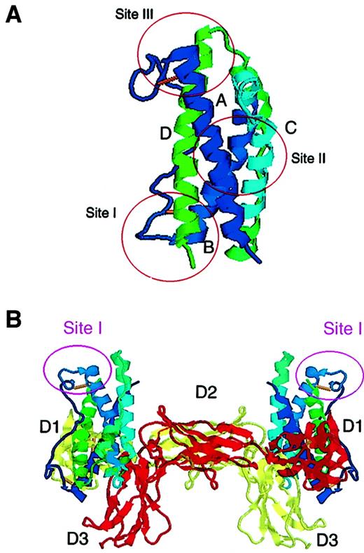
Despite its limited sequence homology, vIL-6 displays many biologic functions of hIL-6.8 Like cellular IL-6, vIL-6 can stimulate the growth of certain factor-dependent cells, can promote hematopoiesis, can act as an angiogenic factor by inducing vascular endothelial growth factor (VEGF),18 and can activate Janus kinase 1, STAT1, and STAT3.19 Compared with hIL-6, however, vIL-6 requires approximately 1000-fold larger amounts of protein for maximal proliferation of IL-6–dependent cell lines.20,21 Recently, the crystal structure of vIL-6 has revealed that vIL-6 shares with other members of the IL-6 family the canonical up-up-down-down, ABCD 4-helix bundle connected by peptide loops22 (Figure 1A). In that study, a soluble tetrameric complex (2:2) of vIL-6 and the 3 NH2-terminal domains (D1D2D3) of gp130 was crystallized in the absence of IL-6R. Based on this vIL-6–gp130 crystal complex, the unused site I face of vIL-6, where hIL-6 would interact with IL-6R, is not occupied by gp130 and is sterically accessible for engagement by other molecules22 (Figure1B). In cell culture systems, the interaction of vIL-6 with the IL-6R chains gp130/IL-6R has been uncertain.4,19,20,23-25 The participation of IL-6R in vIL-6 binding was first deduced from experiments using the IL-6–dependent murine B9 cells,4followed by 2 additional reports from the same group that cotransfection assays involving IL-6R expression vector up-regulated vIL-6 responses in vitro.23,26 Another group reported that anti–IL-6R antibody enhanced the inhibitory effect of anti–gp130 antibody in vIL-6–mediated DNA synthesis by INA-6 human myeloma cells.20 Supportive evidence for a role of IL-6R in mediating vIL-6 function derived from experiments in which anti–IL-6R antibody abrogated vIL-6–induced HIV-1 p24 production in a human monocytic cell line.24 However, other studies showed that IL-6R was not used by vIL-6 in HepG2 human hepatoma cells.19 Further, murine pro-B cells expressing transfected human gp130 but not IL-6R exhibited a strong response to vIL-6 but not to hIL-6.25 Thus, there seems to be a consensus that gp130 is used for vIL-6 signaling, but the usage of IL-6R is uncertain. In this report, we have defined vIL-6 interactions with the IL-6R/gp130 using surface plasmon resonance and a panel of newly generated monoclonal antibodies (mAbs) that recognize the putative vIL-6 site I where hIL-6 would interact with IL-6R.
Schematic cartoon representation of vIL-6 structure.
(A) vIL-6 ABCD 4- helix bundle connected by peptide loops is displayed in different colors. Site I, composed of the N-terminal region of the AB-loop and the C-terminal region of helix D, identifies the epitope recognized by the vIL-6 neutralizing antibodies described here, and corresponds to the presumed site where hIL-6 would interact with IL-6R. Site II on helix A and C, and site III on the initial part of the AB-loop and helix D represent binding surfaces to gp130. (B) vIL-6 is juxtaposed to gp130 in a tetrameric (2:2) signaling model based on the crystal structure of the complex.22 vIL-6 site II is occupied by the D2D3 sites of one gp130 chain (yellow), and site III is occupied by the D1 site of another gp130 chain (red). Site I of vIL-6, comprising the outward helical face, is not occupied by gp130 and is sterically accessible for engagement by other molecules.
Materials and methods
Preparation of recombinant vIL-6 and its deletion mutants
Recombinant vIL-6 was prepared as a fusion protein that has a factor Xa cleavage site between amino terminal tag of maltose-binding protein (MBP) and amino acids 22 to 204 of vIL-6 (MBPvIL-6).18 Deletion mutants of vIL-6 were prepared as follows: vIL-6 M1 and M2 were constructed by inserting a stop codon at positions 37 and 81, respectively. After denaturing, pMALvIL-6 was annealed with the mutagenesis oligonucleotides (M1: ATTTAGATCTTCAATTGGATGCTA; M2: ACTTAGATCTGCGGGTTAATAGGA) that encode a restriction enzyme site for BglII as well as stop codons, and mutant strands were synthesized using the GeneEditor in vitro site-directed mutagenesis system (Promega, Madison, WI). After transformation, positive colonies were screened by GeneEditor andBglII digestion. vIL-6 M3 and M5 were constructed by restriction enzyme digestion of pMALvIL-6 with AatII and EcoRI, respectively, followed by ligation. To construct expression vectors for vIL-6 M4, M6, and M7, C-terminal fragments of vIL-6 were amplified using oligonucleotide primers (M4-BamHI: ACTGGATCCCTTAAAAAGCTCGCCGA T; M6-BamHI: TTTGGATCCTTAACGACGGAGTTTGGA; M7-BamHI: ACGGGATCCAGTCCACCCAA ATTTGAC) and vIL-6-3′-Hind (CCCAAGCTTATTACTTATCGTGGACGT). After digestion with BamHI and HindIII, the polymerase chain reaction products were ligated into pMAL-c2 (New England Biolabs, Beverly, MA). All recombinant protein–expressing clones were analyzed by DNA sequence analysis and expressed in Escherichia coli strain DH5α (Life Technologies, Gaithersburg, MD). Large-scale production and affinity purification of fusion proteins were performed according to the manufacturer's instructions.
Generation of mAbs against vIL-6
To generate mAbs against vIL-6, mice were immunized with MBPvIL-6. After boosting, splenocytes were obtained for hybridoma production by standard procedures. Hybridomas were screened by enzyme-linked immunosorbent assay (ELISA) and Western blotting against recombinant vIL-6. Following bulk culture, mAbs were purified from culture supernatants using protein G columns (Pierce, Rockford, IL). The isotype of each mAb was determined using the Mouse Typer sub-isotyping kit (Bio-Rad, Hercules, CA), according to the manufacturer's instructions.
Immunoblotting of vIL-6
Immunoblotting of vIL-6 was performed as described previously.27 Briefly, 100 ng MBPvIL-6 cleaved with factor Xa (New England Biolabs), recombinant hIL-6 (a kind gift from Sandoz Pharmaceuticals), and recombinant mIL-6 (Peprotech, Rocky Hill, NJ) were loaded into each well of 10% to 20% tricine-sodium dodecyl sulfate gel (NOVEX, San Diego, CA) after boiling for 10 minutes. The separated proteins were transferred onto polyvinylidene difluoride membranes (Immobilon-P; Millipore, Bedford, MA). Immunostaining was performed using rabbit polyclonal antibodies against MBPvIL-6,18 mIL-6 (Peprotech), or mouse anti–vIL-6 mAbs followed by incubation with horseradish peroxidase (HRP)–conjugated antimouse or rabbit IgG antibodies (Amersham, Piscataway, NJ). Immunocomplexes were visualized using the chemiluminescence detection system (Amersham).
Mapping the epitopes recognized by mAbs
Full-length and deletion mutants of vIL-6 fusion proteins were immobilized onto 96-well plates (Immulon 4HBX; Dynex Technologies, Chantilly, VA) in phosphate-buffered saline (PBS) by overnight incubation at 4°C. After blocking nonspecific binding with SuperBlock (Pierce), a standard ELISA protocol was followed,28 using appropriate dilutions of anti–vIL-6 antibody and a 1:500 dilution of goat antimouse polyvalent Ig conjugated to alkaline phosphatase (Sigma, St Louis, MO). Reactions were visualized by usingP-nitrophenyl phosphate (Sigma), and plates were read at 405 nm with λ correction at 550 nm using a microplate reader.
ELISA-based binding assay
The binding of vIL-6 to IL-6R/gp130 was analyzed by ELISA as described previously with some modifications.29 Purified recombinant human soluble IL-6R (sIL-6R; Endogen, Woburn, MA) and human soluble gp130 (sgp130; R & D Systems, Minneapolis, MN) were immobilized on ELISA plate wells (Immunol 4HBX) at 5 μg/mL in PBS. After blocking the plate with SuperBlock, MBPvIL-6 or hIL-6 was applied at 50 ng/mL in 1% bovine serum albumin (BSA)/PBS, and incubated for 5 hours at room temperature. Bound protein was detected with polyclonal rabbit antibodies directed against vIL-6 or hIL-6 (Peprotech) at 1 μg/mL, followed by an HRP-conjugated antirabbit IgG (Bio-Rad) at 1:3000 in PBS/0.05% Tween 20. Reactions were visualized by using tetramethoxybenzene peroxidase substrate (Kirkegaard & Perry Laboratories, Gaithersburg, MD), followed by 2 N H2SO4. The interference of vIL-6 binding to sgp130 by mAbs was analyzed in a similar manner. Biotinylated MBPvIL-621 was incubated at 50 ng/mL in 1% BSA/PBS with anti–vIL-6 mAbs or isotype control murine IgG1 (20 μg/mL) overnight at 4°C. The mixture was applied to the plates coated with sgp130 (2 μg/mL) after SuperBlock treatment. The protein complex was detected by streptavidin-HRP (1:1000; Kirkegaard & Perry Laboratories), and the peroxidase activity was visualized by tetramethoxybenzene peroxidase substrate, followed by 2 N H2SO4. Plates were read at 450 nm with λ correction at 630 nm using a microplate reader.
Surface plasmon resonance
Biospecific interaction analysis was monitored by surface plasmon resonance using BIAcore 2000 system (Biacore, Uppsala, Sweden). The sgp130-Fc (R & D Systems), a fusion protein of sgp130 and human IgG Fc, was immobilized onto the CM5 (carboxymethylated dextran matrix) sensor chip using the amine coupling kit (Biacore), and unreactive groups on the chip were blocked by ethanolamine according to the manufacturer's instructions. A continuous flow (10 μL/mL) of hIL-6 or MBPvIL-6 onto immobilized sgp130-Fc was monitored by passing the analytes across the sensor chip. In parallel experiments, sIL-6R was incubated with hIL-6 and MBPvIL-6 prior to assay. In comparative experiments, a continuous flow (20 μL/mL) of sIL-6R onto immobilized hIL-6 or MBPvIL-6 was monitored in the same manner. The sensor surface was regenerated between assays by 30 seconds of treatment with 10 mM glycine pH 2.2. BIAevaluation 3.0 software (Biacore) was used for all the interaction analyses. The kinetics parameter was calculated according to a 1:1 Langmuir binding model (A + B↔AB) by direct fitting of ligand binding sensorgrams at multiple concentrations. The dissociation equilibrium constant is defined as dissociation constant(Kd) = dissociation rate constant(kd)/association rate constant(ka). The Kd was also determined by Scatchard analysis of equilibrium-state data obtained by a continuous flow (1 μL/mL) of MBPvIL-6 (50-800 μg/mL) onto immobilized sgp130-Fc, according to previously published methods.30
vIL-6 bioassays
The B9 cell proliferation assay was performed essentially as described.31 B9 cells (2 × 10³ cells/well) were incubated in 96-well plates (200 μL/well) with MBPvIL-6 (100 ng/mL) and anti–vIL-6 mAbs (0-10 μg/mL) for 72 hours at 37°C, including a 6-hour terminal pulse with 1 μCi/well of [3H]-thymidine (Amersham, Arlington Heights, IL). Proliferation assays using BAF-130 cells, representing murine BAF-B03 cells expressing transfected human gp130, was performed as described.32 BAF-130 cells were stimulated in 96-well plates (200 μL/well) by MBPvIL-6 or by hIL-6/sIL-6R in the presence or absence of panels of anti–gp130 mAbs (B-R3, B-P8, or B-P4; Cell Sciences, Norwood, MA) for 48 hours at 37°C, including a 6-hour terminal pulse with 1 μCi (0.037 MBq)/well of [3H]-thymidine. [3H]-thymidine incorporation was determined after cell harvesting onto glass fiber filters. The SKW6.4 IgM secretion assay was performed essentially as described.33 SKW6.4 cells (1 × 10⁴cells/well) were incubated in 96-well plates (200 μL/well) with MBPvIL-6 (0-4 μg/mL) or hIL-6 (0-20 ng/mL) with or without anti–vIL-6 mAbs (0-10 μg/mL) for 96 hours at 37°C. IgM levels in the culture supernatants were measured using a human IgM ELISA quantitation kit (Bethyl Laboratories, Montgomery, TX), according to the manufacturer's instructions. This ELISA does not detect murine IgG (data not shown).
Results
Generation of mAbs against KSHV vIL-6
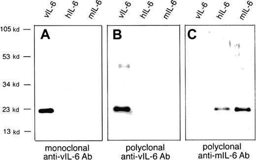
Based on the recently reported crystal structure of vIL-6, vIL-6 site II binds to D2D3 of gp130 and vIL-6 site III binds to D1 of gp130 (Figure 1). By contrast, site I of vIL-6 is not occupied by gp130 and is sterically accessible by engagement for other molecules.22 To investigate further vIL-6 interactions with its receptor(s), we generated vIL-6–specific mAbs by immunizing mice with the fusion protein of vIL-6 and MBP (MBPvIL-6).18 Following a second screening, 6 anti–vIL-6 mAbs with IgG1 isotype were selected. All 6 mAbs recognized by immunoblotting vIL-6 protein that was obtained by cleaving MBPvIL-6 with factor Xa21 (Figure2A). These mAbs also detected vIL-6 in cell lysates of the KSHV+ PEL cell line BCP-127 but not in the control KSHV− Daudi cells by Western blotting (data not shown). We examined whether these mAbs recognize cellular IL-6. Although vIL-6 exhibits 25% amino acid identity to cellular IL-6, none of these mAbs recognized either hIL-6 or mIL-6. Representative results with one of these mAb are shown in Figure 1A. A rabbit polyclonal antiserum raised against vIL-6 recognized vIL-6 but not hIL-6 or mIL-6 (Figure 2B), whereas a polyclonal antiserum against mIL-6 recognized mIL-6 and hIL-6, but not vIL-6 (Figure 2C). These results suggest that the highly immunogenic epitopes on vIL-6 are distinct from those of cellular IL-6. The specific immunogenicity of vIL-6 stands in striking contrast to Epstein-Barr virus–encoded IL-10 homologue, which is recognized by many of the monoclonal and polyclonal antibodies against hIL-10.34
Immunoblots of recombinant hIL-6, mIL-6, and vIL-6.
These immunoblots of recombinant hIL-6, mIL-6, and vIL-6 (each of 100 ng/lane) used mouse monoclonal anti–vIL-6, rabbit polyclonal anti–vIL-6, and rabbit polyclonal anti–mIL-6 antibodies. (A) Representative Western blot analysis using mAb v6m31.2.4. Similar results were obtained with the mAbs v6m12.1.1, v6m13.1.5, v6m17.3.2, v6m24.2.5, and v6m27.1.2 (data not shown). (B,C) Immunoblots of vIL-6, hIL-6, and mIL-6 using rabbit polyclonal antibodies raised against vIL-6 (B) and mIL-6 (C).
Mapping the epitopes recognized by the mAbs
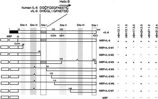
The antigenic epitopes recognized by these vIL-6 mAbs were mapped by ELISA using a panel of vIL-6 deletion mutants. These recombinant proteins were expressed in E coli as MBP fusion molecules and were purified by affinity chromatography (Figure3). Plate-coating efficiency with fusion proteins was confirmed by reacting with polyclonal anti–MBPvIL-6 antibody (data not shown). All 6 antibodies similarly recognized full-length vIL-6. The mAb v6m13.1.5 bound to a 14-amino acid stretch at the N-terminus of vIL-6, and the mAb v6m24.2.5 recognized 5 amino acids at the C-terminus of vIL-6 (Figure 3). The C-terminal end of hIL-6 is involved in site I binding to IL-6R. The mAbs v6m12.1.1, v6m17.3.2, v6m27.1.2, and v6m31.2.4 bound to full-length MBPvIL-6 and to fragment M3 of MBPvIL-6. This fragment M3, but not other mutants, encompasses an internal 13-amino acid vIL-6 fragment at positions 81 to 93, suggesting that these mAbs map to an epitope that includes Asp81 to Cys93 of vIL-6. The hIL-6 fragment corresponding in aa sequence to this vIL-6 portion occurs in the C-terminal part of the AB-loop and the beginning of helix B, which are also involved in hIL-6 site I. Thus, all mAbs, except for v6m13.1.5 recognize a putative site I on vIL-6.
Fusion protein mapping of anti-vIL-6 mAbs.
Schematic representation of deletion mutants of vIL-6 fusion proteins. Numbers above each box represent the amino acid positions relative to the start methionine. Restriction enzyme sites AatII andEcoRI used to construct M3 and M5, respectively, are indicated. The reactivity of each mAb to each fusion protein was determined by ELISA. The reactivity of each mAb to each fusion protein is shown as positive (+) when the absorbance (A405/550) was significantly higher (0.7 absorbance units) than the background or negative (−) when the absorbance was similar (within 0.05 absorbance units) to the background. The black box represents the putative vIL-6 signal peptide portion.5 The dotted box denotes a 13-amino acid peptide of vIL-6 that is recognized by 4 neutralizing mAbs against vIL-6. The bold characters indicate the second conserved cysteine residues. vIL-6 sites I, II, and III are defined based on the crystal structure of the vIL-6/gp130 complex.22
Neutralizing activity of mAbs in IL-6–responsive cell lines
We examined if these mAbs can neutralize vIL-6 bioactivity. vIL-6 has been shown to accelerate the proliferation of murine hybridoma B9 cells but to require approximately 1000-fold larger amounts of protein than hIL-6.18,20,21 The mAbs were cultured at various concentrations with B9 cells in the presence of 100 ng/mL MBPvIL-6. None of these mAbs suppressed the proliferation of B9 cells in response to hIL-6 (data not shown). At the concentrations used, the mAbs v6m13.1.5 and v6m24.2.5 demonstrated no neutralizing activity. By contrast, the mAbs v6m12.1.1, v6m17.3.2, v6m27.1.2, and v6m31.2.4 demonstrated varying degrees of neutralizing activity against vIL-6 (Figure 4A). The mAb v6m31.2.4 exhibited most prominent neutralizing activity in this bioassay. There could be several reasons for the different degrees of neutralizing efficiency, including the possibility that the mAbs recognize somewhat different epitopes within the region of Asp81 to Cys93, that while mapping to the same epitopes, the different mAbs exert variable activity for binding and neutralization,35 or that the differences may be related to the integrity of the antibody molecules. Although the degree of neutralizing activity differed somewhat, all the function-blocking mAbs mapped to the outward helical face of vIL-6 that is not occupied by gp130.
Neutralizing activity of mAbs against vIL-6.
(A) Exponentially growing B9 cells (2 × 10³ cells/well) were cultured in medium supplemented with MBPvIL-6 (100 ng/mL) with or without anti-vIL-6 Ab (0.8-10 μg/mL). [3H]-Thymidine was added during the final 6 hours of culture. The results represent the mean radioactivity of triplicate cultures; SD was within 5% of the mean. Representative data of 2 independent experiments are shown. (B) vIL-6 induces human IgM production in SKW6.4 cells. SKW6.4 cells (1 × 10⁴ cells/well) were incubated in the presence of hIL-6 or MBPvIL-6 at various concentrations, and human IgM production in the supernatants was measured by human IgM ELISA. (C) Effect of mAbs against vIL-6 on IgM secretion by SKW6.4 cells. SKW6.4 cells were incubated in the presence of MBPvIL-6 (2 μg/mL) and mAbs against vIL-6 (0-10 μg/mL). The results represent the mean of triplicate cultures; error bars represent SDs. The asterisk denotes the occurrence of a significant decrease in IgM secretion in cultures containing anti-vIL-6 mAbs (P < .0005) compared to cultures containing MBPvIL-6 (2 μg/mL) alone.
We then confirmed the neutralizing activity of anti–vIL-6 mAbs using the SKW6.4 human B cells that are known to produce IgM in response to hIL-6.33 Like hIL-6, MBPvIL-6 stimulated IgM secretion in SKW6.4 cells in a dose-dependent manner (Figure 4B). Maximal production of IgM was observed with 2 μg/mL MBPvIL-6 and 5 to 10 ng/mL hIL-6. The vIL-6–induced IgM production by SKW6.4 cells was suppressed by mAbs v6m12.1.1, v6m17.3.2, v6m27.1.2, and v6m31.2.4, but only minimally by mAbs v6m13.1.5 and v6m24.2.5 (Figure 4C). The results of these vIL-6 bioassays indicate that clones v6m12.1.1, v6m17.3.2, v6m27.1.2, and v6m31.2.4 have specific neutralizing activity against vIL-6. Lack of neutralizing activity by the mAb v6m24.2.5, which recognizes the C-terminal end of vIL-6 (Figure 3), represents a clear difference from mAb against the C-terminal end of hIL-6 binding, which completely blocked hIL-6 activity by presumably disrupting IL-6R binding.14
Binding of vIL-6 to sgp130 in the absence of sIL-6R
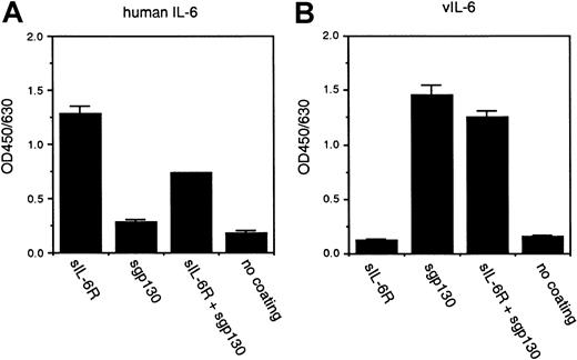
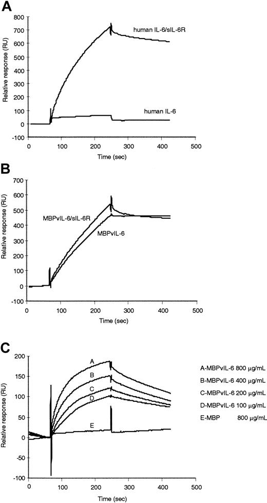
Because our neutralizing mAbs recognize the putative site I face of vIL-6, which corresponds to the putative IL-6R binding site in hIL-6, we examined vIL-6 binding to IL-6R. Using recombinant purified human sIL-6R and sgp130, soluble forms of IL-6R and gp130 lacking the transmembrane and cytoplasmic regions, we tested if vIL-6 could bind to these proteins individually or after they have been incubated together in a cell-free system. Purified sIL-6R or sgp130 or both were immobilized onto a 96-well dish, and vIL-6 binding was detected by polyclonal antibody against vIL-6. In parallel, hIL-6 was used as a control and detected by polyclonal antibody against hIL-6. As expected, hIL-6 bound to sIL-6R but not to sgp130 alone (Figure5). When sIL-6R and sgp130 were first incubated together and then immobilized onto wells, the binding of hIL-6 was reduced, likely reflecting the failure of polyclonal anti–hIL-6 antibody to bind hIL-6 sites II and III that were occupied by sgp130. Noteworthy, MBPvIL-6 bound to wells coated with sgp130 but not to wells coated with sIL-6R alone (Figure 5). The binding of vIL-6 to sgp130 was minimally affected by preincubation of sgp130 with sIL-6R, suggesting that sIL-6R did not interfere with anti–vIL-6 polyclonal antibody attachment to vIL-6 site I. In parallel experiments, sIL-6R and sgp130 immobilized on a plate failed to bind to MBP (data not shown). Lack of IL-6R contribution to vIL-6/gp130 binding was further confirmed by surface plasmon resonance. The CM5 sensor chip immobilized with sgp130 was used for equilibrium binding analysis on the BIAcore system. As shown in Figure6A, hIL-6 showed strong binding to sgp130 only when preincubated with sIL-6R. In contrast, vIL-6 bound to sgp130 without sIL-6R, and vIL-6 preincubation with sIL-6R minimally increased binding affinity for sgp130 (Figure 6B). In accordance with these observations, sIL-6R bound to the CM5 sensor chip immobilized with hIL-6 but not MBPvIL-6 even though MBPvIL-6 was used at up to 2.2-times higher amount than that of hIL-6 (data not shown). A range of MBPvIL-6 concentrations was injected over the immobilized surface of sgp130, and, as a control, MBP (Figure 6C). Control MBP showed minimal signal increase over baseline. Binding of MBPvIL-6 to sgp130 demonstrated concentration dependence and saturability. Using a 1:1 Langmuir binding model, sensograms yielded a ka of 980 1/M · s, kd of 2.2 × 10⁻³ 1/s and Kd of 2.2 μM. Additional BIAcore data (not shown) of MBPvIL-6 binding to immobilized sgp130 at equilibrium was analyzed by Scatchard analysis. Using this method, the Kd for the vIL-6–gp130 interaction was calculated at 2.5 μM, which is comparable to the Kdobtained by the 1:1 Langmuir binding model. Previously, the affinity of hIL-6/sIL-6R complex for gp130 yielded a Kd of 1.7 nM.36 Thus, the binding affinity of vIL-6 for sgp130 is 1000-fold lower than that of hIL-6/sIL-6R complex to gp130. Together, these experiments demonstrate that vIL-6 can bind to gp130 but not IL-6R.
Binding of hIL-6 and vIL-6 to sIL-6R chains as detected by ELISA.
Purified sIL-6R (5 μg/mL) or sgp130 (5 μg/mL) or both were immobilized on an ELISA plate and incubated with hIL-6 (50 ng/mL) or MBPvIL-6 (50 ng/mL). Bound protein was detected with a rabbit polyclonal anti–hIL-6 antibody that recognizes hIL-6 but not vIL-6 (A) or anti–MBPvIL-6 antibody that recognizes vIL-6 but not hIL-6 (B), followed by HRP-conjugated antirabbit IgG antibodies. The results represent the means of triplicate assays; error bars represent SD. Representative data from 3 independent experiments are shown.
Monitoring the binding of hIL-6 or vIL-6 to immobilized sgp130 using the biosensor system BIAcore 2000.
The sgp130 was immobilized at a concentration of 18.2 ng/mm2 on the CM5 biosensor chip. (A) hIL-6 (50 μg/mL) alone or hIL-6 (50 μg/mL) plus sIL-6R (20 μg/mL), (B) MBPvIL-6 (50 μg/mL) alone or MBPvIL-6 (50 μg/mL) plus sIL-6R (20 μg/mL) was passed over the sensor surface at a flow rate of 10 μL/min in PBS. Reagents were incubated for 1 hour prior to assay. (C) Overlay of sensorgrams showing kinetic analysis of vIL-6 with sgp130. The sgp130 was immobilized at a concentration of 1.5 ng/mm2. MBPvIL-6 (100, 200, 400, and 800 μg/mL) was passed over the sensor surface. Control MBP (800 μg/mL) did not show detectable affinity for sgp130. Representative data of 4 independent experiments are shown.
The gp130 binding surfaces are not fully shared by hIL-6, and vIL-6
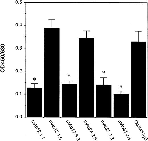
The results from the binding assays strongly suggested that the neutralizing activity by anti–vIL-6 mAbs is not attributable to blocking vIL-6 binding to IL-6R. We therefore examined if the binding of vIL-6 to gp130 in a cell-free system could be inhibited by neutralizing mAbs to vIL-6. Biotinylated vIL-6 (50 ng/mL) was first incubated with control mouse IgG1 (20 μg/mL) or anti–vIL-6 mAbs (20 μg/mL) overnight at 4°C, and then tested for binding to sgp130 bound to ELISA plates. Compared with the isotype control murine IgG1, the 4 anti–vIL-6 neutralizing mAbs interfered with the binding of vIL-6 to sgp130, whereas the 2 nonneutralizing mAbs did not (Figure7). These results provide evidence that the mAbs against vIL-6 exert neutralizing activity by direct inhibition of vIL-6 binding to gp130.
Interference of vIL-6 binding to sgp130 by neutralizing mAbs against vIL-6.
Biotinylated MBPvIL-6 (50 ng/mL) was first incubated with anti–vIL-6 mAbs (20 μg/mL) or isotype control mouse IgG1 (20 μg/mL), and then added onto ELISA wells coated with purified sgp130 (2 μg/mL) in triplicate. Bound protein was detected by streptavidin-HRP. The results represent the means ± SDs. The asterisk indicates the occurrence of a significant decrease in OD450/630(P < .002) compared to control antibody. Representative data of 2 independent experiments are shown.
We then examined if the vIL-6 epitope that our neutralizing mAbs recognize is used as a direct binding surface for gp130. A recent report has shown that vIL-6 induces the expression of hIL-6 in certain cell lines.37 mIL-6 is species specific, whereas hIL-6 binds to both human and murine receptors. To exclude a potential contribution of vIL-6–induced cellular IL-6, we used murine BAF-B03 cells stably transfected with human gp130. This cell line is known to respond to vIL-6 but not to hIL-6 in the absence of sIL-6R.19 32 As expected, hIL-6/sIL-6R induced DNA synthesis in BAF-130 cells (Figure 8A). In agreement with its lower binding affinity, vIL-6 had much lower specific activity than hIL-6/sIL-6R. The vIL-6 deletion mutant MBPvIL-6 M3, which contains the region of Asp81 to Cys93 within site I but lacks full sites II and III comprising the reported vIL-6 interface with gp130 (Figures 1 and 3), did not induce DNA synthesis in BAF-130 cells or compete for vIL-6 or hIL-6/sIL-6R binding to gp130 (Figure8B). This suggested either that this vIL-6 epitope is not part of the vIL-6 binding surface to gp130, as shown by the vIL-6 crystal structure, or that this fragment alone is not sufficient for biologic activity. Neither parental BAF-B03 cells nor mock transfectants responded to vIL-6 (data not shown).
Effects of anti-gp130 mAbs on vIL-6–mediated DNA synthesis.
(A) BAF-130 cells (2 × 10⁴ cells/well) were stimulated with various concentrations of hIL-6/sIL-6R, MBPvIL6, or MBP for 48 hours. [3H]-Thymidine was added during the final 6 hours of culture. (B) Failure of a vIL-6 deletion to stimulate BAF-130 cells or to inhibit the binding of vIL-6 or hIL-6/sIL-6R to gp130. The BAF-130 cells were stimulated with MBPvIL-6 (200 ng/mL) or hIL-6 (25 ng/mL)/sIL-6R (50 ng/mL) in the presence of increasing amounts of MBPvIL-6 M3, which encodes Asp81 to Cys93. The BAF-130 cells were also stimulated with MBPvIL-6 M3 alone (18-40 000 ng/mL). (C) Effects of selected anti-gp130 mAb on hIL-6–induced and vIL-6–induced DNA synthesis. The BAF-130 cells were stimulated with MBPvIL6 (500 ng/mL) or hIL-6 (50 ng/mL)/sIL-6R (100 ng/mL) in the presence of anti-gp130 mAbs (B-R3, B-P4, and B-P8) or control IgG (78-5000 ng/mL). The 100% value corresponds to proliferation in the absence of antibody. The results reflect the mean of counts per minute of triplicate cultures; SD was within 10% of the mean. Representative data of 3 independent experiments are shown.
We then examined if hIL-6 and vIL-6 share the same gp130 binding sites using a panel of anti–gp130 mAbs. These antibodies are known to recognize specific functional sites of gp130 for signal transduction by IL-6–type cytokines.38,39 As reported previously, all 3 anti–gp130 mAbs, B-R3, B-P4 and B-P8, but not an isotype control, inhibited hIL-6–induced DNA synthesis in BAF-130 cells (Figure 8C). By contrast, when stimulated with vIL-6, mAbs B-R3 and B-P4, but not B-P8, suppressed DNA synthesis in BAF-130 cells.39,40 The mAb B-P4 recognizes the membrane-proximal extracellular part of gp130 (D4D5D6) that is critical for homodimerization of gp130.40The B-R3 mAb interferes with gp130 binding by all IL-6 family cytokines, whereas B-P8 mAb interferes with gp130 binding by hIL-6 and ciliary neurotrophic factor.38,40 The mAbs B-R3 and B-P8 recognize the epitopes in gp130 D2 and D3, respectively, which constitute the cytokine-binding module.38 39 These results provide evidence that hIL-6 and vIL-6 share an epitope in D2 of gp130 and that both cytokines induce homodimerization of gp130. The results further suggest that vIL-6 does not bind to the epitope in gp130 D3, which is critical to hIL-6 function.
Discussion
The KSHV vIL-6 is one of the viral proteins implicated in the pathogenesis of AIDS-related malignancies.18,27,41,42vIL-6 can stimulate the growth of PEL cells in an autocrine/paracrine fashion21 and induce VEGF expression, which is critical for the growth of PEL and KS in vivo.18,43-46 A proportion of HIV+ patients have detectable vIL-6 in sera and body fluids,27,41 42 suggesting that this virokine may play a critical role in the pathogenesis of AIDS-associated malignancies by promoting sustained phosphorylation of gp130.
Although vIL-6 shows biologic functions similar to those of cellular IL-6, the interaction between vIL-6 and its receptor(s) is unclear. Cellular IL-6 exerts its actions through a receptor complex consisting of a specific IL-6–binding protein, IL-6R, and a signal-transducing subunit, gp130. The IL-6/IL-6R complex induces the homodimerization of 2 gp130 molecules leading to a number of intracellular signaling events, including activation of the transcription factor NF-IL-6 and STAT3 via phosphorylation of the Janus kinase.6,11,12Cellular IL-6 is unable to transduce signals in the absence of IL-6R. The potential participation of IL-6R in vIL-6 binding was first supported by the experiments using the IL-6–dependent B9 cells.4,20 Moreover, cotransfection assay involving IL-6R expression vector enhanced vIL-6 responses.23,26 However, anti–IL-6R antibody effectively neutralized STAT phosphorylation in HepG2 cells in response to hIL-6 but failed to block STAT activation by vIL-6.19 In contrast, anti–gp130 antibody abrogated signaling of both responses.19 Furthermore, BAF-B03 cells expressing transfected human gp130 but not IL-6R, exhibited a strong response to vIL-6 but not to hIL-6.25 Recently, crystallographic analysis of the vIL-6/sgp130 complex showed that site I of vIL-6 is not occupied by gp130 and is fully accessible within the complex,22 raising again the possibility that this site might engage IL-6R. Here, we provide evidence that vIL-6 does not bind to IL-6R in a cell-free system. By both surface plasmon resonance and ELISA, vIL-6 bound gp130 without a need for IL-6R and failed to bind IL-6R. In addition, mAb v6m24.2.5 recognizing the C-terminal end of helix D of vIL-6 did not show neutralizing activity in bioassays. In contrast to these results with vIL-6, the corresponding C-terminal end of helix D in hIL-6 is included in the binding face to IL-6R, and the binding of mAb to this region totally abrogated hIL-6 activity by blocking the ligand interaction to IL-6R.14 Recently, Mori and coworkers documented that vIL-6 can induce the expression of cellular IL-6 in certain cell types.37 Another report has noted that VEGF stimulates stromal cells to produce hIL-6.47 Because vIL-6 is a potent inducer of VEGF,18 vIL-6 could also stimulate hIL-6 expression through induction of VEGF. Expression of the IL-6 gene is regulated by cis-acting elements that differ depending on the cell type and nature of the activating agent.6 The presence of hIL-6 could be a confounding factor in cell-based systems used to study vIL-6 receptor usage and may explain some of the contrasting results in the literature.
Using the structure of the growth hormone/growth hormone receptor complex as a paradigm for cytokine receptor complex assembly, IL-6–type cytokines are believed to have 3 topologically discrete sites of interactions with their receptors. Site I, if used, is always engaged by a nonsignaling receptor: IL-6R, IL-11R, or ciliary neurotrophic factor receptor. Site II is always engaged by gp130, and site III by a second signaling receptor gp130, OSMR, or LIFR.48 Within gp130, 3 binding epitopes have been identified as critical to its activation by hIL-6/IL-6R: one epitope involves the Ig-like domain (D1); another epitope is located in the cytokine binding module (D2D3); and the other is located in the membrane-proximal extracellular domains (D4D5D6).40According to the crystallographic analysis of the vIL-6/sgp130 (D1D2D3) complex,22 sequence alignment of hIL-6 and vIL-6 shows that contact residues seen in the structure of the vIL-6/gp130 complex are in the same positions as hIL-6-gp130 contact residues previously mapped by mutagenesis.49 In cell culture, the membrane-proximal extracellular part of gp130 (D4D5D6) is critical for vIL-6–mediated signaling, even though vIL-6 can form a tetrameric complex with gp130 (D1D2D3).22 Further, although hIL-6 and vIL-6 share an epitope in gp130 D2, vIL-6 does not appear to use an epitope in gp130 D3 that is critical to hIL-6 function. Together with the evidence that vIL-6 does not bind IL-6R, these results are consistent with the previous observation that vIL-6 did not compete with the hIL-6 superantagonist Sant7 for human myeloma cell stimulation.20 Continued analysis using gp130 mutants and h/vIL-6 chimeras will be important to identify the specificity-determining epitopes that are essential for binding and signaling by hIL-6 and vIL-6.
It is generally believed that receptor conformational changes occur in a sequential process following ligand-receptor engagement.50 In addition, some ligands need allosteric changes to achieve high-affinity binding to their receptors.51-53 On receptor binding, the flexible AB-loop of interferon-γ undergoes a conformational change to facilitate receptor binding.51 Among 4 α-helical bundle proteins, such conformational changes have been reported in IL-4 and granulocyte colony-stimulating factor.52,54 In contrast, although a number of studies have been conducted on the structure-function relationships of IL-6 family cytokines, little is known about potential conformational changes in the ligands preceding receptor engagement. Recent studies addressing functional relationships among integrin domains demonstrated that conformational changes in the extracellular domains of the integrin lymphocyte function-associated (LFA)–1 (CD11a/CD18) regulate binding to its receptor.55 Relevant to the current study, 2 mAbs against LFA-1 blocked receptor binding by interfering with the conformational rearrangement.55Interestingly, those mAbs recognize the regions including the top of helix-5α, close to the conformationally mobile loop.55In our study, the neutralizing mAbs directed against vIL-6 recognize a region of the C-terminal part of the AB-loop and the beginning of helix B. The binding of mAb to this region may change the mobility of helix B and interfere with the ability of sites II and III to achieve a proper orientation. Perhaps the binding of hIL-6 to IL-6R may be necessary to modify a loop-helix interaction into active conformation before binding to gp130. In the case of vIL-6, the absence of a specific receptor chain may result in the low binding affinity of vIL-6 to gp130 by failure to be locked in a high-affinity conformation. Future efforts will be needed to investigate potential conformational changes of hIL-6 and vIL-6 preceding receptor binding. Such structural studies could also be used to advance pharmaceutical development of cytokine-specific gp130 antagonists.
We thank Mike Klutch for help with DNA sequence analysis and Dr Fumihiko Takeshita for providing SKW6.4 cells. We also thank Drs Peter Steinbach, Fred Dyda, Demetrios Braddock, and Motomu Shimaoka for critical discussions, and Dr Issey Okazaki for his technical help with the BIAcore analysis.
The publication costs of this article were defrayed in part by page charge payment. Therefore, and solely to indicate this fact, this article is hereby marked “advertisement” in accordance with 18 U.S.C. section 1734.
References
Author notes
Yoshiyasu Aoki, Medicine Branch, Center for Cancer Research, National Cancer Institute, National Institutes of Health, 10 Center Dr, Rm 12N226 MSC1907, Bethesda, MD 20892; e-mail:aokiy@mail.nih.gov.

![Fig. 4. Neutralizing activity of mAbs against vIL-6. / (A) Exponentially growing B9 cells (2 × 10³ cells/well) were cultured in medium supplemented with MBPvIL-6 (100 ng/mL) with or without anti-vIL-6 Ab (0.8-10 μg/mL). [3H]-Thymidine was added during the final 6 hours of culture. The results represent the mean radioactivity of triplicate cultures; SD was within 5% of the mean. Representative data of 2 independent experiments are shown. (B) vIL-6 induces human IgM production in SKW6.4 cells. SKW6.4 cells (1 × 10⁴ cells/well) were incubated in the presence of hIL-6 or MBPvIL-6 at various concentrations, and human IgM production in the supernatants was measured by human IgM ELISA. (C) Effect of mAbs against vIL-6 on IgM secretion by SKW6.4 cells. SKW6.4 cells were incubated in the presence of MBPvIL-6 (2 μg/mL) and mAbs against vIL-6 (0-10 μg/mL). The results represent the mean of triplicate cultures; error bars represent SDs. The asterisk denotes the occurrence of a significant decrease in IgM secretion in cultures containing anti-vIL-6 mAbs (P < .0005) compared to cultures containing MBPvIL-6 (2 μg/mL) alone.](https://ash.silverchair-cdn.com/ash/content_public/journal/blood/98/10/10.1182_blood.v98.10.3042/5/m_h82211754004.jpeg?Expires=1769099008&Signature=l0Mgc9DQrrERMCXUxAQwhEkmoAFhot4Q9o0-pwUIf1Osx2IHvEEXxhDj-HTD0fgdCh9DqJ1-IIiQFevL4eBCh98~eZ22B0QM2e7Xmds-Lg49FAIxHysq2jPtpvmG76s97xGELZjEE2l9qrYptOroqx6uYmi6Jupqjo2WaFBbFfaPLxhQDpyHR8N6Q0ltAGBNEUKixAKozqXV7kOoRGiwKYxXVeZDsYHkIrHoF03FRa377i~YLY2lbWiiOENv0bVYaFsfpyrEwHz0PQ6oAtdlf-ULKcQHKciunysjxGVR5oHMRvHd~Z8y-~t5Z0EM8gwcvnrc9ZTyqlBeKu5-pSmiPg__&Key-Pair-Id=APKAIE5G5CRDK6RD3PGA)
![Fig. 8. Effects of anti-gp130 mAbs on vIL-6–mediated DNA synthesis. / (A) BAF-130 cells (2 × 10⁴ cells/well) were stimulated with various concentrations of hIL-6/sIL-6R, MBPvIL6, or MBP for 48 hours. [3H]-Thymidine was added during the final 6 hours of culture. (B) Failure of a vIL-6 deletion to stimulate BAF-130 cells or to inhibit the binding of vIL-6 or hIL-6/sIL-6R to gp130. The BAF-130 cells were stimulated with MBPvIL-6 (200 ng/mL) or hIL-6 (25 ng/mL)/sIL-6R (50 ng/mL) in the presence of increasing amounts of MBPvIL-6 M3, which encodes Asp81 to Cys93. The BAF-130 cells were also stimulated with MBPvIL-6 M3 alone (18-40 000 ng/mL). (C) Effects of selected anti-gp130 mAb on hIL-6–induced and vIL-6–induced DNA synthesis. The BAF-130 cells were stimulated with MBPvIL6 (500 ng/mL) or hIL-6 (50 ng/mL)/sIL-6R (100 ng/mL) in the presence of anti-gp130 mAbs (B-R3, B-P4, and B-P8) or control IgG (78-5000 ng/mL). The 100% value corresponds to proliferation in the absence of antibody. The results reflect the mean of counts per minute of triplicate cultures; SD was within 10% of the mean. Representative data of 3 independent experiments are shown.](https://ash.silverchair-cdn.com/ash/content_public/journal/blood/98/10/10.1182_blood.v98.10.3042/5/m_h82211754008.jpeg?Expires=1769099008&Signature=b4yVbTMSiV-OjqzFyqwzHt6jCCpRTyMj7ruCtbIbgSiM4--AnhWMXJf5tRWqMYV9pyo0~9sCcekPAi1z6baqho4w21WFLrF35UQUIAhgN5DZoAqKQk3eViCArNYgt11teHxpBrBhooVqXyCw1-5KC5lIZAzn9IqLFwDmq7BLLrKVYBRnqiQQxCU~3NKprwSLVhboVWDWqO2rB5kxdN1G6hdv3r-BoGVVfM4PX2ckyLHzM~WkwpJ9bDcUGi-y~I-s0mKnTZlJsBFuSWN1l26YGfknx8CFegVJLpR4tfq7v64lGYTPKctmWPlB0WF8VbriIT~pZxtRi6vUt6qkV-e4qQ__&Key-Pair-Id=APKAIE5G5CRDK6RD3PGA)
This feature is available to Subscribers Only
Sign In or Create an Account Close Modal