Abstract
Integrin αvβ3 has been implicated in angiogenesis and other biological processes. However, the ligand-binding sites in αv, a non–I-domain α subunit, remain to be identified. Recently in αIIb, the other partner of the β3 subunit, several discontinuous residues important for ligand binding were identified in the predicted loops between repeats 2 and 3 (W3 4-1 loop) and within repeat 3 (W3 2-3 loop). Based on these findings, alanine-scanning mutagenesis in 293 cells was used to investigate the role of these loops (cysteine [C]142-C155 and glycine [G]172-G181) of αv in ligand binding. Wild-type αvβ3 was able to bind soluble fibrinogen following integrin activation either by 0.5 mM manganese dichloride (MnCl2) or a mutation of β3 threonine (T)562 to asparagine. However, mutation of tyrosine (Y)178 to alanine in the predicted G172-G181 loop of αv abolished fibrinogen binding, and alanine (A) substitutions at adjacent residues phenylalanine (F)177 and tryptophan (W)179 had a similar effect. Cells expressing Y178Aαvalso failed to bind to immobilized fibrinogen. Moreover, the Y178A mutation abolished the binding of WOW-1 Fab, a monovalent ligand-mimetic anti-αvβ3 antibody, and the expression of β3 ligand–induced binding sites (LIBS) induced by arginine-glycine-aspartic acid-tryptophan (RGDW). In sharp contrast to the data obtained with αIIb, none of the mutations in the predicted W3 4-1 loop in αv impaired ligand binding. These results implicate αv Y178 in ligand binding to αvβ3, and they suggest that there are key structural differences in the adhesive ligand-binding sites of αvβ3 and αIIbβ3.
Introduction
Integrins are a large family of αβ heterodimeric adhesion receptors that is often subdivided into groups based on 8 known integrin β subunits.1 The β3-integrin subfamily is composed of αvβ3, originally identified as the vitronectin receptor, and αIIbβ3, a platelet-specific receptor for fibrinogen and von Willebrand factor. αIIbβ3 plays a crucial role in platelet aggregation, normal hemostasis, and pathological thrombus formation.2 On the other hand, αvβ3 is expressed in a number of tissues, including platelets, endothelial cells, vascular smooth muscle cells, and osteoclasts, and it plays a key role in angiogenesis and bone resorption.3 4
The αIIb and αv subunits are homologous and 36% identical in primary amino acid sequence,5 and most ligands that bind to αIIbβ3, including fibrinogen, von Willebrand factor, and vitronectin, also bind to αvβ3. However, there are some distinctive features between these 2 integrins.3 First, the αIIb subunit has been found only in combination with β3, whereas αv can associate with at least 5 β subunits (β1, β3, β5, β6, and β8).6 Second, some ligands, such as osteopontin, matrix metalloproteinase-2, and adenovirus penton base, bind to αvβ3 but not to αIIbβ3. Third, treatment of αIIbβ3 with ethylenediamine tetraacetic acid (EDTA) at 37°C dissociates the complex into its individual subunits; αvβ3 remains a heterodimer. Finally, the ligand-binding function of αvβ3, but not αIIbβ3, is suppressed by calcium (Ca2+).7
Generally, all integrins require divalent cations for ligand recognition, and multiple residues important for ligand binding have been identified on both α and β subunits.8 The N-terminal region of integrin α subunits has 7 repeats of homologous sequences of about 60 amino acid residues. Some integrin α subunits (eg, α2, αL, and αM) contain an inserted domain of about 200 amino acids residues (the I-domain) between the second and the third repeats in the α subunit, which is critically involved in ligand binding.9,10 The crystal structure of the I-domain has been determined, and the metal ion-dependent adhesion site (MIDAS) motif that contributes to cation binding, as well as ligand binding, has been clarified.11Interestingly, a MIDAS-like motif essential for the ligand-binding function was also identified in integrin β subunits.12 13 On the other hand, integrin α subunits, such as αv, αIIb, and α4, do not have the I-domain.
The structural basis for the interaction between non–I-domain α subunits and their ligands remains elusive. For the αIIbsubunit, peptide cross-linking studies have shown that the histamine-histamine-leucine-glycine-glycine-alanine-lysine-glutamine-alanine-glycine-aspartic acid-valine (HHLGGAKQAGDV) sequence derived from the COOH terminus of the γ-chain of fibrinogen interacts with residues 294-314, encompassing the second putative calcium-binding domain of αIIb.14 However, recent characterization of molecular defects in Glanzmann thrombasthenia and mutagenesis studies demonstrated several discontinuous residues important for ligand binding: proline (P)145, D163, L183, G184, tyrosine (Y)189, Y190, phenylalanine (F)191, G193, and D224.15-19 Springer has proposed that the 7 N-terminal sequence repeats of integrin α subunits are folded into a β-propeller domain.20 The proposed domains contain seven 4-stranded β-sheets (W1-W7) arranged in a torus around a pseudosymmetry axis. Interestingly, the discontinuous residues identified in αIIb as important for ligand binding were located in the regions predicted to adopt a β-turn structure on the upper face of the β-propeller model: P145 within the W3 4-1 loop; L183, G184, Y189, Y190, F191, and G193 within the predicted W3 2-3 loop; and D224 within the predicted W4 4-1 loop. Furthermore, the analysis of a Japanese variant of Glanzmann thrombasthenia, KO, and alanine-scanning mutagenesis have shown that D163 in the W3 4-1 loop (cystein [C]146-C167) is essential for ligand binding.16 In contrast, the single available peptide cross-linking study of αvβ3 showed that 2 distinct linear regions in αv, residues 139-167 and 312-349, cross-link to an arganine-glycine-aspartic acid (RGD) peptide.21
In this study, we took advantage of the data regarding ligand-binding sites in αIIb and investigated the role in ligand binding of the predicted loops between repeats 2 and 3 (W3 4-1 loop) and within repeat 3 (W3 2-3 loop) in αv. By performing alanine-scanning mutagenesis of recombinant αvβ3 expressed in 293 cells, we demonstrate a critical role for Y178 within the predicted W3 2-3 loop of αv in ligand binding. In contrast to αIIb, however, no mutations in the W3 4-1 loop affect ligand binding to αvβ3, thereby implying key structural differences in the adhesive ligand-binding sites of the 2 β3 integrins.
Materials and methods
Monoclonal antibodies and peptides
The following monoclonal antibodies (mAbs) were used in the study: LM609,22 a murine mAb specific for αvβ3, and LM142,23 a mAb specific for αv (gift from Dr David Cheresh, The Scripps Research Institute, La Jolla, CA); AP5,24 a mAb specific for β3 (gift from Dr Thomas Kunicki, The Scripps Research Institute); anti-LIBS1 (anti–ligand-induced binding site 1) mAb (specific for β3)25 and anti-LIBS6 mAb (specific for β3)26 (Dr Mark Ginsberg, The Scripps Research Institute); and AP3 (specific for β3)27 (Dr Peter Newman, The Blood Center of Southeastern Wisconsin, Milwaukee, WI). WOW-1 Fab,28 a monovalent ligand-mimetic mAb specific for activated αvβ3, was created by replacing the heavy chain hypervariable region 3 (H-CDR3) of PAC-1 Fab, a ligand-mimetic mAb specific for activated αIIbβ3,29 with a single integrin-binding domain of adenovirus penton base. We also used the RGDW peptide (gift from Dr Jiro Seki, Fujisawa Pharmaceutical Co., Osaka, Japan).30
Construction of αv expression vectors and cell transfection
Wild-type (WT) αv complementary DNA (cDNA) (gift from Dr David Cheresh, The Scripps Research Institute) and WT β3 cDNA (gift from Dr Gilbert White, University of North Carolina, Chapel Hill, NC) were cloned into mammalian expression vector pcDNA3 (Invitrogen Corp, San Diego, CA). To introduce single alanine substitutions into αv, overlap extension polymerase chain reaction (PCR) was carried out as previously described.16For example, to generate the Y178→A (Y178A) αv mutant, we synthesized mismatched sense primer αv178A-s, 5′-GGTCCTGGTAGCTTTGCATGGCAAGGTCAGC-3′ (sense, nucleotides [nt] 648-678; mismatched sequences underlined) and antisense primer αv178A-as, 5′-GCTGACCTTGCCATGCAAAGCTACCAGGACC-3′ (antisense, nt 678-648; mismatched sequences underlined), which were constructed based on the published sequence.31 PCR was performed by using αv cDNA as a template and primers αv258-s, 5′-GCAAACACCACCCAGCC-3′ (sense, nt 258-274) and αv178A-as, or primers αv178A-s and αv952-as, 5′-GCAGCCATCTGCTCGCCAG-3′ (antisense, nt 952-934).
The 2 individually amplified PCR products were mixed and used as a template for PCR using primers αv258-s and αv952-as. The amplified PCR products were digested with PpuMI and AflIII. The fragments digested with AflIII and XbaI were isolated from the full-length αv cDNA cloned into pcDNA3. These 2 fragments were introduced together into the pcDNA3 that had been digested with PpuMI and XbaI. The nucleotide sequences of the inserts were confirmed by sequence analysis. In a selected experiment, the C142-C155 loop in αv was swapped with the corresponding sequence of αIIb C146-C167. The WT or mutant αvconstruct was cotransfected into 293 cells with WT β3construct by the calcium-phosphate method as previously described.32 The cells were cultured in Dulbecco's modified Eagle's medium (DMEM) with 10% heat-inactivated fetal calf serum (FCS) and analyzed 2 days after transfection.
Immunoprecipitation
Immunoprecipitation was performed as previously described with slight modification.33 In brief, cells were surface-labeled with sulfo-NHS-biotin (Pierce, Rockford, IL) and lysed in a buffer containing 1% Triton X-100, 25 mM Tris-HCl (tris[hydroxymethyl] aminomethane–hydrochloride), 100 mM sodium chloride (NaCl) (pH 7.4), 0.1 mg/mL leupeptin, 4 μg/mL pepstatin A, 1 mM phenylmethylsulfonyl fluoride, and 10 mM benzamide. Then 200 μg protein from each sample was immunoprecipitated with the mAb LM609, and precipitated bands were identified on Western blots using peroxidase-conjugated avidin.
Ligand-binding studies
Fibrinogen (Kabi, Stockholm, Sweden) was labeled with fluorescein isothiocyanate (FITC) as previously described34 and stored at −80°C until use. FITC-fibrinogen binding to 293 cells was assessed by flow cytometry as described.33 Briefly, 50-μL aliquots of 1.5 × 10⁵ washed cells in Ca2+-free–Tyrode–HEPES (4-(2-Hydroxyethyl)-1-piperazineethanesulfonic acid) buffer containing 1 mM magnesium dichloride (MgCl2) were incubated with mAb LM142, specific for αv (5 μg/mL), for 30 minutes on ice. After washing, 0.5 mM manganese dichloride (MnCl2) was added into the cell suspension to induce a high-affinity state of αvβ3. Cells were then incubated with 150 μg/mL FITC-fibrinogen in the presence or absence of 1 mM RGDW peptide and phycoerythrin (PE)-conjugated antimouse immunoglobulin G (IgG) (1:5 dilution) for 25 minutes at 22°C and then incubated with propidium iodine (PI) (Sigma Chemical Co., St Louis, MO) for 5 minutes at 22°C. After washing, fibrinogen binding (FL1) was analyzed on the gated subset of single high αvβ3-expressing (FL2) live cells (PI-negative, FL3). Specific fibrinogen binding was defined as that inhibited by 1 mM RGDW peptide.
For WOW-1 Fab binding to αvβ3, cells in Ca2+-free–Tyrode–HEPES buffer containing 1 mM MgCl2 were incubated with 5 μg/mL WOW-1 Fab in the presence of 0.5 mM MnCl2 for 30 minutes at 22°C. After washing, cells were incubated with 5 μg/mL Alexa-conjugated goat antimouse IgG F(ab′)2 (Molecular Probes, Eugene, OR) for 25 minutes on ice and then incubated with PI for 5 minutes at 22°C. After washing, WOW-1 binding (FL1) was analyzed on the gated subset of single, living cells. WOW-1 binding to untransfected 293 cells was routinely taken as a measure of nonspecific binding because this value was similar to that obtained for WOW-1 binding to αvβ3 transfected cells in the presence of 1 mM RGDW.
For the induction of LIBS on αvβ3, washed cells in Tyrode-HEPES buffer containing 1 mM MgCl2 and 1 mM calcium dichloride (CaCl2) were incubated with 1 mM RGDW for 30 minutes at 22°C. The cells were then incubated for 30 minutes with AP5, anti-LIBS1, or anti-LIBS6 at a final concentration of 5 μg/mL. After washing, cells were incubated with FITC-conjugated goat F(ab′)2 antimouse IgG for 25 minutes. The mixtures were incubated with PI for an additional 5 minutes and washed, and mAb binding was analyzed on the gated subset of single, living cells.
Adhesion assays
Adhesion assays were performed as described by Faull et al.35 Wells of 96-well microtiter plates were coated with up to 1 μg fibrinogen per well in 100 μL phosphate-buffered saline (PBS) and incubated at 4°C overnight. After washing with PBS, wells were blocked with PBS containing 1% bovine serum albumin (BSA) (Sigma) for 90 minutes at 22°C. To determine background adhesion, control wells were coated with 1% BSA. Cells were washed twice with PBS and resuspended in DMEM containing 0.1% BSA at a concentration of 1 × 10⁶ cells per mL. Then, 100-μL aliquots of cell suspension were added to wells in triplicate. The plate was incubated in a humidified 37°C incubator for 60 minutes. After washing with PBS, the adherent cells were checked by visual inspection, and adhesion was quantified by measuring endogenous cellular acid phosphatase activity in an enzyme-linked immunosorbent assay.
Results
Expression of αvβ3 mutants in 293 cells
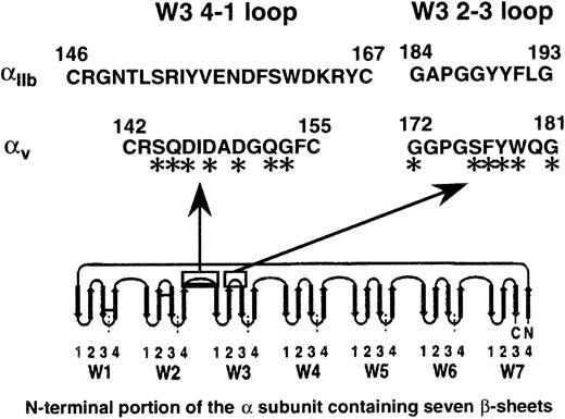
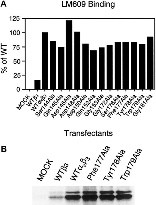
Recent studies have demonstrated that several residues within the C146-C167 loop and the G184-G193 loop in the αIIbsubunit, which correspond to the W3 4-1 loop and W3 2-3 loop in the proposed β-propeller model, respectively, play a critical role in ligand binding. Figure 1 shows the corresponding regions (C142-C155 and G172-G181) in the αvsubunit and the residues we replaced with alanine. The WT or mutant αv construct was transiently cotransfected into 293 cells with the WT β3 construct, and αvβ3 surface expression was examined by flow cytometry using the αvβ3complex-specific mAb LM609. As shown in Figure2A, the surface expression of mutant αvβ3 was 70% to approximately 123% of WT αvβ3. Similar results were obtained when mAb LM142 was used to quantify αv and mAb AP3 was used to quantify β3 (data not shown).
Amino acid sequences of the predicted W3 4-1 loop between the N-terminal repeats 2 and 3 and the predicted W3 2-3 loop within repeat 3 in the α subunits of β3 integrins.
Both loops are located on the upper face of the β-propeller model.20 The asterisks indicate that the residues were substituted by alanine in this study. This figure is adapted from a β-propeller model proposed by Springer,20 and the arrows indicate β strands.
Surface expression of αvβ3mutants in transiently transfected 293 cells.
(A) The surface expression of transfected αvβ3 was analyzed 2 days after transfection by flow cytometry. Cells expressing WT or mutant αvβ3 were incubated with 5 μg/mL αvβ3 complex-specific mAb LM609 for 30 minutes on ice and then washed once. Bound antibodies were detected by FITC-conjugated goat F(ab′)2 antimouse IgG. Relative amounts of the binding were normalized to a 100% value for LM609 binding to cells expressing WT αvβ3. The results are representative of 3 separate experiments. (B) Immunoprecipitation showing the surface expression of transfected αvβ3. Transiently transfected cells were surface-labeled with sulfo-NHS-biotin and lysed in the lysing buffer containing 1% Triton X-100. WT or mutant αvβ3 was precipitated with LM609 (specific for the αvβ3 complex) and separated on a 6% sodium dodecyl sulfate polyacrylamide gel under reducing conditions. After transfer, a membrane was incubated with peroxidase-conjugated avidin and developed with chemiluminescence. The results are representative of 3 separate experiments.
Because 293 cells normally express αvβ1 but not αvβ3,36 we were concerned that endogenous αv might associate with transfected β3 and contribute to the total αvβ3 expressed on these cells. However, by comparing αvβ3 transfectants to β3 transfectants, we found that endogenous αv contributed no more than approximately 17% to the total αvβ3 expressed. Immunoprecipitation experiments employing LM609 further showed that the expression of endogenous αv associated with transfected β3 in 293 cells is low, and that the surface expression levels of mutant F177A, Y178A, and W179Aαvβ3 are comparable to those of WT αvβ3 (Figure 2B). These results indicate that transfected αv, not endogenous αv, was the major contributor toward αvβ3expression in 293 cells. Moreover, none of the alanine-scanning mutants of αv adversely affected surface expression of αvβ3.
The Y178A mutation in αv abolishes soluble ligand binding to αvβ3
To analyze the ligand-binding function of each mutant αvβ3, we initially examined the binding of FITC-conjugated soluble fibrinogen to αvβ3. Because αvβ3 expressed on 293 cells is present in a low-affinity state and does not bind soluble ligands, cells were incubated with 0.5 mM MnCl2, which induces a high-affinity state of integrins by a direct effect on the extracellular domain (Figure3A).37 To avoid even a minimal contribution of endogenous αv in 293 cells to fibrinogen binding, we selectively analyzed the subset of transfectants expressing high levels of exogenous αvβ3monitored by LM142. As shown in Figure 3B, a D119Y mutation within the ligand-binding site of β3 abolished fibrinogen binding to αvβ3 as expected,38 confirming the specificity of the ligand-binding assay in this system. Moreover, a Y178A mutation within the predicted G172-G181 loop of αvabolished fibrinogen binding, and both F177A and W179A mutations adjacent to the Y178 also moderately impaired binding. However, none of the mutations within the C142-C155 loop of αv (S144A, Q145A, D146A, D148A, D150A, Q152A, and G153A) disturbed fibrinogen binding to the receptor.
Ligand-binding function of αvβ3 mutants.
(A) The binding of soluble fibrinogen and a ligand-mimetic mAb, WOW-1 Fab, to WT or mutant αvβ3 were examined in the presence or absence of 0.5 mM MnCl2 by flow cytometry. For fibrinogen binding, washed cells were first incubated with 5 μg/mL mAb LM142 (specific for αv) for 30 minutes on ice. After washing, cells with 0.5 mM MnCl2 were incubated with 150 μg/mL FITC-conjugated fibrinogen and PE-conjugated antimouse IgG (1:5 dilution) for 25 minutes at 22°C and then incubated with PI for 5 minutes at 22°C. After washing, fibrinogen binding (FL1) was analyzed on the gated subset of single, high αvβ3 expression (FL2) and live cells (PI-negative, FL3) as indicated. (B) Relative amounts of fibrinogen binding are normalized to a 100% value for the binding to cells expressing WT αvβ3 (% of WT). Fibrinogen binding in the presence of 1 mM RGDW was used as a negative control. Data represent the mean ± SE of 3 experiments. (C) WOW-1 Fab binding. For WOW-1 binding, cells with 0.5 mM MnCl2 were first incubated with 5 μg/ml WOW-1 Fab for 30 minutes at 22°C. After washing, cells were incubated with 5 μg/mL Alexa-conjugated antimouse IgG for 25 minutes on ice and then incubated with PI for 5 minutes at 22°C. After washing, bound antibodies were analyzed. WOW-1 binding to 293 cells was used as a negative control. Relative amounts of WOW-1 Fab binding are expressed by the following formula: % binding of WOW-1 Fab to WT αvβ3/% binding of LM609 to WT αvβ3. The αvαIIb mutant represents a chimera in which the C142-C155 loop in αv was swapped with the corresponding sequence of αIIb C146-C167. Data represent the mean ± SE of 3 experiments.
To further examine the ligand-binding function of mutant αvβ3, we next examined the binding of WOW-1 Fab, a monovalent ligand-mimetic anti-αvβ3antibody. The Y178A mutation in αv, as well as the D119Y mutation in β3, markedly inhibited WOW-1 binding to MnCl2-treated cells. On the other hand, swapping of the C142-C155 region of αv with the corresponding region of αIIb (C146-C167) did not show a marked inhibition of WOW-1 Fab binding. Because WOW-1 Fab is sensitive to changes in αvβ3 affinity rather than avidity,28 these results suggest that the Y178A mutation disrupts the conformation of the ligand-binding pocket in αvβ3.
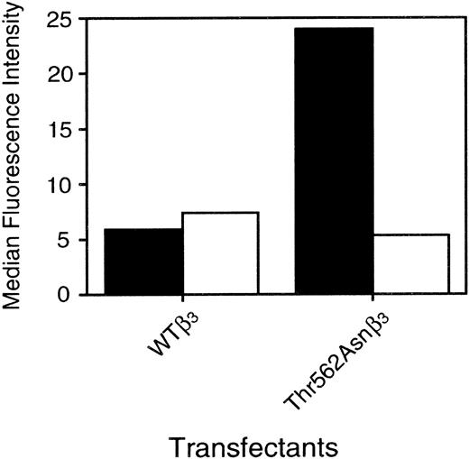
Because the integrin activator MnCl2 may have additional effects on cells, we cotransfected the WT or the mutant αv with the T562Nβ3 mutant, which constitutively activates β3 integrins and eliminates the need for MnCl2.33 As shown in Figure4, T562Nβ3 augmented fibrinogen binding when complexed with the WT αv, but it failed to induce fibrinogen binding when complexed with the Y178Aαv. These results indicate that Y178 in the αv subunit is critical for soluble ligand binding to αvβ3.
Effects of β3-activating mutant (T562N) on fibrinogen binding.
WT (▪) or Y178Aαv (■) construct was transiently cotransfected with WT or T562Nβ3 construct into 293 cells. Fibrinogen binding to transfected αvβ3 was determined by flow cytometry. Cells were first incubated with 5 μg/mL LM142 (specific for αv) for 30 minutes on ice. After washing, cells were incubated with 150 μg/mL FITC-conjugated fibrinogen and PE-conjugated antimouse IgG for 25 minutes at 22°C in the presence or absence of 1 mM RGDW and then incubated with PI for 5 minutes at 22°C. After washing, cells expressing high levels of transfected αvβ3 were analyzed. In these experiments, all were performed in the absence of MnCl2. The results are representative of 3 separate experiments.
The Y178A mutation in αv prevents the induction of LIBS epitopes by RGDW peptide
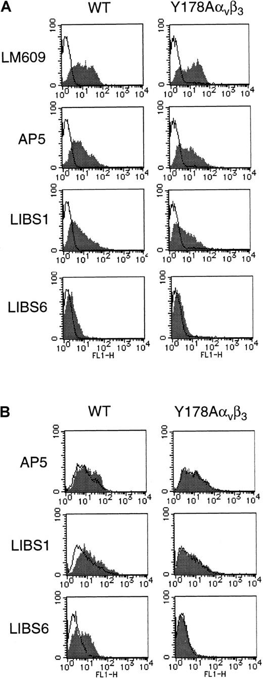
To further clarify the effects of the Y178A mutation on the structure of αvβ3, the reactivities of several mAbs (AP5, LIBS1, and LIBS6) specific for LIBS on β3 were examined in the absence of ligands. These LIBS epitopes are believed to be outside the ligand-binding pocket of the receptor. As shown in Figure 5A, there was no apparent difference in the reactivities of these mAbs between WT αvβ3 and Y178Aαvβ3, suggesting that this mutation does not grossly alter the conformation of αvβ3. The binding of activation-independent ligands, such as RGD peptides to αvβ3, has been shown to induce LIBS expression on the receptor.39Indeed, 1 mM RGDW increased the binding of all 3 LIBS mAbs to WT αvβ3. However, it failed to induce LIBS expression on the Y178Aαvβ3 mutant. These data suggest that the Y178A mutation in αv disturbs the binding of small and macromolecular RGD ligands to αvβ3.
LIBS expression on Y178Aαvβ3mutant.
WT or Y178Aαv construct was transiently cotransfected with WT β3 construct into 293 cells. Three different mAbs specific for β3 LIBS (AP5, anti-LIBS1, and anti-LIBS6) were employed to assess the LIBS expression, and LM609 (specific for αvβ3) was employed to monitor the surface expression of transfected αvβ3. (A) LIBS expression in the absence of RGDW peptide. Closed and open histograms represent the binding of anti-LIBS antibodies and control mouse IgG1, respectively. The results are representative of 2 separate experiments. (B) LIBS expression in the presence (▪) or absence (■) of 1 mM RGDW peptide. Closed and open histograms represent the binding of anti-LIBS antibodies in the presence and absence of RGDW, respectively. The results are representative of 2 separate experiments.
The Y178A mutation of αv inhibits cell adhesion to immobilized fibrinogen
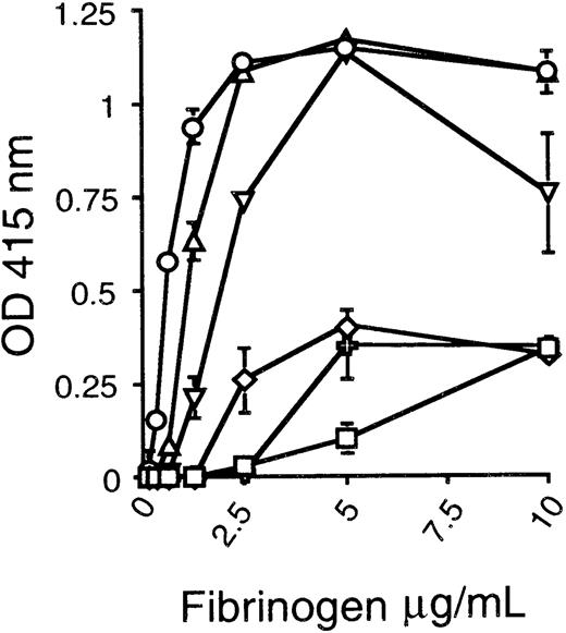
Because immobilized vitronectin and fibrinogen are activation-independent ligands for αvβ3, we further examined cell adhesion to immobilized ligands in the absence of integrin activation. Parent 293 cells showed marked adhesion to vitronectin probably via endogenous αvβ1(data not shown),36 whereas they showed only modest adhesion to fibrinogen even at a concentration of 10 μg/mL. Therefore, we examined the effect of the Y178Aαvβ3 on the cell adhesion to immobilized fibrinogen but not to vitronectin. Transfection of both WT αv and β3 markedly increased the adhesion of the transfectants; transfection of WT β3 alone induced only a modest increase in adhesion at concentrations of 2.5 and 5 μg/mL (Figure 6). As compared with the WT β3 transfectant, Y178Aαvβ3 failed to increase cell adhesion to immobilized fibrinogen. However, the adhesion of F177A and W179Aαvβ3 mutants was only slightly impaired, especially at a relatively low concentration of fibrinogen (1.25 μg/mL). Thus, the effect of Y178A on binding of αvβ3 to soluble fibrinogen is also observed with immobilized fibrinogen.
Adhesion of αvβ3 mutants to immobilized fibrinogen.
WT or mutant αv construct was transiently cotransfected with WT β3 construct into 293 cells. In WT β3 cells (⋄), only WT β3 construct was transfected into cells. WT (○) or mutant αvβ3-transfected cells were incubated for 60 minutes at 37°C with immobilized fibrinogen at serial concentrations. After washing with PBS, the adherent cells were quantified with a colorimetric reaction using endogenous cellular acid phosphatase activity. ▵, Phe177Ala; ✙, Tyr178Ala; ▿, Trp179Ala; ■, untransfected cells. Data represent the mean ± SE of triplicate measures of optical density at 415 nm.
Discussion
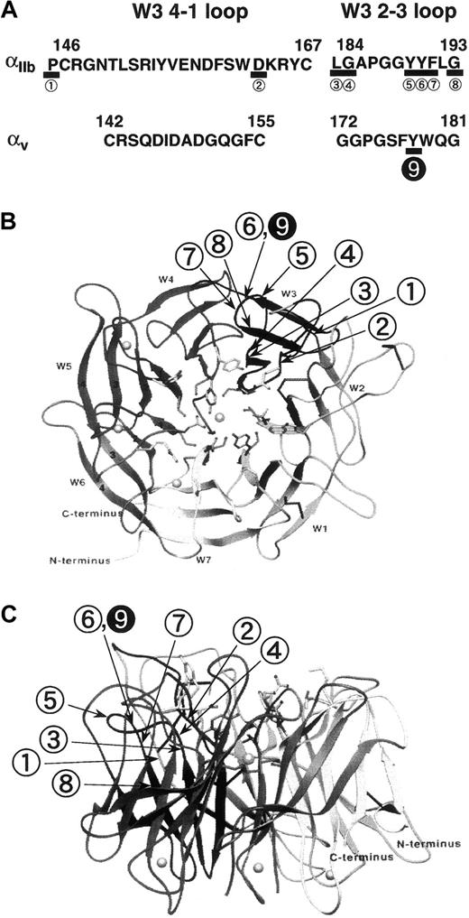
The aim of this study is to reveal the structural basis for the interaction between αvβ3 and its ligands, especially ligand-binding sites in the αv subunit of αvβ3. In non–I-domain integrin α subunits, particularly the αIIb subunit, several ligand-binding sites have been identified by the characterization of naturally occurring mutations in patients with Glanzmann thrombasthenia as well as mutagenesis analyses.15-19 However, ligand-binding regions of αv remain elusive. To clarify the critical regions for ligand binding in the αvsubunit, we focused on the predicted W3 4-1 loop (C142-C155) and W3 2-3 loop (G172-G181) and investigated their role in ligand binding by alanine-scanning mutagenesis. The results demonstrate that Y178 in the W3 2-3 loop in αv is one of the critical residues for ligand binding. In sharp contrast to αIIb, none of the mutations in the predicted W3 4-1 loop impaired ligand binding, which suggests that there are key structural differences in the adhesive ligand-binding sites of αvβ3 and αIIbβ3. The differences in the locations of the ligand-binding sites between αv and αIIb in the β-propeller model are summarized and illustrated in Figure 7.
Critical residues for ligand binding in the α subunits of β3 integrins.
(A) Comparison of critical residues for ligand binding in αv with those in αIIb. In αIIb multiple residues (underlined) critical for ligand binding have been identified in both the W3 4-1 and W3 2-3 loops. In sharp contrast, in αv only Y178 within the W3 2-3 loop is critical for ligand binding. The figures in panels B and C are adapted from a β-propeller model proposed by Springer,20 and they show the location of these critical residues. The view is shown from the top (B) and from the side (C).
There is mounting evidence that the predicted loops between N-terminal repeats 2 and 3 (W3 4-1 loop) and within repeat 3 (W3 2-3 loop) are important for ligand binding in non–I-domain αsubunits.18,40-43 Previous studies have shown that the N-terminal one-third of the α subunit regulates ligand-recognition specificity of β3 integrins44 and that residues 139-167 in αv corresponding to the W3 4-1 loop are a cross-linking site for RGD peptides.21 However, the αv Y178 identified in this study is located in the predicted W3 2-3 loop, and none of the alanine substitutions within the W3 4-1 loop examined impaired ligand binding. The inhibition of ligand binding by alanine substitutions at the residues adjacent to Y178 (F177 and W179) and the failure of the induction of β3 LIBS with RGDW, even at high concentrations, provide further support for the critical role of Y178 in ligand binding. Although the W3 4-1 loop and W3 2-3 loop are separated in the primary structure, the proposed propeller model suggests that these regions are close to each other in 3-dimensional space and may explain the apparent discrepancy between the previous cross-linking study and the present study (Figure 7).
Interestingly, many critical residues identified in non–I-domain α subunits have aromatic side chains, suggesting that in addition to oxygenated residues such as D163 in αIIb,16 aromatic residues are important for ligand binding.45 The corresponding residues to the αv Y178 in αIIb(Y190),18 α4 (Y187),42,43 and α5 (F187),43 and the adjacent residues Y186 and W188 in α340 have been demonstrated to be critical for ligand binding, although the role of W3 2-3 loop in α3 is still controversial.41 In contrast, the role of W3 4-1 loop appears to be different between integrin α subunits. D163 in αIIb and threonine (T)162 and G163 in α3, which are located in the predicted W3 4-1 loop, appear critical for ligand binding, whereas the W3 4-1 loop in α4 and in αv (this study) do not. More recently it has been demonstrated that the replacement of the W3 4-1 loop in αv with the corresponding loop in α5 did not disturb ligand binding but changed ligand-recognition specificity.46 Our swapping mutagenesis of the W3 4-1 loop of αv with the corresponding region of αIIb did not abolish WOW-1 Fab binding, suggesting that the W3 4-1 loop is not critical for ligand-recognition specificity between αvβ3 and αIIbβ3.
WOW-1, a monovalent ligand-mimetic antibody, was created by replacing the H-CDR3 of PAC-1 Fab with a single integrin-binding domain of the adenovirus penton base, a viral coat protein that consists of 5 subunits, each containing an integrin-binding RGD motif. Penton base is known to facilitate adenovirus internalization through αvintegrins, particularly αvβ3 and αvβ5.47 WOW-1–like penton base recognizes that activated state of αvβ3 and αvβ5. Using mAb B5-IVF2 specific for β5, we have determined that 293 cells express β5 as well as αv (S.H. and Y.T., unpublished data, June 1999). Thus, WOW-1 Fab might bind to αvβ3-transfected 293 cells through αvβ5 as well as αvβ3. Nonetheless, the bulk of WOW-1 Fab binding to transfected cells appeared to be through αvβ3 because antibody binding was largely abolished by the Y178A substitution in αv or the D119Y substitution in β3. Integrin activation encompasses at least 2 events: (1) modulation of receptor affinity through conformational changes and (2) modulation of receptor avidity through facilitation of lateral diffusion and/or clustering of heterodimers. The binding of monovalent ligand WOW-1 Fab to αvβ3 likely reflects affinity modulation, whereas the binding of multivalent ligand, such as fibrinogen, likely reflects both affinity and avidity modulation.28
Our data with WOW-1 Fab suggest that the Y178A substitution disturbs integrin affinity modulation rather than avidity modulation. In addition to ligand binding, cell adhesion to immobilized ligand can be strongly influenced by post–ligand-binding events. Indeed, in spite of the moderate inhibition of soluble fibrinogen binding by F177A as well as W179A substitution, these substitutions induced only a modest inhibition of cell adhesion to immobilized fibrinogen. One could argue the possibility that the Y178A substitution in αv may disturb conformational changes from resting to activated states of integrin because of the failure of the induction of β3LIBS with RGDW.48 However, this possibility is unlikely because the Y178A substitution in αv completely abolished the interaction with immobilized fibrinogen, an activation-independent ligand. Thus, this residue is likely involved in direct contact with the ligand. It would be interesting to know whether the Y178A substitution may affect divalent cation binding. Recently, employing human-to-mouse chimeras, Puzon-McLaughlin et al49localized binding sites for ligand-mimetic murine mAbs against αIIbβ3 and demonstrated that the involvement of several discontinuous sites in both αIIband β3 is unique to ligand-mimetic antibodies. Involvement of certain residues in both αv (Y178) and β3 (D119) subunits in WOW-1 binding is consistent with their data, and these residues may participate in a ligand-binding pocket in αvβ3.
The αvβ3 is expressed in a number of cell types: endothelial cells, arterial smooth muscle cells, platelets, subpopulation of leukocytes, osteoclasts, and tumor cells. The αvβ3 is involved in cell adhesion, proliferation, and migration and has been shown to play a crucial role in tumor angiogenesis, intimal hyperplasia after arterial injury, wound healing, and osteoporosis in the adult organism. Recently, human clinical trials are in progress to evaluate the effects of the humanized anti-αvβ3 mAb in patients with late-stage cancer.50 The present results provide new information concerning the interaction between RGD ligands and the non–I-domain αv subunit as well as key structural differences between αvβ3 and αIIbβ3 with regard to ligand binding. This information may facilitate the development of novel antagonists specific for αvβ3.
Acknowledgments
We thank Dr David Cheresh for mAbs LM142 and LM609 and the vector containing WT αv cDNA; Dr Mark Ginsberg for mAbs anti-LIBS1 and anti-LIBS6; Dr Thomas Kunicki for a mAb AP5; Dr Peter Newman for a mAb AP3; Dr Gilbert White for the vector containing WT β3 cDNA; Dr Martin Hemler for a mAb B5-IVF2; and Dr Jiro Seki for RGDW peptide.
Supported by a grant from the Ministry of Education, Science and Culture, Tokyo, Japan; a grant from the Japan Society for the Promotion of Science, Tokyo, Japan; a grant from the Senri Life Science Foundation, Osaka, Japan; a grant from the Yamanouchi Foundation for Research on Metabolic Disorders, Tokyo, Japan; Welfide Medical Research Foundation, Osaka, Japan; and grant HL56595 from the National Institutes of Health, Bethesda, MD.
Submitted July 6, 2000; accepted September 14, 2000.
The publication costs of this article were defrayed in part by page charge payment. Therefore, and solely to indicate this fact, this article is hereby marked “advertisement” in accordance with 18 U.S.C. section 1734.
References
Author notes
Yoshiaki Tomiyama, Department of Internal Medicine and Molecular Science, Graduate School of Medicine, Osaka University, 2-2 B5, Yamadaoka, Suita, Osaka 565-0871, Japan; e-mail:yoshi@hp-blood.med.osaka-u.ac.jp.


This feature is available to Subscribers Only
Sign In or Create an Account Close Modal