

Key Points
A novel MPLR464G variant induces CAMT due to defective trafficking to the cell surface.
The CALR mutant renders MPLR464G responsive to ELT.

Abstract
Congenital amegakaryocytic thrombocytopenia (CAMT) is a severe inherited thrombocytopenia due to loss-of-function mutations affecting the thrombopoietin (TPO) receptor, MPL. Here, we report a new homozygous MPL variant responsible for CAMT in 1 consanguineous family. The propositus and her sister presented with severe thrombocytopenia associated with mild anemia. Next-generation sequencing revealed the presence of a homozygous MPLR464G mutation resulting in a weak cell-surface expression of the receptor in platelets. In cell lines, we observed a defect in MPLR464G maturation associated with its retention in the endoplasmic reticulum. The low cell-surface expression of MPLR464G induced very limited signaling with TPO stimulation, leading to survival and reduced proliferation of cells. Overexpression of a myeloproliferative neoplasm–associated calreticulin (CALR) mutant did not rescue trafficking of MPLR464G to the cell surface and did not induce constitutive signaling. However, it unexpectedly restored a normal response to eltrombopag (ELT), but not to TPO. This effect was only partially mimicked by the purified recombinant CALR mutant protein. Finally, the endogenous CALR mutant was able to restore the megakaryocyte differentiation of patient CD34+ cells carrying MPLR464G in response to ELT.
Introduction
Congenital amegakaryocytic thrombocytopenia (CAMT; OMIM 604498) is a rare autosomal-recessive bone marrow failure disorder that can evolve into severe aplastic anemia and leukemia.1 Usually, patients with CAMT exhibit high thrombopoietin (TPO) serum levels, and a majority of cases carry heterozygous compound or homozygous nonsense, missense, and splicing mutations in the MPL gene encoding the TPO receptor.2,3 The cell-surface expression of mature MPL necessary for TPO-induced signaling is mainly achieved through the conventional endoplasmic reticulum (ER)-Golgi pathway dependent on MPL association with JAK2 as a chaperone protein.4 However, an unconventional, autophagy-dependent manner of delivery of immature MPL to the cell surface has also been described.5 TPO/MPL signaling controls hematopoietic stem cell survival and megakaryocyte (MK) differentiation.6 Interestingly, TPO and MPL play a more important role in humans than in mice. Indeed, Thpo7 and Mpl8 knockout mice are viable, with a residual platelet production preventing hemorrhage, and do not develop bone marrow failure.
Study design
Patients
Blood samples from patients and healthy subjects were collected after informed written consent and obtained in accordance with the Declaration of Helsinki. The study was approved by the Comité de Protection des Personnes CPP N°2020T2-02.
Samples and cell lines
Peripheral blood CD34+ cells, megakaryoblastic UT-7 cells, HEK293T cells, and Ba/F3 cells were transduced, sorted, and cultured as described in supplemental Methods (available on the Blood Web site).
Statistics
Statistical analyses were performed using PRISM software (GraphPad). Statistical significance was determined using a Mann-Whitney test or 2-way analysis of variance with Bonferroni posttest. Differences were considered significant at P < .05.
More details are provided in supplemental Methods.
Results and discussion
We report here 1 French family (supplemental Figure 1A) with a novel homozygous MPL mutation in 2 children with severe thrombocytopenia and high TPO levels. The propositus is a 3-year-old female (II.3) born to first-cousin parents of Turkish origin and diagnosed with a severe thrombocytopenia (34 × 109 platelets per L) at the age of 3 months. At the age of 2 years, due to the decrease in platelet counts (<20 × 109/L) associated with a slight anemia, she was first unsuccessfully treated with romiplostim, then with eltrombopag (ELT) with only a weak response. Her 8-year-old sister (II.2) was also severely thrombocytopenic (39 × 109 platelets per L), while both parents had normal platelet counts (supplemental Figure 1B).
Next-generation sequencing identified MPL p.R464G (variant allele frequency [VAF], 100%) and ZFPM1 p.A137G (VAF, 50%) variants for the propositus and MPL p.R464G (VAF, 100%) variant for her sister. The MPL p.R464G mutation at a VAF of 50% in both parents was confirmed by Sanger sequencing (data not shown). Interestingly, the same variant was recently reported in 1 unrelated heterozygous CAMT patient.9 The R464 residue is located on the extracellular domain of MPL.
To tease out the mechanism by which this mutant could lead to severe thrombocytopenia, we first investigated its expression on the surface of platelets from the propositus sister (II.2), presenting higher platelet levels throughout the follow-up. As shown in Figure 1A, an almost-complete absence of MPL at the platelet surface was observed.
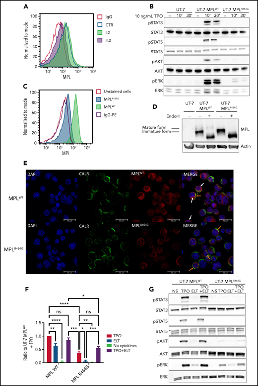
MPLR464G is only weakly expressed on the cell surface and induces very weak signaling compared with MPLWT. (A) Flow cytometric analyses of MPL expression on platelets from 1 patient homozygous for MPLR464G (II.2), her heterozygous mother (I.2), and 1 healthy control (CTR). (B) Western blot analysis of TPO-induced signaling in UT-7 cells without MPL expression (UT-7), overexpressing the WT form of MPL (UT-7 MPLWT), and mutant MPL (UT-7 MPLR464G). Cells were starved overnight and stimulated with 10 ng/mL TPO for 10 or 30 minutes. No signal is detected in parental UT-7 cells, and a weak ERK signal is detected in the presence of MPLR464G in UT-7 cells. (C) Flow cytometric analyses of MPL expression on the surface of Ba/F3 cells overexpressing MPLWT and MPLR464G. (D) Western blot analysis of the mature (85 kDa) and immature (80 kDa) forms of MPL. The mature form of MPL that is resistant to endoglycosidase H (Endo H) digestion and able to reach the cell membrane is not detectable in UT-7 cells overexpressing MPLR464G. (E) Immunofluorescence staining for the expression of MPL and CALR in UT-7 cells overexpressing MPLWT or MPLR464G. The orange arrows indicate MPL colocalization with CALR in the ER, and the white arrows indicate diffuse cytoplasmic/cell-surface MPL expression. Scale bars, 30 μm. An anti-MPL coupled with phycoerythrin (PE; A,C) and uncoupled anti-MPL antibodies (D-E) were used. (F-G) Proliferation curves and western blot analysis of signaling of UT-7 cells overexpressing MPLWT and MPLR464G in presence of TPO (10 ng/mL), ELT (2 μg/mL), TPO (10 ng/mL) + ELT (2 μg/mL), or without cytokines (no cytokines). (F) 5 × 10⁴ cells were plated in triplicate in 24-well plates and counted every day for 4 days. The counts were reported to the condition of UT-7 MPLWT+TPO (in blue color). Shown are averages of 3 independent experiments at day 4, each performed in triplicate ± standard error of the mean (error bars). *P < .05; ns, not significant; Mann-Whitney unpaired, nonparametric 1-tailed test. (G) Cells were starved overnight and stimulated for 10 minutes. A weak ERK signaling is detected in the presence of MPLR464G in all 3 conditions (TPO, ELT, and TPO + ELT). No signaling is detected in nonstimulated (NS) cells. **P < .01; ***P < .005; ****P < .001.
MPLR464G is only weakly expressed on the cell surface and induces very weak signaling compared with MPLWT. (A) Flow cytometric analyses of MPL expression on platelets from 1 patient homozygous for MPLR464G (II.2), her heterozygous mother (I.2), and 1 healthy control (CTR). (B) Western blot analysis of TPO-induced signaling in UT-7 cells without MPL expression (UT-7), overexpressing the WT form of MPL (UT-7 MPLWT), and mutant MPL (UT-7 MPLR464G). Cells were starved overnight and stimulated with 10 ng/mL TPO for 10 or 30 minutes. No signal is detected in parental UT-7 cells, and a weak ERK signal is detected in the presence of MPLR464G in UT-7 cells. (C) Flow cytometric analyses of MPL expression on the surface of Ba/F3 cells overexpressing MPLWT and MPLR464G. (D) Western blot analysis of the mature (85 kDa) and immature (80 kDa) forms of MPL. The mature form of MPL that is resistant to endoglycosidase H (Endo H) digestion and able to reach the cell membrane is not detectable in UT-7 cells overexpressing MPLR464G. (E) Immunofluorescence staining for the expression of MPL and CALR in UT-7 cells overexpressing MPLWT or MPLR464G. The orange arrows indicate MPL colocalization with CALR in the ER, and the white arrows indicate diffuse cytoplasmic/cell-surface MPL expression. Scale bars, 30 μm. An anti-MPL coupled with phycoerythrin (PE; A,C) and uncoupled anti-MPL antibodies (D-E) were used. (F-G) Proliferation curves and western blot analysis of signaling of UT-7 cells overexpressing MPLWT and MPLR464G in presence of TPO (10 ng/mL), ELT (2 μg/mL), TPO (10 ng/mL) + ELT (2 μg/mL), or without cytokines (no cytokines). (F) 5 × 10⁴ cells were plated in triplicate in 24-well plates and counted every day for 4 days. The counts were reported to the condition of UT-7 MPLWT+TPO (in blue color). Shown are averages of 3 independent experiments at day 4, each performed in triplicate ± standard error of the mean (error bars). *P < .05; ns, not significant; Mann-Whitney unpaired, nonparametric 1-tailed test. (G) Cells were starved overnight and stimulated for 10 minutes. A weak ERK signaling is detected in the presence of MPLR464G in all 3 conditions (TPO, ELT, and TPO + ELT). No signaling is detected in nonstimulated (NS) cells. **P < .01; ***P < .005; ****P < .001.
Next, we overexpressed the wild-type (WT) and mutant MPLR464G in both the human granulocyte-macrophage colony-stimulating factor (GM-CSF)–dependent UT-7 and the interleukin-3–dependent murine Ba/F3 cell lines. As expected, in UT-7 cells, TPO stimulation of STAT3, STAT5, AKT, and ERK pathways was observed only when MPLWT was expressed. In both UT-7 and Ba/F3 cells overexpressing MPLR464G, TPO induced a slight but significant activation of ERK and a barely detectable phosphorylation of STAT and AKT that were not observed in the absence of cytokines (Figure 1B; supplemental Figure 2A-B). No defect in signaling was detected in presence of GM-CSF (supplemental Figure 2C). Similarly, a dual luciferase assay in HEK293T cells for STAT5 activity showed a discrete response to TPO (supplemental Figure 3), suggesting that the mutation affects either the interaction with TPO or the cell-surface expression of MPL. Low levels of MPLR464G were detected at the cell surface of Ba/F3 cells by flow cytometry (Figure 1C), and even lower levels were found on the membrane of UT-7 cells (not shown). Western blot analysis showed that MPLR464G was in an immature, incompletely glycosylated form, but total MPLR464G compared with MPLWT cell amounts were not different (Figure 1D). A defect in trafficking, rather than an increased degradation, was corroborated by the colocalization of MPLR464G with the calreticulin (CALR) ER chaperone, suggesting its retention in the ER (Figure 1E; supplemental Figure 4). The proliferation rate of UT-7 MPLR464G cells in the presence of TPO was profoundly decreased compared with UT-7 MPLWT cells (Figure 1F). A complete absence of proliferation was detected in the presence of ELT, a small-molecule agonist of MPL that binds in proximity to the mutation10 (Figure 1F). Furthermore, the combination of TPO and ELT did not achieve a significant synergism on proliferation and signaling (Figure 1F-G; supplemental Figure 5). Overall, these results indicate that due to a trafficking defect, only a small amount of MPLR464G reaches the cell surface, inducing weak signaling in response to TPO stimulation, as previously described for cell lines expressing limited levels of MPLWT.11 CAMT can be classified in 2 groups; type I presents a more severe phenotype and a total loss of cell-surface MPL expression due to deletions and nonsense and frameshift mutations, whereas the less severe type II may have residual receptor function due to missense MPL mutations.12 Our results indicate the MPLR464G variant belongs to the type II CAMT.
Trafficking of the previously described type I CAMT–associated MPLR102P is totally blocked in the ER; however, both its traffic and signaling can be restored by the expression of 2 CALR mutants, CALRdel52 and CALRins5, identified in myeloproliferative neoplasms.13 We investigated whether a similar rescue was possible for MPLR464G. As shown in Figure 2A, CALRWT overexpression in UT-7 MPLR464G cells was not able to induce constitutive signaling or signaling in the presence of ELT. In contrast, a slight constitutive signaling was detected with CALRdel52 overexpression, and, unexpectedly, this signaling was enhanced by ELT, but not by TPO (Figure 2A; supplemental Figure 6). However, CALRdel52 was not able to rescue the trafficking defect of MPLR464G (Figure 2B). CALRdel52 has been shown to activate MPLWT, 14-16 but it only slightly activates MPLR464G. Interestingly, CALRdel52 renders MPLR464G cells responsive to ELT and increases the response of MPLWT cells to ELT (Figure 2A; supplemental Figure 6), suggesting that CALRdel52 may induce conformation changes in MPL and a better accessibility of the ELT-binding site or/and improve MPL dimerization. In contrast, the interaction between CALRdel52 and MPLR464G on the cell surface may prevent TPO binding and may explain the modest additional effect of TPO on signaling in CALRdel52-expressing cells (Figure 2A). In agreement with these results, the proliferation of UT-7 MPLR464G cells expressing CALRdel52 was almost completely restored in the presence of ELT, but not TPO, which also did not increase the effects of ELT. This is consistent with our previous findings that CALRdel52 protein binds to MPL and sterically hinders the TPO-binding site,15 although the TPO-binding site per se is not required for activation by CALR mutants of MPL.14 In contrast, overexpression of CALR WT elicited a synergism between TPO and ELT (Figure 2C). Furthermore, proliferation was completely dependent on JAK2 activation, suggesting a normal interaction between MPLR464G and JAK2 (supplemental Figure 7). As CALRdel52 activates MPL on the cell surface,13 and since secreted soluble CALRdel52 was reported to potentially act as a ligand for cell-surface MPL17 (S.N.C., manuscript in preparation), we investigated the effects of exogenously added soluble CALRdel52 protein on the proliferation of MPLR464G-expressing UT-7 cells. As shown in Figure 2D, the soluble CALRdel52 only partially restored the response to ELT of UT-7 MPLR464G cells, suggesting that endogenous CALRdel52 may bind more efficiently to MPLR464G than exogenous CALRdel52. Lastly, we wondered whether CALRdel52 could restore MK differentiation of patient CD34+ cells in vitro. To this end, the II.2 patient CD34+ cells were transduced with a CALRdel52 retrovirus and cultured in presence of stem cell factor and ELT (supplemental Figure 8). As expected, only the cells expressing CALRdel52 were able to differentiate into MKs (Figure 2E).
![CALR mutant (CALRdel52) restores the response of MPLR464G to ELT. UT-7 and Ba/F3 cells were transduced with retroviruses to overexpress WT MPL (MPLWT) or mutant MPL (MPLR464G) together with GFP, and retroviruses overexpressing WT CALR (CALRWT) or mutant CALR (CALRdel52) with mCherry, and sorted for GFP+mCherry+ cells. Parental UT-7 cells were used as negative control. (A) Western blot analysis of signaling induced by TPO or ELT. UT-7 cells were cultured in presence of GM-CSF, starved overnight, and stimulated with TPO (10 ng/mL) or ELT (2 μg/mL) for 10 minutes. CALRdel52 enhances signaling in presence of ELT both in MPLWT- and MPLR464G- expressing cells. (B) Flow cytometric analysis of MPL expression on Ba/F3 cells using an allophycocyanin-coupled MPL antibody. CALRdel52 does not induce MPLR464G traffic to the cell membrane. IgG, immunoglobulin G. (C) Proliferation assay in the presence of TPO, ELT, or TPO + ELT or without cytokines (no cytokines). (D) Proliferation assay in presence of ELT and recombinant CALRdel52 (recCALRdel52) (20 μg/mL). In panels C and D, 5 × 10⁴ UT-7 cells were plated in triplicate in 24-well plates and counted every day for 4 days. Shown are averages of 3 independent experiments, each performed in triplicate ± standard error of the mean (error bars). In panel C, the counts are reported for the condition UT-7 MPLWT/CALRWT (purple) + TPO and are shown only at day 4 of culture. *P < .05, Mann-Whitney unpaired, nonparametric 1-tailed test. (E) Control (CTRL) and patient CD34+ cells (II-2) isolated from peripheral blood were transduced with retrovirus encoding CALRdel52, sorted at day 2 on CD34+, and cultured in presence of stem cell factor and ELT for 12 days. At least 20 mCherry+ and 20 mCherry− cells were analyzed. All mCherry+ cells expressed MK markers, while no mCherry− cells differentiated into MKs (not shown). Representative pictures of mCherry+ CTRL and II-2 MK (CD41+ and von Willebrand factor [vWF]+) are shown. White arrows indicate individual MKs; 2 different plans of the same view are shown (i,ii). Scale bars, 30 μm.](https://ash.silverchair-cdn.com/ash/content_public/journal/blood/138/6/10.1182_blood.2020010567/4/m_bloodbld2020010567f2.png?Expires=1769094088&Signature=AsM0FreDPpNlX3XFzZQpgFL5vJNCetulDo73tjalS3KKZT1zPhXYDvC9L6V4GyLZtVnJ4nsmcd30SiktDTsTRXba4hUK77A9-eIQh1tSza22FNG3Mv67Pn365hvgSXA8JRRJFwLnCUJDbJhqHlI-7RnyP1ad2L~dEpr2zTQJ7R09PTdfouewqdY68xBaGioq~6p2t8g83ClLpGpsalHZq2ZZulrfC7gEmrVyGMtUWMNs6VNQ3L2wQMx7dMZsrUcFrCzP06rD2vmzrx1iSnFyBeYstiWhw4RH8QS2T7xmk-x~l2I8CfUx4pP4U1mj3lRGh8tDKR4fEps3n~8npTJOhg__&Key-Pair-Id=APKAIE5G5CRDK6RD3PGA)
CALR mutant (CALRdel52) restores the response of MPLR464G to ELT. UT-7 and Ba/F3 cells were transduced with retroviruses to overexpress WT MPL (MPLWT) or mutant MPL (MPLR464G) together with GFP, and retroviruses overexpressing WT CALR (CALRWT) or mutant CALR (CALRdel52) with mCherry, and sorted for GFP+mCherry+ cells. Parental UT-7 cells were used as negative control. (A) Western blot analysis of signaling induced by TPO or ELT. UT-7 cells were cultured in presence of GM-CSF, starved overnight, and stimulated with TPO (10 ng/mL) or ELT (2 μg/mL) for 10 minutes. CALRdel52 enhances signaling in presence of ELT both in MPLWT- and MPLR464G- expressing cells. (B) Flow cytometric analysis of MPL expression on Ba/F3 cells using an allophycocyanin-coupled MPL antibody. CALRdel52 does not induce MPLR464G traffic to the cell membrane. IgG, immunoglobulin G. (C) Proliferation assay in the presence of TPO, ELT, or TPO + ELT or without cytokines (no cytokines). (D) Proliferation assay in presence of ELT and recombinant CALRdel52 (recCALRdel52) (20 μg/mL). In panels C and D, 5 × 10⁴ UT-7 cells were plated in triplicate in 24-well plates and counted every day for 4 days. Shown are averages of 3 independent experiments, each performed in triplicate ± standard error of the mean (error bars). In panel C, the counts are reported for the condition UT-7 MPLWT/CALRWT (purple) + TPO and are shown only at day 4 of culture. *P < .05, Mann-Whitney unpaired, nonparametric 1-tailed test. (E) Control (CTRL) and patient CD34+ cells (II-2) isolated from peripheral blood were transduced with retrovirus encoding CALRdel52, sorted at day 2 on CD34+, and cultured in presence of stem cell factor and ELT for 12 days. At least 20 mCherry+ and 20 mCherry− cells were analyzed. All mCherry+ cells expressed MK markers, while no mCherry− cells differentiated into MKs (not shown). Representative pictures of mCherry+ CTRL and II-2 MK (CD41+ and von Willebrand factor [vWF]+) are shown. White arrows indicate individual MKs; 2 different plans of the same view are shown (i,ii). Scale bars, 30 μm.
CALR mutant (CALRdel52) restores the response of MPLR464G to ELT. UT-7 and Ba/F3 cells were transduced with retroviruses to overexpress WT MPL (MPLWT) or mutant MPL (MPLR464G) together with GFP, and retroviruses overexpressing WT CALR (CALRWT) or mutant CALR (CALRdel52) with mCherry, and sorted for GFP+mCherry+ cells. Parental UT-7 cells were used as negative control. (A) Western blot analysis of signaling induced by TPO or ELT. UT-7 cells were cultured in presence of GM-CSF, starved overnight, and stimulated with TPO (10 ng/mL) or ELT (2 μg/mL) for 10 minutes. CALRdel52 enhances signaling in presence of ELT both in MPLWT- and MPLR464G- expressing cells. (B) Flow cytometric analysis of MPL expression on Ba/F3 cells using an allophycocyanin-coupled MPL antibody. CALRdel52 does not induce MPLR464G traffic to the cell membrane. IgG, immunoglobulin G. (C) Proliferation assay in the presence of TPO, ELT, or TPO + ELT or without cytokines (no cytokines). (D) Proliferation assay in presence of ELT and recombinant CALRdel52 (recCALRdel52) (20 μg/mL). In panels C and D, 5 × 10⁴ UT-7 cells were plated in triplicate in 24-well plates and counted every day for 4 days. Shown are averages of 3 independent experiments, each performed in triplicate ± standard error of the mean (error bars). In panel C, the counts are reported for the condition UT-7 MPLWT/CALRWT (purple) + TPO and are shown only at day 4 of culture. *P < .05, Mann-Whitney unpaired, nonparametric 1-tailed test. (E) Control (CTRL) and patient CD34+ cells (II-2) isolated from peripheral blood were transduced with retrovirus encoding CALRdel52, sorted at day 2 on CD34+, and cultured in presence of stem cell factor and ELT for 12 days. At least 20 mCherry+ and 20 mCherry− cells were analyzed. All mCherry+ cells expressed MK markers, while no mCherry− cells differentiated into MKs (not shown). Representative pictures of mCherry+ CTRL and II-2 MK (CD41+ and von Willebrand factor [vWF]+) are shown. White arrows indicate individual MKs; 2 different plans of the same view are shown (i,ii). Scale bars, 30 μm.
Overall, these data demonstrate that CALRdel52/MPLR464G complex leads to a conformation that is very permissive to the activating effect of ELT, which binds at the outset of the transmembrane domain (H499). CALRdel52 binds to the extracellular domain of MPL involving glycosylated N117 in D1, likely exposing the H499-containing region where ELT binds. The combination of TPO and ELT seems to have an effect similar to that observed with exogenous CALRdel52 and ELT. Taken together, these data may reveal novel approaches to modify the response of MPL to ELT and suggest that a dual activation of MPL may be efficient in cases of low MPL trafficking to the cell membrane.
Acknowledgments
The authors thank the patients who participated in this study; M.C. Alessi, who coordinated the “Centre de Référence des Pathologies Plaquettaire” (Paris, France); and P. Rameau and C. Catelain from the Imaging and Cytometry Platform (Plate-Forme Imagerie et Cytométrie), Unité Mixte de Service Analyse Moléculaire, Modélisation et Imagerie de la aladie Cancéreuse (PFIC, UMS AMMICA), Gustave Roussy Villejuif, France for their expertise in cytometry.
F.B.-V. was supported by the Université Sorbonne Paris Cité/Université Paris Diderot and French Society of Hematology. The work of H.R.’s team is supported by the Ligue Nationale contre le Cancer (Equipe Labellisée 2016 and 2019 [H.R.]) and Institut Naitonal du Cancer Institut Naitonal du Cancer (INCa PLBIO); 2015, 2017, and 2018) (I.P.). S.C. was supported by funding from Ludwig Institute for Cancer Research, Fondation contre le Cancer of Belgium, Salus Sanguinis Foundation (Bruxelles, Belgium), and Fondation “Les Avions de Sébastien”, Projects Action de Recherché Concertée 16/21-073 and WelBio F 44/8/5 - MCF/UIG - 10955, is acknowledged.
Authorship
Contribution: F.B.-V., G.L., and L.N.V. designed and performed experiments, analyzed data, and contributed to the manuscript draft; M.O., P.B., and N.B. performed experiments and analyzed data; B.N. provided clinical and biological follow-up of patients; S.N.C. provided bacterial recombinant CALRdel52; C.M., I.P., W.V., and S.N.C. discussed results and contributed to manuscript editing; R.F. and H.R. designed and supervised the work and wrote the paper; and all authors contributed to the final approval of the manuscript.
Conflict-of-interest disclosure: The authors declare no competing financial interests.
Correspondence: Hana Raslova, INSERM UMR 1287, Gustave Roussy, 114 rue Edouard Vaillant, 94805 Villejuif, France; e-mail: hana.raslova@gustaveroussy.fr.
For original data, e-mail the corresponding author.
The online version of this article contains a data supplement.
The publication costs of this article were defrayed in part by page charge payment. Therefore, and solely to indicate this fact, this article is hereby marked “advertisement” in accordance with 18 USC section 1734.
