Abstract
Dickkopf-1 (DKK1), a soluble inhibitor of Wnt signaling secreted by multiple myeloma (MM) cells contributes to osteolytic bone disease by inhibiting the differentiation of osteoblasts. In this study, we tested the effect of anti-DKK1 therapy on bone metabolism and tumor growth in a SCID-rab system. SCID-rab mice were engrafted with primary MM cells expressing varying levels of DKK1 from 11 patients and treated with control and DKK1-neutralizing antibodies for 4 to 6 weeks. Whereas bone mineral density (BMD) of the implanted myelomatous bone in control mice was reduced during the experimental period, the BMD in mice treated with anti-DKK1 increased from pretreatment levels (P < .001). Histologic examination revealed that myelomatous bones of anti-DKK1–treated mice had increased numbers of osteocalcin-expressing osteoblasts and reduced number of multinucleated TRAP-expressing osteoclasts. The bone anabolic effect of anti-DKK1 was associated with reduced MM burden (P < .04). Anti-DKK1 also significantly increased BMD of the implanted bone and murine femur in nonmyelomatous SCID-rab mice, suggesting that DKK1 is physiologically an important regulator of bone remodeling in adults. We conclude that DKK1 is a key player in MM bone disease and that blocking DKK1 activity in myelomatous bones reduces osteolytic bone resorption, increases bone formation, and helps control MM growth.
Introduction
Multiple myeloma (MM), a tumor of terminally differentiated plasma cells that home to and expand in the bone marrow (BM), is associated with osteolytic bone disease. This debilitating condition is caused by an uncoupling of bone remodeling as a result of increased activity of osteoclasts and decreased activity of osteoblasts.1,2 Much of the research on the mechanisms of osteolysis in MM has focused on the role of osteoclasts in shifting the uncoupling process.2,3 Although bone resorption can be blocked by bisphosphonates, which inhibit osteoclastogenesis,4,5 the inability of these compounds to induce bone formation and repair lytic lesions indicates that a functional defect of osteoblasts is also involved. Indeed, alterations in the number and function of osteoblasts is a primary event in MM.6,7
Recent studies have revealed that Wnt signaling is involved in both normal skeletogenesis8,9 and cancer-related bone disease.10,11 The first link between Wnt signaling and human bone disease came from the observations that inactivating mutations in the Wnt coreceptor, LRP5, causes the osteoporosis-pseudoglioma (OPPG) syndrome.12 Subsequently it was shown that in the syndrome of hereditary high bone density13 mutations in LRP5, distinct from those seen in OPPG, prevent binding of Dickkopf-1 (DKK1), a soluble inhibitor of Wnt and high-affinity ligand for LRP5.14
The importance of DKK1 in normal skeletal development has also been demonstrated by the extra digits in DKK1 null mice and loss of bony structures in chicken and mice exposed to elevated levels of DKK1.15.16 To determine the role of DKK1 in vivo and overcome the embryonic lethality of homozygous deletion, Morvan and colleagues17 showed that mice lacking a single allele of DKK1 have a marked increase in bone mass. In contrast, transgenic overexpression of DKK1 under the control of Col1A1 promoters caused severe osteopenia.18
We have shown that whereas plasma cells from normal healthy donors and those from patients with monoclonal gammopathy of undetermined significance (MGUS) do not express DKK1, plasma cells from virtually all patients with MM express this protein19 (J.D.S., unpublished data, October 2006). Moreover, the expression of DKK1 in plasma cells and serum levels of this soluble protein were positively correlated with the presence of bone lesions in patients with myeloma.19,20 Our study also showed that serum taken from patients with MM inhibited osteoblast differentiation in vitro, an effect that was blocked by a neutralizing antibody to DKK1.19
The role of DKK1 in promoting the development of bone lesions is not limited to MM but has also recently been expanded to prostate cancer. The osteolytic prostate cancer line PC-3, when transfected with shRNA targeting DKK1, reverted to an osteoblastic phenotype.21 In addition, transfection of DKK1 into the osteoblastic prostate cancer cell line C4-2B, which normally induces a mix of osteoblastic and osteolytic lesions, caused the cells to develop osteolytic tumors in SCID mice.21
Recent evidence suggests that in addition to inhibiting osteoblastogenesis, elevated DKK1 levels may enhance osteoclastogenesis. The balance between the levels of receptor activator of the NF-κB ligand (RANKL) and osteoprotegerin (OPG), a soluble receptor and antagonist of RANK signaling, controls osteoclastogenesis.22,23 Immature, but not mature, osteoblasts are rich sources of RANKL.24 Wnt signaling in osteoblasts up-regulates expression of OPG25 and down-regulates the expression of RANKL,26 suggesting a mechanism by which Wnt signaling in osteoblasts indirectly regulates osteoclastogenesis. Taken together, these studies indicate that DKK1 is a key regulator of bone remodeling in physiologic and pathologic conditions and that blocking this factor may result in stimulation of osteoblastogenesis and inhibition of osteoclastogenesis in myelomatous bone.
We have recently developed the SCID-rab mouse model for human primary MM.27 These mice are constructed by implanting a nonfetal rabbit bone into which primary human MM cells are directly injected. Similar to our extensively tested and validated SCID-hu system, which uses a human fetal bone,28–31 MM cells from the majority of patients grow exclusively in the implanted bone and produce typical myeloma manifestations including stimulation of osteoclastogenesis, suppression of osteoblastogenesis, and induction of severe osteolytic bone disease. Here we report that treatment of myelomatous SCID-rab mice with anti-DKK1 antibody can stimulate bone formation in myelomatous bone and reduce bone loss and tumor growth in vivo.
Materials and methods
Myeloma cells
MM cells were obtained from heparinized BM aspirates from 11 patients with active myeloma during scheduled clinic visits. Signed Institutional Review Board–approved informed consent forms are kept on record. Informed consent was obtained in accordance with the Declaration of Helsinki. Pertinent patient information is provided in Table 1. The BM samples were separated by density centrifugation using Ficoll-Paque (specific gravity, 1.077 g/mL; Amersham Biosciences, Piscataway, NJ), and the proportion of MM plasma cells in the light-density cell fractions was determined by CD38/CD45 flow cytometry. Aliquots of BM cells were taken for in vivo studies. The rest of the BM cells were used for isolation of plasma cells by CD138 immunomagnetic bead selection (Miltenyi Biotec, Auburn, CA).32 The isolated plasma cells were subjected to global gene expression profiling as previously described.32,33
Patient characteristics and changes in BMD of the implanted bone and hIg levels in SCID-rab mice during the experiment
| Patient no. . | Stage* . | Prior therapy . | Isotype . | MRI focal lesions, no. . | MBS, no.† . | DKK1 expression, signal‡ . | BMD, % change§ . | hIg, % change‖ . | ||
|---|---|---|---|---|---|---|---|---|---|---|
| IgG . | DKK1 AB . | IgG . | DKK1 AB . | |||||||
| 1 | II | No | IgGκ | 1 | 0 | 1 392 | 93 | 107 | 144 | 94 |
| 2 | IIIb | No | IgGλ | 2 | 1 | 4 469 | 78 | 88 | 379 | 57 |
| 3 | IIIb | No | IgGλ | 100 | 100 | 9 755 | 99 | 102 | 129 | 89 |
| 4 | II | No | IgGλ | 2 | 0 | 1 448 | 104 | 99 | 1151 | 528 |
| 5 | IIIa | No | IgGκ | 12 | 7 | 12 948 | 98 | 145 | 243 | 153 |
| 6 | III | No | IgGλ | 0 | 0 | 4 896 | 102 | 114 | 119 | 75 |
| 7 | IIIa | No | IgGκ | 1 | 1 | 1 681 | 98 | 109 | 149 | 143 |
| 8 | IIIa | No | IgGκ | 21 | 11 | 16 152 | 68 | 83 | 284 | 181 |
| 9 | IIIb | No | IgGλ | 18 | 2 | 331 | 64 | 101 | 550 | 270 |
| 10 | IIIb | Yes | IgGλ | 26 | 13 | 1 361 | 98 | 110 | 180 | 228 |
| 11 | III | No | IgGλ | 9 | 0 | 257 | 78 | 102 | 1163 | 1155 |
MRI indicates magnetic resonance imaging; MBS, metastatic bone survey; AB, antibody.
Stage at diagnosis, according to the Durie-Salmon staging system.
Metastatic bone survey, examined lytic lesions by standard x-rays.
Signal intensity detected in CD138-selected myeloma plasma cells using microarray.19,32,33 For comparison, median intensity level in normal plasma cells is 800.
Analyzed using PIXImus dual-energy x-ray absorptiometry (DEXA) and calculated as percent change from pretreatment level.
Determined by ELISA and calculated as percent change from pretreatment level.
Construction of primary myelomatous SCID-rab mice
SCID-rab mice were constructed as previously described.27 Briefly, 6- to 8-week-old CB.17/Icr-SCID mice were obtained from Harlan Sprague Dawley (Indianapolis, IN) and pregnant New Zealand rabbits from Myrtle Rabbitry (Thompson Station, TN). The mice, female rabbits, and their offspring were housed and monitored in our animal facility. The Institutional Animal Care and Use Committee approved all experimental procedures and protocols. The 4-week-old rabbits were deeply anesthetized with a high-dose of phenobarbital sodium and killed by cervical dislocation. The femora and tibiae were cut into 2 pieces, with the proximal and distal ends kept closed. The bone was inserted subcutaneously through a small (5-mm) incision. The incision was then closed with sterile surgical staples, and engraftment of the bones was allowed to take place for 6 to 8 weeks. For each experiment, 3 to 10 × 106 unseparated myeloma BM cells containing more than 20% plasma cells in 100 μL phosphate-buffered saline (PBS) were injected directly into the implanted rabbit bone. Mice were periodically bled from the tail vein and changes in levels of circulating human immunoglobulin (hIg) of the M-protein isotype were used as an indicator of MM growth. When hIg levels reached 50 μg/mL or higher, 2 mice injected with cells from the same patient were used for study. Usually, the hosts with the higher hIg levels, indicative of higher tumor burden, were selected for treatment, whereas the others served as controls.
Drug treatment
Anti–human DKK1–neutralizing antibody and control IgG antibody were purchased from R&D Systems (Minneapolis, MN) and were diluted in PBS. According to the manufacturer, the anti-DKK1 antibody blocks human DKK1 and shows 50% cross-reactivity with mouse DKK1 and no cross-reactivity with DKK2, DKK3, and DKK4. Treatment consisted of subcutaneous injection of 100 μg antibody in 100 μL PBS into the surrounding area of the implanted bone. Mice received treatment 5 days a week for 4 to 6 weeks.
Determination of hIg levels
Radiographic and bone mineral density evaluations
Mice were anesthetized with ketamine plus xylazine. Radiographs taken with an AXR Minishot-100 beryllium source instrument (Associated X-Ray Imaging, Haverhill, MA) used a 10-second exposure at 40 kV. Changes in bone mineral density (BMD) of the implanted bone and mouse femur were determined using a PIXImus DEXA (GE Medical Systems LUNAR, Madison, WI).31
Immunohistochemistry and histochemistry
Mice were deeply anesthetized with ketamine plus xylazine and killed by cervical dislocation. Bones were fixed in 10% phosphate-buffered formalin for 24 hours. Rabbit and murine bones were further decalcified with 10% (wt/vol) EDTA, pH 7.0. The bones were embedded in paraffin for sectioning. Sections (5 μm) were deparaffinized in xylene, rehydrated with ethanol, and rinsed in PBS, and then underwent antigen retrieval using microwave. After peroxidase quenching with 3% hydrogen peroxide for 10 minutes, sections reacted with 5 μg/mL mouse anti–bovine osteocalcin monoclonal antibody and mouse IgG control antibody (QED Bioscience, San Diego, CA) and the assay was completed with the use of the Dako immunoperoxidase kit (Dako, Carpinteria, CA). Sections were lightly counterstained with hematoxylin.27,31 According to the manufacturer, the osteocalcin antibody cross-reacts with human and rabbit but not with mouse tissues. Tartrate-resistant acid phosphatase (TRAP) staining of deparaffinized bone sections was performed with an acid phosphatase kit (Sigma, St Louis, MO).30 Osteocalcin-expressing osteoblasts and TRAP+ multinucleated osteoclasts in 4 nonoverlapping, millimeter-square areas were counted.
Statistical analysis
All values are expressed as mean ± SEM. The Student paired t test was used to test the effect of treatment on BMD, myeloma burden, and osteoblast and osteoclast numbers.
Results
The effect of anti-DKK1–neutralizing antibody on bone parameters
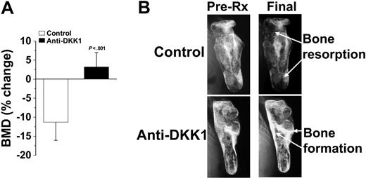
MM cells from 11 patients were successfully engrafted in SCID-rab mice and used for this study. As previously reported, MM growth in this system was restricted to the implanted bone and characterized by increased level of hIg in mice sera and induction of MM bone disease. As shown in Table 1, MM cells were taken from patients with various clinical stages and bone disease and had different expression level of DKK1 as assessed by microarray. When SCID-rab mice had established MM as indicated by hIg level of more than 50 μg/mL,30 11 hosts engrafted with MM cells from 11 different patients were treated with DKK1-neutralizing antibody and additional 11 matching SCID-rab hosts served as control and treated with irrelevant IgG antibody for 4 to 6 weeks. Anti-DKK1 did not appear to cause gross toxicities in all experiments. To test the effect of treatment on myeloma-induced bone disease, x-ray radiographs and BMD measurements were taken prior to treatment initiation and at the end of each experiment. In control hosts, the implanted BMD was reduced by 0.1% to 36% from pretreatment levels in 9 experiments and was elevated by 2% and 4% in 2 experiments (Table 1). In contrast, the implanted BMD in DKK1 antibody-treated hosts was elevated in 8 experiments by 1% to 45% and was reduced in 3 experiments by 1% to 17% from pretreatment levels (Table 1). Overall, whereas the implanted BMD in control hosts was reduced by 10.9% ± 4.3%, the implanted BMD in DKK1 AB-treated hosts was increased by 3.5% ± 3.4% from pretreatment levels (P < .001; Figure 1).
DKK1-neutralizing antibody promotes bone formation in myelomatous bones. SCID-rab mice engrafted with myeloma cells from 11 patients were treated with anti-DKK1 or control IgG antibody. (A) Changes in BMD (mean ± SEM) of the implanted bones from pretreatment levels. (B) X-ray radiographs of the implanted bones at pretreatment and end of experiment. Note that although BMD in control bones was reduced, anti-DKK1 treatment resulted in increased BMD during the experimental period.
DKK1-neutralizing antibody promotes bone formation in myelomatous bones. SCID-rab mice engrafted with myeloma cells from 11 patients were treated with anti-DKK1 or control IgG antibody. (A) Changes in BMD (mean ± SEM) of the implanted bones from pretreatment levels. (B) X-ray radiographs of the implanted bones at pretreatment and end of experiment. Note that although BMD in control bones was reduced, anti-DKK1 treatment resulted in increased BMD during the experimental period.
In certain experiments, the bone anabolic effect of DKK1 antibody could be visualized on radiographs; as demonstrated in Figure 1, before treatment, osteolytic bone lesions were evident in implanted bones of both control and DKK1 antibody-treated hosts. Whereas bone resorption continued in the implanted control bone, DKK1 antibody treatment resulted in increased bone mass and prevention of further lytic lesions.
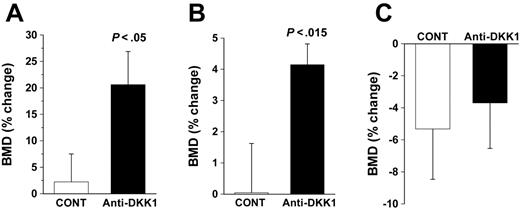
We also looked for the effect of DKK1 antibody treatment on BMD of implanted bones and murine femurs in nonmyelomatous SCID-rab mice (n = 18) and the uninvolved murine femur of myelomatous SCID-rab hosts (n = 9). Treatment with DKK1 antibody resulted in a significant increase in BMD of the nonmyelomatous implanted bone relative to controls (19% ± 6% versus 3% ± 5%; P < .05; Figure 2A) and in the murine femur (4.4% ± 0.6% versus 0.1% ± 1.4%; P < .015; Figure 2B). The BMD of uninvolved mouse femurs from myelomatous hosts was insignificantly reduced from pretreatment levels and was not changed following treatment with DKK1 antibody (4.0% ± 3.2% versus 3.4% ± 2.5%; Figure 2C).
DKK1-neutralizing antibody promotes bone formation in nonmyelomatous bones. SCID-rab mice were engrafted with primary myeloma cells through a direct injection of tumor cells into the implanted rabbit bone (myelomatous hosts)27 or left uninjected (nonmyelomatous hosts). We measured BMD of the experimental SCID-rab mice femur and implanted rabbit bones. (A-B) Changes in BMD (mean ± SEM) of the implanted rabbit bones (A) and mouse femur (B) in nonmyelomatous SCID-rab mice treated with control IgG and anti-DKK1–neutralizing antibody for 4 weeks. (C) Changes in BMD of the uninvolved mouse femur in myelomatous SCID-rab mice treated with control IgG and anti-DKK1 neutralizing antibody.
DKK1-neutralizing antibody promotes bone formation in nonmyelomatous bones. SCID-rab mice were engrafted with primary myeloma cells through a direct injection of tumor cells into the implanted rabbit bone (myelomatous hosts)27 or left uninjected (nonmyelomatous hosts). We measured BMD of the experimental SCID-rab mice femur and implanted rabbit bones. (A-B) Changes in BMD (mean ± SEM) of the implanted rabbit bones (A) and mouse femur (B) in nonmyelomatous SCID-rab mice treated with control IgG and anti-DKK1–neutralizing antibody for 4 weeks. (C) Changes in BMD of the uninvolved mouse femur in myelomatous SCID-rab mice treated with control IgG and anti-DKK1 neutralizing antibody.
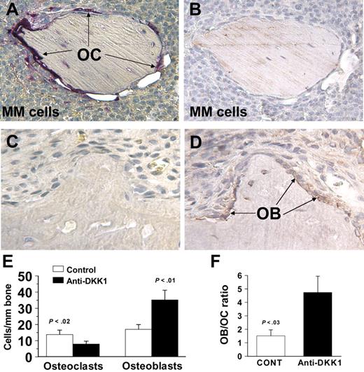
To further study the effect of DKK1 antibody on bone cells, sequential myelomatous bone sections from control and DKK1 antibody-treated SCID-rab mice were stained for TRAP and immunohistochemically stained for osteocalcin, and the numbers of multinucleated osteoclasts and osteocalcin-expressing osteogenic cells were counted (Figure 3). Relative to controls, DKK1 antibody treatment resulted in increased numbers of osteoblasts per millimeter bone (35 ± 6 versus 17 ± 3; P < .01) and reduced numbers of multinucleated TRAP+ osteoclasts (8 ± 2 versus 14 ± 3 osteoclasts/mm P < .02). Overall, the ratio of osteoblasts to osteoclasts shifted from 1.5 ± 0.4 in controls to 6.2 ± 1.7 in DKK1 antibody-treated bones (P < .03; Figure 3).
DKK1-neutralizing antibody stimulates osteoblastogenesis and reduces osteoclastogenesis in myelomatous bones. (A-D) Decalcified, sequential, myelomatous bone sections from control (A-B) and anti-DKK1–treated hosts (C,D) were stained for TRAP (A,C) and immunohistochemically stained for osteocalcin (B,D). Whereas in control bones TRAP-expressing osteoclasts covered the whole bone surface and no osteocalcin-expressing osteoblasts were detected, anti-DKK1 treatment resulted in marked reduction in number of osteoclasts and increased number of osteoblasts. OB indicates osteoblasts; OC, osteoclasts. (E-F) Numbers of multinucleated, TRAP-expressing osteoclasts and osteocalcin-expressing osteoblasts (E) and ratio of osteoblasts to osteoclasts in myelomatous bones (F). Data are expressed as mean ± SEM. Original magnification, 200×. Images were obtained using an Olympus BH2 microscope equipped with a 160 ×/0.17 numerical aperture objective (Olympus, Melville, NY). Images were acquired using a SPOT2 digital camera (Diagnostic Instruments, Sterling Heights, MI), and were processed using Adobe Photoshop version 10 (Adobe Systems, San Jose, CA).
DKK1-neutralizing antibody stimulates osteoblastogenesis and reduces osteoclastogenesis in myelomatous bones. (A-D) Decalcified, sequential, myelomatous bone sections from control (A-B) and anti-DKK1–treated hosts (C,D) were stained for TRAP (A,C) and immunohistochemically stained for osteocalcin (B,D). Whereas in control bones TRAP-expressing osteoclasts covered the whole bone surface and no osteocalcin-expressing osteoblasts were detected, anti-DKK1 treatment resulted in marked reduction in number of osteoclasts and increased number of osteoblasts. OB indicates osteoblasts; OC, osteoclasts. (E-F) Numbers of multinucleated, TRAP-expressing osteoclasts and osteocalcin-expressing osteoblasts (E) and ratio of osteoblasts to osteoclasts in myelomatous bones (F). Data are expressed as mean ± SEM. Original magnification, 200×. Images were obtained using an Olympus BH2 microscope equipped with a 160 ×/0.17 numerical aperture objective (Olympus, Melville, NY). Images were acquired using a SPOT2 digital camera (Diagnostic Instruments, Sterling Heights, MI), and were processed using Adobe Photoshop version 10 (Adobe Systems, San Jose, CA).
Inhibition of bone disease and increased bone formation by anti-DKK1 is associated with reduced myeloma burden
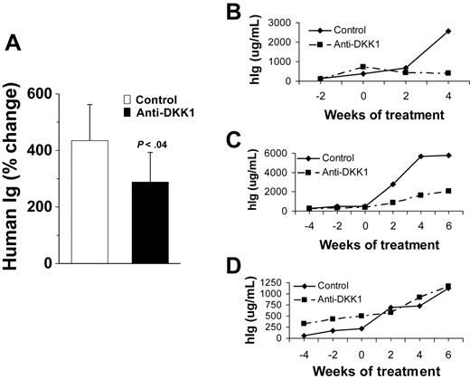
We next looked at whether the effect of DKK1 antibody on myeloma bone disease was associated with changes in MM tumor growth. Treatment resulted in reduced tumor burden (defined as reduced hIg from pretreatment levels), growth rate inhibition (defined as slower increased in hIg from pretreatment level by > 25% than control hosts), and no effect (defined as < 25% difference in increased hIg levels from pretreatment). Whereas in control AB-treated hosts tumor burden was increased in all experiments, DKK1 antibody resulted in reduction of tumor burden in 4 of 11 experiments (patients 1, 2, 3, and 6), inhibition of growth rate in 4 of 11 experiments (patients 4, 5, 8, and 9), and no effect in 3 experiments (patients 7, 10, and 11; Table 1). Three representative experiments are demonstrated in Figure 4. Overall, tumor burden in control antibody- and DKK1 antibody-treated hosts was increased from pretreatment level by 408% ± 118% and 270% ± 95%, respectively (P < .04; Figure 4). Intriguingly, anti-DKK1 treatment resulted in increased BMD of the myelomatous bone regardless of antitumor effect (Table 1), suggesting that growth of MM cells from certain patients is more susceptible to microenvironmental changes by DKK1 inhibition.
DKK1-neutralizing antibody reduces primary myeloma burden in SCID-rab mice. (A) Changes in levels of light-chain immunoglobulin (mean ± SEM) from pretreatment in control IgG and anti-DKK1 neutralizing antibody treated hosts. (B-D) Three representative experiments demonstrating heterogeneous response (A, tumor reduction; B, growth delay; C, no response) of anti-DKK1 in SCID-rab mice.
DKK1-neutralizing antibody reduces primary myeloma burden in SCID-rab mice. (A) Changes in levels of light-chain immunoglobulin (mean ± SEM) from pretreatment in control IgG and anti-DKK1 neutralizing antibody treated hosts. (B-D) Three representative experiments demonstrating heterogeneous response (A, tumor reduction; B, growth delay; C, no response) of anti-DKK1 in SCID-rab mice.
Discussion
In this study, we provide proof of the concept that anti-DKK1 therapy may be an effective adjunct in the clinical management of MM bone disease. In addition, we found that this treatment had bone anabolic effects on nonmyelomatous bones suggesting that DKK1 neutralization may have broad applications in the treatment disorders related to low bone mass. Here we demonstrated that daily subcutaneous injections of a neutralizing DKK1 antibody in the area surrounding myelomatous bone ameliorated bone turnover presumably through increased osteoblastogenesis and reduction of osteoclastogenesis. Interestingly, this effect was often associated with reduced tumor burden as well.
In addition to its high production by MM cells,19 DKK1 is normally produced by BM mesenchymal cells (eg, certain MSCs, osteoblasts, and osteocytes) and directly regulates osteoblastogenesis8,10,12,13,15–18 and osteoclastogenesis.25,26 This may explain the efficacy of anti-DKK1 in bones engrafted with MM cells expressing low and high levels of DKK1 and also in nonmyelomatous bones. Conversely, and more likely, the heterogeneous and partial effects of anti-DKK1 suggests that other factors, in addition to DKK1, may also be involved in this process. Indeed, in addition to stimulation of osteoclastogenesis through alteration of RANKL/OPG ratio22,23 and production of osteoclast activating agents such as MIP1α34 MM cells actively suppress osteoblastogenesis via production of interleukin 7 (IL-7)35 and other inhibitors of Wnt signaling including sFRP-236 and FRZB/sFRP-3,10 and through stimulation of IL-3 secretion by other BM cells.37 The heterogeneous effect of anti-DKK1 may also be related to disease stage and the dose, schedule, and metabolism of the drug, and other confounding factors associated with model systems.
Growing evidence suggests that bone disease alters the hemostasis in the BM microenvironment in a manner that promotes tumor growth.38 Thus, restoring bone remodeling may not only prevent skeletal complications, but also restrain medullary tumor growth. Blocking bone resorption with RANK-Fc,22,30 OPG,39 or bisphosphonates30,40 in experimental animals inhibited growth of medullary but not extramedullary MM. We and others have demonstrated that cultured osteoclasts support survival and proliferation of MM cells through a process requiring cell-to-cell contact.31,41–43 In contrast, osteoblasts inhibit survival and proliferation of MM cells and interfere with the ability of osteoclasts to stimulate growth of MM cells.31 Moreover, infusing myelomatous bones with MSCs results in increased bone formation and inhibition of MM progression.31 Collectively, these studies suggest that MM bone disease and tumor growth are interdependent,38,44 at least at the medullary stage, thus supporting results from this study in which increased osteoblast activity and bone formation in myelomatous bones by blocking DKK1 activity also controlled MM progression.
Lytic bone disease represents a serious life-threatening complication in MM. Current standard management is limited to reducing tumor burden and treatment with bisphosphonates, which can be associated with adverse effects such as renal failure and osteonecrosis of the jaw.3,45–47 Clinically, bisphosphonates, which impair osteoclast activity, reduce but do not completely prevent skeletal complications nor promote the repair of large osteolytic lesions, indicating that a functional defect of osteoblasts is also involved in MM osteolysis. In addition to secretion of various inhibitors of osteoblastogenesis by MM cells, patients with MM are often treated with dexamethasone, an important steroidal component known to induce osteoporosis by reducing the life span of osteoblasts.48 Recent studies have shown that treatment of osteoblasts with dexamethasone results in a time- and dose-dependent increase in DKK1 expression,49 suggesting that this drug blocks bone formation also through prevention of osteoblast differentiation in a mechanism involving up-regulation of DKK1. Dexamethasone thus may exacerbate bone disease phenotype in MM. In contrast, bortezomib, the first clinically approved proteasome inhibitor for treatment of MM,50 was shown to stimulate bone formation in myelomatous bones,51 to inhibit IL-1–stimulated bone resorption, and down-regulate DKK1 expression in 3 human cell lines of mesenchymal origin.52 Garrett and colleagues53 have shown that other proteasome inhibitors led to increased osteoblast differentiation and bone formation in vitro and in vivo through the regulation of BMP-2 transcription and protein expression in osteoblasts. Consistent with these preclinical data, we and others have recently shown that bortezomib increases markers of osteoblast differentiation in serum of patients with MM.54,55 These studies suggest that the primary therapeutic drugs used in the management of MM may negatively (eg, dexamethasone) or positively (eg, bortezomib) affect MM bone disease through regulation of DKK1 production in myelomatous bones. Thus, combining current treatments with anti-DKK1 may abrogate dexamethasone-induced osteoporosis and act synergistically with proteasome inhibitor to stimulate bone formation and repair bone lesions in MM.
Taken together, we have demonstrated that anti-DKK1 stimulated osteoblast activity, reduced osteoclastogenesis, and promoted bone formation in myelomatous and nonmyelomatous bones. The bone anabolic effect of anti-DKK1 was associated with reduced MM burden. Therefore, similar to successful treatment with other neutralizing antibodies directed against soluble targets such as VEGF in cancer (avastin56,57 ) and RANKL in osteoporosis (denosumab58 ), DKK1-neutralizing antibody holds promise as a potential new treatment of various disorders related to low bone mass and perhaps osteoporosis.
Authorship
Contribution: S.Y. performed research, analyzed and interpreted data, and wrote the paper; W.L. assisted with animal studies; F.Z. analyzed all microarray data; R.W. evaluated bone disease in patients whose myeloma cells were tested in this study; B.B. contributed resources including patient material and clinical data; and J.D.S. conceptualized the work, analyzed and interpreted data, and wrote the paper.
Conflict-of-interest disclosure: The authors declare no competing financial interests.
Correspondence: John D. Shaughnessy Jr or Shmuel Yaccoby, Myeloma Institute for Research and Therapy, University of Arkansas for Medical Sciences, 4301 W Markham, Little Rock, AR 72205; e-mail: shanghnessyjohn@uams.edu or yaccobyshmuel@uams.edu.
An Inside Blood analysis of this article appears at the front of this issue.
The publication costs of this article were defrayed in part by page charge payment. Therefore, and solely to indicate this fact, this article is hereby marked “advertisement” in accordance with 18 USC section 1734.
Acknowledgments
This work was supported by grants CA-93897 (S.Y.), CA55819 (B.B. and J.D.S.), and CA97513 (J.D.S.) from the National Cancer Institute and by Senior and Translational Research Awards from the Multiple Myeloma Research Foundation (S.Y.).
We would like to recognize the efforts of the members of the Lambert Laboratory of Myeloma Genetics; Bob Kordsmeier, Christopher Randolph, Owen Stephens, David R. Williams, Yan Xaio, and Hongwei Xu as well as Rinku Saha and Paul Perkins from S.Y.'s laboratory. The authors also wish to thank the faculty, staff and patients of the Myeloma Institute for Research and Therapy for their support.


This feature is available to Subscribers Only
Sign In or Create an Account Close Modal