Chronic myelogenous leukemia (CML) is a clonal stem cell disease caused by the BCR-ABL oncoprotein and is characterized, in its early phase, by excessive accumulation of mature myeloid cells, which eventually leads to acute leukemia. The genetic events involved in CML's progression to acute leukemia remain largely unknown. Recent studies have detected the presence of theNUP98-HOXA9 fusion oncogene in acute leukemia derived from CML patients, which suggests that these 2 oncoproteins may interact and influence CML disease progression. Using in vitro purging of BCR-ABL–transduced mouse bone marrow cells, we can now report that recipients of bone marrow cells engineered to coexpressBCR-ABL with NUP98-HOXA9 develop acute leukemia within 7 to 10 days after transplantation. However, no disease is detected for more than 2 months in mice receiving bone marrow cells expressing either BCR-ABL orNUP98-HOXA9. We also provide evidence of high levels ofHOXA9 expressed in leukemic blasts from acute-phase CML patients and that it interacts significantly on a genetic level withBCR-ABL in our in vivo CML model. Together, these studies support a causative, as opposed to a consequential, role forNUP98-HOXA9 (and possibly HOXA9) in CML disease progression.
Introduction
During the last 3 decades, cytogenetic studies have revealed recurrent chromosomal translocations in human leukemias.1,2 These translocations result in fusion genes that frequently encode for chimeric proteins that participate in leukemic transformation of bone marrow–derived cells. Past and ongoing studies from many laboratories, including our own, have shown that these chimeric oncoproteins are incapable of inducing complete transformation of myeloid precursors and that they unquestionably require coactivation of other oncogenes and/or inactivation of tumor suppressor genes3 in order to do so.4-15 Defining the full complement of oncoproteins that are necessary for leukemic transformation is relevant to our understanding of leukemogenesis and might offer new molecular targets for the development of more specific antileukemic drugs.16-18 Thus, it is both fundamentally and clinically relevant to identify sets of oncogenes that participate in human leukemias, in particular in the progression of chronic myelogenous leukemia (CML) from the chronic to the acute phase.
As an initial step in defining oncogenes involved in CML progression, we focused on recent cytogenetic studies that describe additional chromosomal translocations in acute leukemia derived from CML patients, suggesting that these translocations potentially collaborated withBCR-ABL in disease progression. Specifically, theNUP98-HOXA9 fusion gene was detected in blast cells in 3 patients with typical Philadelphia-positive (Ph+) CML.19,20 However, there have also been patients who developed NUP98-HOXA9–induced myeloproliferation and subsequently acquired a Ph chromosome in their leukemic cells when acute leukemia was diagnosed,21suggesting that NUP98-HOXA9 and BCR-ABL may genetically interact in human leukemia.
There have also been blast crisis specimens from CML patients that express high levels of the HOXA9 oncogene when compared with cells isolated from chronic phase patients, raising the possibility that HOXA9 may also directly interact with BCR-ABLin the transformation of bone marrow cells.22 These results were extended in our current studies, where we found that cells from blast crisis patients express higher levels of HOXA9than those found in mononuclear cells from accelerated phase CML patients (Figure 2).
Several other additional translocations, typically observed in acute human leukemia, have been manifested by CML cells undergoing blast transformation (ie, at diagnosis of acute leukemia in patients previously in chronic phase CML). These include the t(11;17), which involves the MLL/ALL/HRX gene23; the t(8;21) with the AML1 gene24; the t(3;21) translocation, which results in a fusion between the AML1, MDS1, and EVI1 genes25; and the t(9;16)(q34;p11) found in one case in blast crisis and severe disseminated intravascular coagulopathy.26 We also recently found a patient with acute leukemia with a typical t(1;19) involving the E2a-PBX1 fusion oncoprotein and a Philadelphia chromosome, which also raises the possibility that these 2 elements might interact in leukemic transformation (G.S. and A. Hendrick, unpublished observation, 1999).
Together, these results suggest that several leukemic oncoproteins in the family of Hox genes and their cofactors (eg, PBX1), including NUP98-HOXA9, HOXA9, andE2a-PBX1, might genetically interact with BCR-ABLand lead to acute leukemic transformation of human bone marrow cells.
Transduction of mouse bone marrow cells by BCR-ABLretroviral vectors was recently demonstrated to accurately reproduce human CML with the caveat that mice die of acute myeloproliferation with pulmonary hemorrhage within 2 to 3 weeks following reconstitution.27-29 This early death from myeloproliferation precluded any studies aimed at defining genetic interactions that are mostly based on shortening the time to acute leukemia produced by the collaborating oncogenes.
In the studies reported here, we have improved upon the mouse model ofBCR-ABL–induced chronic leukemia by circumventingBCR-ABL–induced early lethality. With our mouse model, it was possible to identify 2 oncogenes that genetically interact withBCR-ABL to generate acute leukemia. The system described should open avenues for testing collaborator oncogenes toBCR-ABL using mouse bone marrow (BM) cells.
Materials and methods
Animals
All mice were bred and maintained as previously reported.30 Donor (C57BL/6Ly-Pep3b × C3H/HeJ)F1 [(PepC3)F1] and recipients (C57BL/6J × C3H/HeJ)F1[(B6C3)F1] are phenotypically distinguishable by their cell-surface expression of different allelic forms of the Ly5 locus: (B6C3)F1 mice are homozygous for the Ly5.2 allotype, and (PepC3)F1 mice are heterozygous for the Ly5.1/Ly5.2 allotypes.
Recombinant retroviral vectors
The retroviral vectors used in this study,MSCV-pgk-EGFP (no. 652) and MSCV-HOXA9-pgk-EGFP(no. 654), were described previously.7,MSCV-BCR-ABL-ires-EGFP (no. 802), a kind gift of Dr R. van Etten, is essentially identical to the Mig210 virus described by Pear and colleagues.27 TheMSCV-pgk-DSRed (no. 930) and MSCV-HOXA9-pgk-DSRed(no. 931) were constructed by subcloning anNheI/HpaI fragment from pDSRedI-CI (from Clontech, Palo Alto, CA) into blunted NcoI/ClaIMSCV-pgk-EGFP vector (no. 652) andMSCV-HOXA9-pgk-EGFP (no. 654).MSCV-NUP98-HOXA9-pgk-RFP (no. 946) was generated by digesting plasmid no. 662 (previously described in Kroon et al7) with EcoRI, releasing the full-lengthNUP98-HOXA9, and subcloning into plasmid no. 930 linearized with EcoRI. MSCV-NUP98-HOXA9-pgk-neo (no. 619),MSCV-HOXA9-pgk-neo (no. 412), and MSCV-pgk-neo(no. 225) were also previously described.7,30 The production of a functional protein for either BCR-ABL, NUP98-HOXA9, or HOXA9 from the integrated proviruses has previously been described for all of these constructs.6,7 29
Retroviral generation, infection, and transplantation of primary bone marrow cells
High-titer helper-free recombinant retroviruses were generated and tested as previously described.5 Bone marrow cells were obtained from C57BL/6Ly-Pep3b or (PepC3)F1 mice injected 4 days earlier with 5-fluorouracil (5-FU) (150 mg/kg body weight), prestimulated, and cocultivated with irradiated viral producer cells, and then loosely adherent and nonadherent cells were recovered and injected immediately (t = 0), or following a 7-day culture (Figure 1), into sublethally irradiated (850 cGy) 7- to 12-week-old (B6C3)F1 recipient mice, as previously described.6 Gene transfer efficiencies to donor BM cells (BMCs) were determined by flow cytometry 24 hours following harvesting from viral producers at between 32% to 77% (highest, BCR-ABL) or by G418 resistance as reported.7
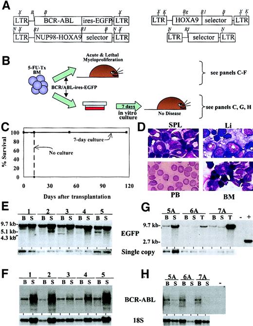
A 7-day culture system that eliminates acute lethal myeloproliferation induced by the transplantation of bone marrow cells engineered to overexpress BCR-ABL.
(A) Schematic representation of the different retroviruses used in these studies. Selector genes were the enhanced green fluorescent protein (EGFP), the red fluorescent protein (RFP), or the neomycin-resistance gene. B indicates BamHI; Bg,BglII; RI, EcoRI; K, KpnI; N,NheI; X, XbaI. RFP in the selector gene shown for the restriction mapping of the NUP98-HOXA9 andHOXA9 retroviruses. (B) Description of the 7-day culture protocol used to eliminate the acute myeloproliferative syndrome induced by transplanting BM cells engineered to overexpressBCR-ABL. (C) Survival curve of recipients of 2 × 10⁵BCR-ABL–transduced BM cells grown (solid line) or not (dashed line) for 7 days in vitro with hemopoietic growth factors. (D) Cytological analysis of bone marrow (BM), liver (Li), spleen (SPL), and peripheral blood (PB) specimens showing the acute and lethal myeloproliferative disease developing in recipients of BCR-ABL–transduced cells transplanted immediately following coculture on viral producers. The spleen is infiltrated by myeloid precursors (EGFP+) that are barely detectable in the PB. Original magnification × 40; stain, Wright Giemsa. (E) Southern blot analyses demonstrating the integrated provirus (top blots) in the DNA isolated from the BM (B) or spleen (S) of recipients of BCR-ABL–transduced cells (nos. 1-5). DNA was digested with XbaI (panels E and G). The bands smaller than 9.7 kb probably represent several clones that harbor rearranged proviruses. Panels F and H depict Northern blot analyses to demonstrate expression level of BCR-ABL in the various samples as detailed in panels E and G. T indicates thymus. Exposure times are 12 hours for panel F and 8 days for panel H. 18S RNA probe is exposed for the same time for all lanes. The “minus” lane indicates 10 μg RNA isolated from spleen-derived cells from an unmanipulated syngenic mouse. Panel G shows Southern blot analysis of genomic DNA isolated from recipients that received 2 × 10⁵(day “0”) BCR-ABL–transduced BM cells grown for 7 days in vitro prior to transplantation. Note the large variation in exposure time from 12 hours for mice 1 to 5 to 3 days for mice 5A to 7A. The mouse identification numbers in this figure correspond to those shown in Table 1. Single copy is from the endogenous actingene: exposure times for membranes exposed to actin are identical. The “−” lane indicates 10 μg DNA isolated from the spleen of a syngenic control mouse, and the “+” sign is a positive control for hybridization consisting of 20 pg KpnI-digested retroviral plasmid no. 652 (“Materials and methods”) generating a 2.7-kb fragment.
In vitro cultures
Bone marrow cells harvested from coculture on viral producers were seeded in liquid cultures at 1.2 × 10⁵ cells per milliliter and expanded in vitro for 7 days in Iscove modified Dulbecco medium (IMDM) containing 10% fetal calf serum (FCS), 5 ng/mL murine interleukin-3 (IL-3), 10 ng/mL human IL-6, 25 ng/mL murine Steel factor and 3 U/mL erythropoietin, 192 ng/mL transferrin, 2% deionized bovine serum albumin (BSA), 2 mM glutamine, and 5 × 10⁻⁵ M β-mercaptoethanol.
RT-PCR studies
Semiquantitative studies for detecting the expression ofHOXA9 were essentially performed as previously reported.31 Briefly, total cDNA was amplified with the use of an oligo deoxythymidine (dT)–based oligonucleotide global polymerase chain reaction (PCR) amplification method. Amplified cDNA was transferred to nylon membranes, which were later hybridized to a probe specific to HOXA9 orβ-actin. Semiquantitative BCR-ABL expression studies were done as previously described,32 with the following modifications: single-step reverse transcriptase (RT) and first PCR amplification and primers for internal control: ABL Fc1 external (ext) (5′-TTCAGCGGCCA-GTAGCATCTGACT-3′, sense) and Fc3 ext (5′-GCAGTGTTGATCCTGTAATGG-3′, antisense). Nested PCR was then performed with one fifth of the first RT-PCR reaction with ABL internal primers: Fc2 internal (int) 5′-TTGTGGCCA-GTGGAGATAACA-3′sense, and Fc3 int 5′-TATCTCAGCGA-GATGGACCT-3′ antisense. Amplification was carried out for 35 cycles.
Clinical specimens
Leukemia samples representing greater than 80% infiltration by leukemia cells (all confirmed by D.-C.R. and/or G.S.) were collected in preservative-free heparin, and mononuclear cells were separated by ficoll-hypaque density gradient centrifugation. Samples were obtained with the informed consent of the patients under protocols approved by the Human Subjects Protection Committee of the Maisonneuve-Rosemont Hospital (Montreal, QC, Canada) and the internal review board of Institut de recherches cliniques de Montréal (IRCM). All cell samples were cryopreserved in 10% dimethylsulfoxide (DMSO) by means of standard techniques and stored in the vapor phase of liquid nitrogen until used as described.33
DNA and RNA analysis
Southern blot analysis was performed as described previously.5 Expression of appropriate proviral mRNAs was confirmed by Northern blot analysis. The probes used for RNA and DNA analyses were random primer 32P-labeled fragments ofHOXA9 (HindIII fragment of pBSNUP98-HOXA9, no. 727); BCR (BamHI fragment of MSCV-BCR-ABL-ires-EGFP, no. 802);EGFP (NcoI/ClaI fragment ofMSCV-pgk-EGFP, no. 652); RFP(HpaI/NheI fragment of pDsRed-CI, from Clontech); and actin (PstI fragment as described31). Northern blots were performed as follows: membranes were stripped and rehybridized by means of an end-labeled oligonucleotide, 5′-ACG GTA TCT GAT CGT CCT CGA ACC-3′, specific for 18S rRNA to evaluate the relative amounts of total RNA loaded in each lane.
Results
The development of a mouse model to study oncogenes that interact with BCR-ABL in leukemic transformation
Retroviral-mediated expression of BCR-ABL in primary mouse bone marrow (BM) cells previously exposed to the cytotoxic drug 5-FU induces an aggressive CML-like disease that kills mice in less than 2 to 3 weeks after bone marrow transplantation. This early demise precluded analysis of genetic interaction betweenBCR-ABL and other oncogenes chosen for these studies (Figure 1A-F; description of experimental protocols, and of the early myeloproliferative disease occurring in recipients of BCR-ABL–transduced BM cells). Since in vitro cultures appear to selectively deplete BCR-ABL–expressing primitive bone marrow progenitors in humans,34 we reasoned that it might similarly purge BCR-ABL–transduced cells in mice and potentially eliminate this acute myeloproliferative disease, thereby allowing us to study in vivo genetic interactions betweenBCR-ABL and the other oncogenes.
The culture conditions chosen were the same as those used for retroviral gene transfer, except that cells were maintained for 7 days in vitro following retroviral gene transfer (Figure 1B). The effect of this in vitro culture on the leukemogenic potential of transduced bone marrow cells was determined by comparing the time to leukemia occurence in recipients of 2 × 10⁵ BM cells transplanted immediately following retroviral gene transfer with recipients of the same number of day-0 cells grown in vitro for 7 days. This comparison was done for all oncogenes used in these studies, that is,BCR-ABL, NUP98-HOXA9, and HOXA9. While the 7-day culture period had little impact on the occurrence of leukemia onset for recipients of NUP98-HOXA9 orHOXA9–transduced cells (52-223 days after transplantation, n = 7), it completely abrogated the acute myeloproliferative disease in recipients of BCR-ABL–transduced cells (Figure 1C,G-H; Table 1).
Characteristics of recipients of BCR-ABL–transduced BM cells transplanted immediately after retroviral gene transfer (nos. 1-11) or after 1 week (nos. 1A-14A) of culture in vitro
| Mouse ID . | Cell dose, × 104* . | Day mouse killed . | Spleen size, g† . | Pulmonary hemorrhage . | Liver infiltration . | Reconstitution, % Ly5.1 BM-SPL-THY . | Reconstitution, % GFP BM-SPL-THY . |
|---|---|---|---|---|---|---|---|
| BMT immediately afterBCR-ABL gene transfer | |||||||
| 1 | 20 | 10 | 0.46 | ND | Yes | ND | 45-49-4 |
| 2 | 20 | 10 | 0.38 | ND | Yes | ND | 39-49-5 |
| 3 | 20 | 10 | 0.48 | ND | Yes | ND | 44-47-1 |
| 4 | 20 | 10 | 0.48 | ND | Yes | ND | 45-48-1 |
| 5 | 20 | 10 | 0.44 | ND | Yes | ND | 41-41-1 |
| 6 | 20 | 11 | 0.28 | Yes | Yes | ND | ND |
| 7 | 20 | 11 | 0.29 | Yes | Yes | ND | ND |
| 8 | 20 | 12 | 0.33 | Yes | Yes | ND | ND |
| 9 | 20 | 12 | 0.21 | Yes | Yes | ND | ND |
| 10 | 20 | 11 | 0.48 | Yes | Yes | ND | 71-77-ND |
| 11 | 20 | 12 | 0.66 | Yes | Yes | ND | 70-68-ND |
| BMT 7 days after BCR-ABL gene transfer | |||||||
| 1A | 500 | 106 | 0.1 | No | No | ND | ND |
| 2A | 200 | 175 | 0.17 | No | No | ND | 0-0-0 |
| 3A | 20 | 175 | 0.16‡ | No | No | ND | 0-0-0 |
| 4A | 20 | 175 | 0.19 | No | No | ND | 0-1-0 |
| 5A | 20 | 52 | 0.12 | No | No | ND | 1-9-75 |
| 6A | 20 | 52 | 0.08 | No | No | ND | 0-0-2 |
| 7A | 20 | 52 | 0.12 | No | No | ND | 0-0-11-153 |
| 8A | 2 | 97 | 0.07 | No | No | 44-68-81 | 0-0-0 |
| 9A | 0.2 | 96 | 0.04 | No | No | 78-99-97 | 3-1-5 |
| 10A | 0.2 | 96 | 0.07 | No | No | 63-99-96 | 2-1-1 |
| 11A | 0.2 | 96 | 0.08 | No | No | 79-98-99 | 3-1-0 |
| 12A | 0.02 | 97 | 0.07 | No | No | 1-2-0 | ND |
| 13A | 0.02 | 117 | 0.06 | No | No | 2-2-1 | ND |
| 14A | 0.02 | 117 | 0.1 | No | No | 3-2-0 | ND |
ID indicates identification; THY, thymus; BMT, bone marrow transplantation; and ND, not done.
Cell dose adjusted to day 0, that is, independent of the expansion during the 7-day culture. BCR-ABL–transduced cells expand by 30-fold in 7 days under the condition used (ie, these cultures were initiated with 2 × 10⁵ cells per culture).
Normal size for adult mice is approximately 0.1 g.
Spleen was infiltrated by immature myeloid cells in this mouse.
As previously reported, recipients of BCR-ABL–transduced BM cells transplanted immediately after viral transduction developed acute myeloproliferation within 11 ± 1 days after transplantation (dotted line, Figure 1C, shows survival; Figure 1D-F describes the myeloproliferative disorder). A description of each mouse dying from acute myeloproliferation is provided in Table 1 (mice 1 to 11). Note that these myeloproliferative disorders were characterized by pulmonary hemorrhage, splenomegaly, and BM infiltration by immature myeloid cells, but low white blood cell (WBC) counts (Table 1; Figure 1D). This acute disease was highly polyclonal, as indicated by the analysis of proviral integration sites into DNA isolated from hemopoietic organs of these animals (data not shown). In contrast, recipients of cells maintained in culture for 7 days thrive normally (solid line, Figure1C) and were killed at various times for analysis. At 52 days after transplantation, 3 mice were analyzed (mice 5A, 6A, and 7A; Table 1; Figure 1G-H); all mice were either normal (n = 2) or showed signs of a mild chronic myeloproliferative disorder characterized by slight increase in mature neutrophils in the bone marrow and spleen and by the presence of a megakaryocyte in the lungs (data not shown). The 3 mice analyzed were reconstituted with low levels ofBCR-ABL–transduced cells in their bone marrow and spleens (mice 5A to 7A), but thymic reconstitution was high for 2 of the 3 mice (mice 5A and 7A; Table 1, Figure 1G). BCR-ABL was expressed at very low levels in these cells, as shown by Northern blot analysis (Figure 1H; exposure time was 8 days, compared with 12 hours for the blot shown in Figure 1F). Clonal analysis indicated that in contrast to the highly polyclonal nature of the acute myeloproliferative disease, recipients of cells grown in vitro for 7 days were reconstituted with very few clones, none of which had lymphoid and myeloid potential (data not shown).
The low level of repopulation by BCR-ABL–transduced cells in the 3 mice analyzed at 52 days after transplantation suggested that these cells died in the culture conditions since, according to fluorescence-assisted cell sorter (FACS) analysis ofEGFP expression, they represented 70% of the cells that initiated these cultures.
A series of limiting dilution experiments were performed to determine the range of depletion of BCR-ABL–transduced long-term repopulating cells (detected by either GFP positivity [Table 1] or by Southern and Northern blot analyses [Figure 1G-H]) in these cultures versus that of untransduced cells (detected as Ly5.1+GFP−). The results indicated that recipients of expanded BCR-ABL–transduced cells were poorly reconstituted by GFP+ cells when analyzed at between 52 to 175 days after transplantation. This was true for all mice tested except for mouse 5A and mouse 7A whose thymuses and (in the case of mouse 5A only) spleens were significantly reconstituted (percentage of GFP; Table 1, Figure 1G). Interestingly, mouse 7A had only 1% GFP+ cells in its thymus but had a moderately strong signal by Southern blot analysis (lane 8, Figure 1G), indicating that promoter shutdown might have occurred in these cells. Southern and Northern blot analyses of the hemopoietic tissues of all other mice in this group confirmed that reconstitution byBCR-ABL–transduced orBCR-ABL–expressing cells did not occur at significant levels and thus that promoter shutdown was not frequent (data not shown). In addition, and further confirming low levels of reconstitution by BCR-ABL–transduced cells, between 0% and 5% of the myeloid progenitors isolated from their bone marrow generated colonies that expressed detectable levels of GFP by epifluorescence (compared with approximately 70% GFP+colonies at the time the cultures were initiated). Although not analyzed for CD4 or CD8 expression, there was no evidence of thymic or lymph node enlargement that might have suggested the occurrence of lymphoproliferation in any of these mice.
While the reconstituting ability of BCR-ABL–transduced cells harvested from our 7-day culture was poor, untransduced cells (Ly5.1+GFP−), which represented approximately 30% of the cells at the time these cultures were initiated, remained competent to provide between 44% and 99% repopulation of the hemopoietic organs of these same recipients that received transplants of only 0.2 × 10⁵ “expanded” cells (column 7, mice 8A through 14A; Table 1) or nearing the frequency of competitive repopulation units (CRUs) in the inoculum, which was shown in previous experiments to be within this range.35 36Thus, while untransduced CRUs seemed to be maintained when grown in the presence of BCR-ABL–transduced cells, it appears that the latter were undetectable in our recipients, for a net loss nearing 3 logs (potentially higher) of these cells during our 7-day culture.
Our culture conditions thus significantly and preferentially depletedBCR-ABL–transduced repopulating cells, which possibly explains the lack of acute myeloproliferation in the subject mice. Interestingly, however, limiting dilution analysis also showed that our culture conditions greatly supported the ex vivo expansion of leukemia-repopulating cells (the myeloid leukemia was generated from BM cells overexpressing HOXA9 plus Meis1 as described5 30). These results suggested that our 7-day culture system was purgingBCR-ABL–transduced long-term repopulating cells while only mildly influencing the untransduced cells, but at the same time providing a good environment to expand “fully transformed” myeloid cells. As tested below, this provided an opportunity to evaluate possible collaboration between BCR-ABL and theNUP98-HOXA9 or the HOXA9 oncogenes.
NUP98-HOXA9 and HOXA9 genetically interact with BCR-ABL to generate acute myeloid leukemia (AML) in vivo
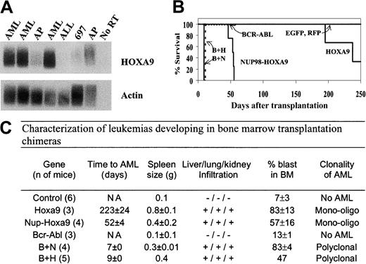
As previously mentioned, cytogenetic studies have reported the association between NUP98-HOXA9 and BCR-ABL in myeloid leukemic blasts, suggesting that both oncoproteins genetically interact in human leukemias. In addition, blast phase CML cells were previously reported to express higher levels of HOXA9 than cells isolated from chronic phase patients, again suggesting thatHOXA9 might also interact with BCR-ABL to transform BM cells. These observations were confirmed by semiquantitative RT-PCR studies using mononuclear cells from patients in blast (n = 3) versus accelerated phase CML (n = 2) (Figure2A).
NUP98-HOXA9 and HOXA9genetically interact with BCR-ABL to acutely transform bone marrow cells.
(A) Semiquantitative RT-PCR analysis demonstrates high levels ofHOXA9 in cells obtained from CML patients at diagnosis of myeloid blast transformation (lanes 1, 2, 4; AML) and low levels in cells from patients in the accelerated phase (AP; pre-AML) of their disease (lanes 3 and 7). Note that HOXA9 is not expressed in acute lymphoblastic leukemias (ALLs); 697 is a pre-B ALL cell line. “No RT” indicates the absence of reverse transcriptase in the reaction. Probe used as indicated, exposure time: 4½ hours for HOXA9 and 2½ hours for actin. (B) Survival curve of recipients of 2 × 10⁵ (day-0 equivalent) BM cells transduced with the indicated retrovirus and grown prior to transplantation for 7 days in vitro with hemopoietic growth factors; “B” refers toBCR-ABL; “H,” to HOXA9; and “N,” toNUP98-HOXA9. (C) Characteristics of leukemias developing in the various bone marrow transplantation chimeras described in panel B. AML, acute myeloid leukemia; % blast evaluated on 100 cells per each mouse. NA indicates data not available.
Recipients of cells transduced with BCR-ABL plusNUP98-HOXA9 or BCR-ABL plus HOXA9 that were not grown in vitro for 7 days died within 12 days of an aggressive myeloproliferative disease that was very difficult to distinguish from acute myeloproliferation as seen in recipients of cells engineered to express only BCR-ABL. Taking advantage of the in vitro “purging culture” described in the previous section, the interaction between the NUP98-HOXA9 (or HOXA9) and BCR-ABL oncoproteins was tested as illustrated in Figure 1B. As expected from our previous studies,5,6,30 recipients ofHOXA9-transduced BM cells started to die of AML at approximately 3 months after transplantation, and recipients ofNUP98-HOXA9–transduced cells at 1½ to 2 months (Figure 2B).7
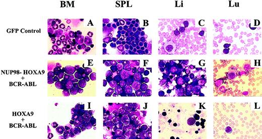
As detailed in Table 1, recipients ofBCR-ABL–transduced cells killed at 52 to 175 days after transplantation showed no signs of acute leukemia. In contrast, mice that received BM cells infected with BCR-ABL plusHOXA9 or BCR-ABL plus NUP98-HOXA9retroviruses and grown in vitro for 7 days died within 9 days of acute leukemia (see B plus H and B plus N in Figure 2B). In all cases, the leukemias were myeloid as evaluated by morphological criteria (Figure 3E-H shows NUP98-HOXA9plus BCR-ABL, and Figure 3I-L shows HOXA9plus BCR-ABL). High proportions of the BM cells in these mice were myeloid blasts, and the remaining cells were myeloid precursors at various stages (Figures 2C,3E). All leukemic animals had very elevated WBC counts (estimated by blood-smear evaluation at greater than 50 000/μL; G.S.); had enlarged spleens (Figure2C); and presented liver, lung, and kidney infiltration by leukemic blasts (Figures 2C,3E-L). In contrast to the acute myeloproliferative disease, no evidence of pulmonary hemorrhage was detected in these leukemic animals.
Morphological analysis of leukemic and myeloproliferative diseases occurring in the various transplantation chimeras.
Cytological analysis of bone marrow (BM), spleen (SPL), liver (Li), and lung (Lu) from recipients of bone marrow cells infected with the indicated retroviruses. Cytospins were prepared and stained as described.6 Original magnifications × 40.
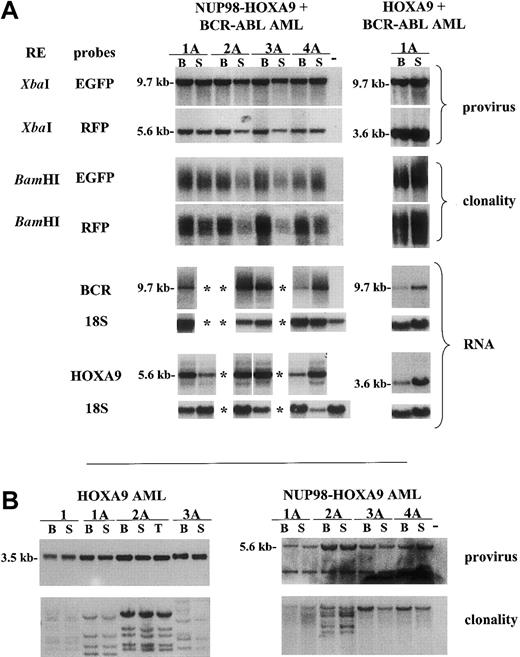
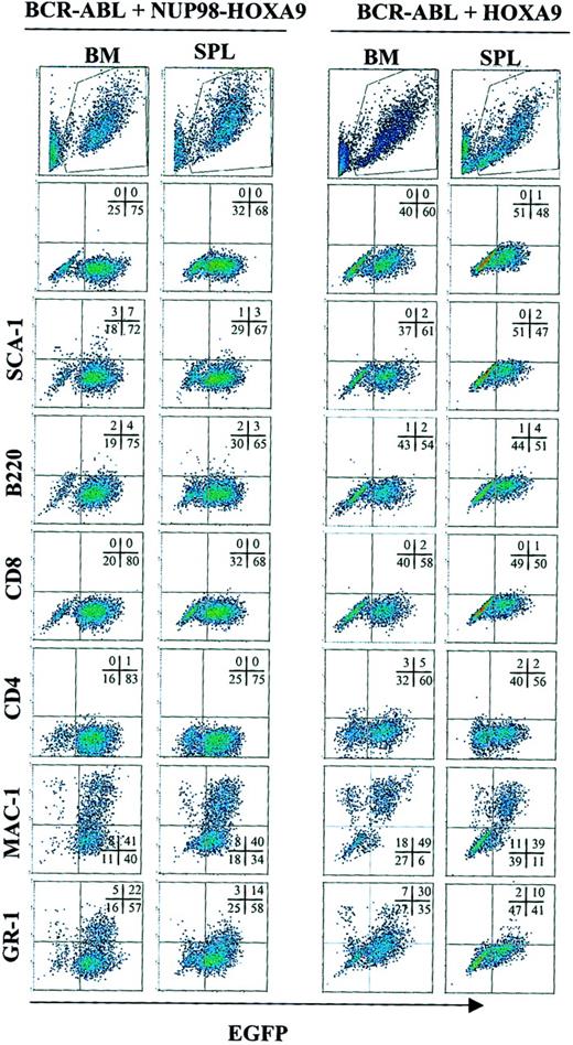
FACS analysis showed that the majority of the leukemic blasts were GFP+ (from BCR-ABL provirus) but did not express (fewer than 1%) the red fluorescent protein (RFP) (from either theNUP98-HOXA9 or the HOXA9 proviruses) although both of these proviruses were easily detected in the leukemic cells by Southern blot analysis (Figure 4A, second panel from top), and expression of NUP98-HOXA9 orHOXA9 was very high in the leukemic blasts as indicated in Figure 4A (lower panels; see RNA). This suggests that the pgk-RFP cassette was inactive in our leukemic cells but that the promoter and enhancer elements in the MSCV long terminal repeat (LTR) were active and driving the expression of the oncogenes. More detailed phenotypical analysis of these leukemias could not be performed with these cells, but when the analysis was repeated with leukemic cells from another experiment, in which the RFP selector gene was replaced by neor,leukemic blasts transduced with NUP98-HOXA9 (orHOXA9) plus BCR-ABL expressed Mac1 and to a lesser extent Gr1 (Figure 5). The presence of a functional neor gene in theNUP98-HOXA9 or in the HOXA9 integrated provirus and of EGFP in the BCR-ABL provirus made it possible to demonstrate the nature of the interaction betweenBCR-ABL and NUP98-HOXA9 or HOXA9.While at the time of BM transplantation (t = 0; Figure6), 10% to 16% of the colony-forming cells were resistant to G418 (NUP98-HOXA9 andHOXA9, respectively) and also expressed EGFP(BCR-ABL), a much higher proportion (43% to 70%) of the leukemic progenitors derived from the recipients suffering from overt leukemia were both EGFP+ and neomycin resistant (t = AML; Figure 6), indicating that cells expressing both oncogenes were positively selected in vivo.
Sufficiency of NUP98-HOXA9 andBCR-ABL for full transformation of mouse bone marrow cells.
(A) Top 4 rows: Southern blot analyses of DNA isolated from the bone marrow (B) and spleen (S) of recipients of NUP98-HOXA9 plusBCR-ABL (left) or HOXA9- plusBCR-ABL–transduced cells (right). Mice were killed when acute leukemia was apparent (ie, at 7 and 9 days after transplantation, respectively). DNA was digested with the indicated restriction enzyme (RE; Figure 1A shows a schematic representation of the integrated provirus). Note the smears in the 3rd and 4th rows (from top) indicating the polyclonal nature of the different leukemias. Bottom 4 rows: Northern blot analyses of RNA isolated for the same mice and hybridized to probes specific to BCR-ABL (BCR)and HOXA9 as indicated. Exposure times were 12 hours(BCR-ABL) and 4 days (HOXA9). (B) Southern blot analyses of DNA isolated from the bone marrow (B) and spleen (S) of recipients of HOXA9-transduced (left) orNUP98-HOXA9–transduced (right) cells killed when acute leukemia was apparent. For HOXA9, DNA was digested withKpnI (for proviral integrity) and with BglII (for clonal analysis). For NUP98-HOXA9, DNA was digested withXbaI and BamHI to test proviral integrity and clonal analysis, respectively. EGFP and RFP probes were used to hybridize DNA isolated from HOXA9- andNUP98-HOXA9–induced leukemias, respectively. Minus signs are as indicated in Figure 1. *Indicates data not available.
Phenotypical analysis of acute myeloid leukemias developing in recipients of BCR-ABL– andNUP98-HOXA9– or HOXA9-transduced cells.
Ten thousand cells were analyzed per specimen. Each scattergram is a representative of 4 mice similarly analyzed.
Phenotypical analysis of acute myeloid leukemias developing in recipients of BCR-ABL– andNUP98-HOXA9– or HOXA9-transduced cells.
Ten thousand cells were analyzed per specimen. Each scattergram is a representative of 4 mice similarly analyzed.
Genetic interaction of NUP98-HOXA9 orHOXA9 with BCR-ABL to transform mouse BM cells.
The proportion of doubly transduced (ie, G418-resistant and EGFP-expressing) colony-forming cells (CFCs) increased from between 10% and 16% at the time of transplantation to between 46% and 67% at the time leukemia occurred in primary recipients. The selector gene was neor forNUP98-HOXA9 or HOXA9 provirus (Figure 1A) and EGFP for BCR-ABL (n = 3 mice for NUP98-HOXA9plus BCR-ABL; n = 2 mice for HOXA9plus BCR-ABL). At least 30 colonies were examined per mouse. Untransduced BM cells failed to grow in the presence of G418.
In agreement with our previous studies, leukemia that developed in recipients of NUP98-HOXA9–transduced (Figure 4B, right lower panel) or HOXA9–transduced (Figure 4B, left lower panel) cells were monoclonal or oligoclonal, clearly indicating the requirement for additional genetic events for leukemic transformation of these cells. In contrast, clonal analysis of leukemias from recipients that received transplants of cells coexpressing BCR-ABL plus NUP98-HOXA9 orHOXA9 exposed the highly polyclonal nature of these leukemic cells for all mice that were studied (presence of smear in Figure 4A, third and fourth panels).
This clearly indicated that the combination of these oncogenes was sufficient for the full leukemic transformation of mouse BM cells and, at least for NUP98-HOXA9, strongly suggests its involvement in the progression of CML to AML in selected patients.
Together, these studies indicate that our in vitro purging system (1) was effective at eliminating the acute myeloproliferative disease caused by BCR-ABL–transduced 5-FU–treated BM cells even when high cell doses were transplanted; (2) did not accelerate the occurrence of leukemia onset in recipients of NUP98-HOXA9–or HOXA9-transduced cells; (3) allowed the expansion of leukemia-repopulating cells of myeloid origin ex vivo; and (4) was capable of maintaining (and perhaps potentially expanding)BCR-ABL–transduced cells that coexpressed eitherNUP98-HOXA9 or HOXA9, thus indicating the presence of a strong genetic interaction between BCR-ABL and these genes in AML.
Discussion
In these studies, we developed an in vitro/in vivo model that allows the purging of BCR-ABL–induced acute myeloproliferative disease. With this model, it was possible to demonstrate a potent genetic interaction between BCR-ABL and 2 oncoproteins: namely, NUP98-HOXA9 and HOXA9. In particular, the interaction between BCR-ABL andNUP98-HOXA9 is potentially relevant to human leukemias as their paired presence has been observed in several leukemic specimens (see “Introduction”). Significantly, the strength of the interaction between BCR-ABL and the other oncoproteins analyzed in our studies was such that, in all cases, a highly polyclonal leukemia occurred in vivo. The polyclonal nature of the leukemias combined with the extremely short time required to develop full-blown AML strongly suggests that these oncoproteins are sufficient to fully transform at least a subset of mouse BM cells. Our findings also demonstrate the importance of the cytogenetic studies describing the presence of more than one recurrent translocation in leukemic cells since, at least for the genes studied here, the correlation between coexpression (detected cytogenetically) and genetic interaction was established. Supporting this, a recent study suggested that BCR-ABL andAML1/MDS1/EVI1 (AME) also genetically interact.37 The availability of additional results, especially with the use of more sensitive tools such as spectral karyotyping (SKY),38 should facilitate the design of functional studies like this one that should help establish the potential number of complementation groups involved inBCR-ABL–induced transformation of BM cells.
Since both NUP98-HOXA9 and HOXA9 collaborate withBCR-ABL and with Meis1,7 30 we speculated that NUP98-HOXA9 and HOXA9 belong to the same complementation group. This hypothesis was tested in the course of these studies where mouse BM cells were engineered to coexpress these 2 genes and transplanted into primary recipients together with cells expressing either HOXA9 orNUP98-HOXA9. Leukemia onset was not accelerated in mice that received transplants of cells coexpressing both oncogenes, which indicates the absence of oncogenic collaboration betweenHOXA9 and NUP98-HOXA9 (data not shown). It remains to be demonstrated whether Meis1 andBCR-ABL belong to the same complementation group.
Although the involvement of NUP98-HOXA9 in the progression of CML seems unambiguous, the role of HOXA9 is, in this context, less clear. As shown here, 3 of 3 cases of blast crisis CML expressed high levels of HOXA9 when compared with levels detected in cells from accelerated phase CML. Although we used the mononuclear fractions to eliminate mature cells in our samples, and morphological analysis showed that cells in both groups consisted mainly of blasts and promyelocytes, the possibility of selection of cells that naturally express high levels of HOXA9 in samples from patients in blast phase cannot be eliminated. However, this proves that overexpressed proteins (eg, HOXA9) can reproduce the oncogenic effect of fusion oncoproteins (ie, NUP98-HOXA9). Therefore, it will be important to evaluate misexpression of oncogenes (in addition to fusion oncogenes) when seeking genes involved in the progression of CML to acute leukemia. Recent studies done with human large-cell lymphomas support this argument.39
By exploiting both the results of cytogenetic studies made with human leukemic specimens and our in vitro purging system, we were able to demonstrate the presence of a strong genetic interaction betweenBCR-ABL and NUP98-HOXA9 or HOXA9.These results also suggest that these oncogenes could change the course of CML from an indolent chronic disorder to an aggressive acute leukemia. It is hoped that the reported system will be effective enough to permit a functional screen of other collaborators toBCR-ABL, a subject of ongoing investigation in our laboratory.
Note.
While this paper was being revised, the oncogenic collaboration between BCR-ABL and NUP98-HOXA9 was demonstrated by Dash et al.40 Significantly different from our study, these authors used a diluted retroviral preparation to avoid the myeloproliferative disease normally occurring in recipients ofBCR-ABL–transduced cells.
Dr Neal Copeland and Dr Trang Hoang's critical review of this manuscript is greatly acknowledged. Dr Julie Rousseau is also acknowledged for helping with the identification of leukemic specimens used in this study. We thank Ms Theresa Holst for editing and proofreading of this paper. Ms Marie-Eve Leroux and Mr Stéphane Matte are also thanked for their expertise and help regarding maintenance and manipulation of animals kept at the specific pathogen-free (SPF) facility. The support of Ms Nathalie Tessier and Mr Eric Massicotte is acknowledged for flow cytometric analyses and of Mr Christian Charbonneau for digital imaging services at the IRCM.
Prepublished online as Blood First Edition Paper, August 1, 2002; DOI 10.1182/blood-2002-04-1244.
Supported by a grant from the National Cancer Institute of Canada (grant no. MOP-15064) to G.S. and a grant from the Research Cancer Society to D.-C.R. D.-C.R is a Scholar of the Fonds de la Recherche en Santé du Québec, and G.S. is a Canadian Institutes of Health Research Clinician-Scientist Scholar.
The publication costs of this article were defrayed in part by page charge payment. Therefore, and solely to indicate this fact, this article is hereby marked “advertisement” in accordance with 18 U.S.C. section 1734.
References
Author notes
Guy Sauvageau, Clinical Research Institute of Montreal, 110 Pine Ave W, Montreal, QC, Canada, H2W 1R7; e-mail:sauvagg@ircm.qc.ca.


This feature is available to Subscribers Only
Sign In or Create an Account Close Modal