Richter syndrome (RS) is defined as the transformation of chronic lymphocytic leukemia (CLL) into an aggressive lymphoma, most commonly diffuse large B-cell lymphoma (DLBCL). RS occurs in approximately 2% to 10% of CLL patients during the course of their disease, with a transformation rate of 0.5% to 1% per year. A combination of germline genetic characteristics, clinical features (eg, advanced Rai stage), biologic (ζ-associated protein-70+, CD38+, CD49d+) and somatic genetic (del17p13.1 or del11q23.1) characteristics of CLL B cells, and certain CLL therapies are associated with higher risk of RS. Recent studies have also identified the crucial role of CDKN2A loss, TP53 disruption, C-MYC activation, and NOTCH1 mutations in the transformation from CLL to RS. An excisional lymph node biopsy is considered the gold standard for diagnosis of RS; a 18F-fluorodeoxyglucose positron emission tomography scan can help inform the optimal site for biopsy. Approximately 80% of DLBCL cases in patients with CLL are clonally related to the underlying CLL, and the median survival for these patients is approximately 1 year. In contrast, the remaining 20% of patients have a clonally unrelated DLBCL and have a prognosis similar to that of de novo DLBCL. For patients with clonally related DLBCL, induction therapy with either an anthracycline- or platinum-based regimen is the standard approach. Postremission stem cell transplantation should be considered for appropriate patients. This article summarizes our approach to the clinical management of CLL patients who develop RS.
Clinical vignette
A 58-year-old man with no significant medical history was diagnosed with Rai stage IV chronic lymphocytic leukemia (CLL) after evaluation of progressive lymphadenopathy. The immunoglobulin variable heavy chain (IGHV) gene mutation status was unmutated and the CLL B cells expressed ζ-associated protein-70 and CD38. Fluorescence in situ hybridization (FISH) showed trisomy 12 in 25% nuclei. He was treated with 6 cycles of fludarabine, cyclophosphamide, and rituximab, and achieved a complete response. Eighteen months after the completion of therapy, he presented with fever, night sweats, and rapidly increasing lymphadenopathy. 18F-fluorodeoxyglucose (FDG) positron emission tomography/computed tomography (PET/CT) scan demonstrated several FDG-avid lymph nodes in the cervical, axillary, and retroperitoneal areas, with standardized uptake value (SUV) ranging from 5 to 15. An excisional lymph node biopsy of a 6 × 3 cm cervical lymph node with the highest SUV showed diffuse large B-cell lymphoma (DLBCL), consistent with Richter syndrome (RS).
Introduction
Each year approximately 16 000 individuals in the United States are diagnosed with CLL.1 Although classified as a low-grade lymphoproliferative disorder, 2% to 10% CLL patients will experience transformation to a more aggressive lymphoma during the course of their disease, with a transformation rate of 0.5% to 1% per year.2,,,,-7 The first description of transformation was proposed by Maurice Richter in his seminal 1928 article describing the pathologic appearance of a “generalized reticular cell sarcoma” in a patient with CLL.8 Forty years later, Lortholary et al introduced the term “Richter syndrome” to describe DLBCL occurring in CLL patients.9 The 2008 World Health Classification of hematopoietic tumors defines RS as the transformation of CLL into a more aggressive lymphoma.10 The vast majority of these transformations are DLBCL; although rarely, transformation to Hodgkin lymphoma (HL) is documented. According to the Hans-Choi algorithm (based on immunohistochemistry),11 90% to 95% of DLBCL transformations in patients with CLL are of the more aggressive activated B-cell subtype (ABC).5,12 Recent studies have subcategorized DLBCL arising in the context of CLL as either (1) DLBCL that is “clonally related” to CLL (representing ∼80% of cases) or (2) development of DLBCL that is “clonally unrelated” to the underlying CLL (remaining 20% cases).5,12 As discussed in the following section, the distinction between these entities has critical implications for both prognosis and management.
The rubric “Richter syndrome” has also been used to describe transformation of other low-grade B-cell lymphoproliferative disorders to an aggressive lymphoma.13 The rate of transformation to DLBCL in such patients varies by histology, occurring in 11% to 30% of patients with in follicular lymphoma (∼2% to 3% risk per year),14,15 13% of patients with lymphoplasmacytic lymphoma,16 and 11% in splenic marginal zone lymphoma.17 Because the biology and risk factors for transformation vary by histologic subtype, this review is primarily focused on the approach to the diagnosis and management of RS in patients with CLL.
How common is RS in CLL patients?
Several retrospective studies have estimated that the prevalence of RS in CLL patients ranges from 1% to 11% (Table 1).2,4,7,18,,-21 The large variation in prevalence in these historical series is due to heterogeneity in the patient populations studied, varying length of follow up, how aggressively biopsies were pursued in patients with rapidly progressive lymphadenopathy, and whether the analysis was restricted to biopsy-proven cases or included individuals with “clinically suspected” transformation.7 Although the incidence of RS among CLL patients in specific clinical trials has also been reported,22,,-25 these rates must be interpreted with caution because they are derived from selected CLL patients who progressed to require treatment, fit the eligibility criteria for trial participation, and in which the therapy used may have influenced the risk of transformation. More accurate estimations of the risk transformation can be derived from prospective cohort studies of newly diagnosed CLL patients. In our study of all newly diagnosed CLL patients seen at Mayo Clinic between 2000 and 2011, 37/1641 (2.3%) patients developed biopsy-proven RS at a rate of 0.5%/year.2 Nearly half of these patients (n = 17; 46%) experienced RS before requiring treatment of CLL.2 A common misperception is that RS in CLL patients is a late event, and that the majority of cases do not occur until 5 to 10 years after CLL diagnosis.26 Studies that report the median time to occurrence of RS challenge this notion, with a median time from CLL diagnosis to RS of 1.8 to 5 years.2,19,-21
Data from hospital discharges provide insight into the substantial individual and societal cost of RS. A retrospective cohort study using the Agency for Healthcare Research and Quality’s database (which captures approximately 96% of hospital discharges in the United States) recorded 695 080 inpatient admissions for CLL from 2001 to 2010, of which 46 613 (6.7%) were secondary to RS. The mean length of stay for RS patients was 8 days, the inpatient mortality rate 9.3%, and the average cost per admission approximately $60 000 (total inpatient cost $2.74 billion for individuals with RS patients over a 10-year period).27
Who develops RS?
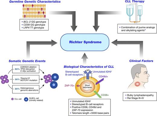
Germline genetic characteristics, clinical characteristics, biologic and genetic features of the CLL B-cell clone, and therapy for progressive CLL influence the risk of RS (Figure 1).
Risk factors associated with development of Richter syndrome in patients with CLL. *Data on the role of CLL therapy are controversial, with some studies suggesting they are contributory to the development of RS in CLL patients, and other studies suggesting not (see text for details). ZAP-70, ζ-associated protein-70.
Risk factors associated with development of Richter syndrome in patients with CLL. *Data on the role of CLL therapy are controversial, with some studies suggesting they are contributory to the development of RS in CLL patients, and other studies suggesting not (see text for details). ZAP-70, ζ-associated protein-70.
Inherited predisposition to RS
Heritable germline polymorphisms may predispose certain individuals who develop CLL to later experience transformation. Specifically, germline polymorphisms in B-cell lymphoma 2 (BCL-2; rs4987852),28 CD38 (rs6449182),29 and low-density lipoprotein receptor-related protein 4 (LRP4; rs2306029)30 genes have been reported to confer increased risk of RS. The precise functional consequences of these germline polymorphisms in the pathogenesis of RS remain to be elucidated.
Clinical and laboratory features at CLL diagnosis
Advanced Rai stage (III-IV) at CLL diagnosis and lymph nodes >3 cm on physical examination are the only clinical features associated with a higher risk of future RS.2,3,6,20 Other clinical features commonly used to predict CLL progression, including rapid lymphocyte doubling time, pattern and percentage of bone marrow involvement, splenomegaly, elevated β-2 microglobulin, and elevated lactate dehydrogenase (LDH), have not been found to reliably predict subsequent RS.
Biological characteristics of the CLL B cell
Several characteristics of the B-cell receptor are strongly linked with RS. CLL patients with leukemic B cells that are IGHV-unmutated are at an approximate 4-fold risk of RS relative to IGHV-mutated patients.2,5,21 Approximately 30% of all CLL patients have a high degree of similarity in their B-cell receptor structure because of homology of the heavy chain complementarity determining region 3.31,32 These patients with “stereotyped” B-cell receptors have a higher risk of subsequent RS, independent of IGHV mutation status.2,21,33 Although specific IGHV family usage, IGHV 4-39, was reported to be associated with a higher risk of RS,21 this observation was not confirmed in other studies.2,33
Other biologic characteristics of the leukemic B cell that are associated with the risk of CLL progression such as ζ-associated protein-70, CD38, and CD49d status have also been found to be associated with risk of future RS.2,5,20 Shorter telomere length, which is a marker of genetic instability, is also associated with increased risk of RS.34 Whether the associations between these biologic characteristics of CLL B cells are mechanistically involved in RS or are simply markers of patients who are more likely to acquire genetic events that lead to RS is not yet determined.
Somatic genetic characteristics
Early studies suggested that FISH-detectable genetic abnormalities associated with more aggressive CLL (ie, del(11q23.1) and del(17p13.1)), are also associated with RS.2,5,20 More recent studies indicate that CLL patients harboring NOTCH1 mutations have a significantly higher probability of transformation to RS (45%) compared with those without this defect (4%), suggesting an important role of this ligand-dependent transcription factor in the pathogenesis of RS.35,-37
In a seminal study, Chigrinova and colleagues performed genome\wide DNA profiling using single nucleotide polymorphism arrays on samples from 315 CLL patients without transformation after median follow up of 6.25 years, 28 patients with CLL who later developed RS, 59 CLL patients at the time of RS, and 127 patients with de novo DLBCL.38 Several novel observations were made:
The CLL phase preceding RS had similar genomic complexity to CLL cases that did not develop RS, but was enriched for specific genetic lesions (higher frequency of del(17p13.1), del(15q21.3), and add(2p25.3)).
Inactivation of tumor suppressor p53 (TP53, by loss or mutation; typically acquired before transformation) or inactivation of cyclin-dependent kinase inhibitor 2A (CDKN2A, typically acquired at the time of transformation) appeared to contribute to development of RS in 50% of cases.
A v-myc myelocytomatosis viral oncogene homolog (C-MYC) activation primarily occurred in the subset of patients with RS who also harbor TP53 inactivation.
Trisomy 12 (typically in combination with NOTCH1 mutations) was present in an additional ∼30% of RS cases and was mutually exclusive to cases with inactivation of TP53/CDKN2A.
The genomic complexity of RS was intermediate to that of CLL and DLBCL38 ; RS patients with inactivation of TP53 and/or CDKN2A experienced shorter survival than RS patients without these defects, a finding consistent with the poor prognosis associated with CDKN2A loss in de novo DLBCL.39,-41
In another recent article, Fabbri and colleagues reported similar observations using whole-exome sequencing to compare the CLL and RS phases of 9 patients with RS.42 Potential epigenetic events contributing to the pathogenesis of RS in CLL patients have also been explored. Oncostatin M and spingosine-1-phosphatase receptor 4, genes involved in cell proliferation, migration, and inhibition of apoptosis, were reported to be differentially methylated in paired analysis of RS and CLL tissue samples from the same patients. Increased methylation and transcriptional silencing was also noted for CDKN2A and Wilms tumor 1 genes.43
Collectively, these results suggest mutations in specific genetic pathways are frequently involved in transformation of CLL to clonally related RS. The first involves the acquisition of TP53 disruption, C-MYC activation, or CDKN2A deletion that occurs in ∼50% cases. The second involves trisomy 12 and NOTCH1 mutations that occur in ∼30% cases and are mutually exclusive to cases with inactivation of TP53/CDKN2A. Heterogenous genomic aberrations contribute to RS transformation in the remaining 20% cases (Figure 1).
CLL therapy and transformation risk
Although the link between CLL therapy and subsequent myelodysplasia is well established,44 the link between CLL treatment and RS is less clear. In a cohort study of 1641 newly diagnosed CLL patients seen at Mayo Clinic and followed prospectively, the rate of transformation increased from 0.5% per year before therapy to 1% per year after receiving treatment of CLL.2 Specifically, the risk of RS increased 3-fold (odds ratio = 3.26; P = .0003) among patients who received the combination of alkylating agents (which induce DNA damage) and purine nucleoside analogs (which impair DNA repair), but did not increase for those who received only 1 of these drug classes. In contrast, in the Cancer and Leukemia Group B 9011 study that compared fludarabine and chlorambucil monotherapy with fludarabine and chlorambucil in combination, no difference in the 10-year rate of transformation was observed between treatment arms (7% fludarabine monotherapy, 5% chlorambucil monotherapy, and 8% combination arm).25 Similarly, after a median follow up of 3.5 years, there was no difference in the rates of RS in patients treated with either chlorambucil alone (2%), fludarabine alone (1%), or the combination of fludarabine and cyclophosphamide (1%) in Leukemia Research Foundation Chronic Lymphocytic Leukaemia 4 (LRF CLL4) Trial.22 Thus, the impact of conventional CLL therapy alone or in combination on risk of RS is unclear at this time.
Although novel agents such as ibrutinib (a Bruton’s tyrosine kinase inhibitor) and idelalisib (a phosphatidylinositide 3-kinase δ inhibitor) have shown remarkable efficacy in CLL, little is known about their relationship to RS.45,-47 In a report on 85 heavily pretreated patients with relapsed/refractory CLL who received ibrutinib, 7 (8%) patients developed RS.46 Many of these events developed shortly after starting therapy, suggesting unrecognized RS likely predated initiation of ibrutinib treatment. Extended follow up among larger cohorts of CLL patients will be necessary to fully understand the risk of RS in CLL patients treated with these therapeutic agents.
Immunosuppression and RS
Infection with Epstein-Barr virus (EBV), detected by staining for latent membrane protein 1 or by in situ hybridization of EBV-encoded RNA transcripts, has been reported in 0% to 15% of RS tissue samples.2,5,7,20,48 Whether the EBV is causative in the disease process of RS in these patients or is merely a manifestation of the underlying immunosuppressed state associated with CLL is incompletely understood. Several cases of EBV-positive aggressive lymphomas following T-cell depleting therapies such as alemtuzumab have also been described in CLL patients.49,-51 This type of alemtuzumab-associated aggressive lymphoma is typical clonally unrelated to the underlying CLL, and thus it is debatable whether or not to consider such events true RS.52
How do we diagnose RS?
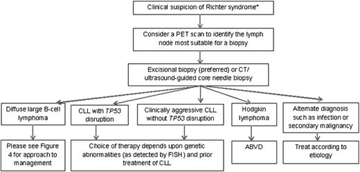
Our diagnostic approach to a CLL patient with suspicion of RS is shown in Figure 2. Although nonspecific, several signs and symptoms should alert the CLL provider of a possible transformation. These include high-grade fevers, rapidly enlarging lymph nodes, weight loss, hypercalcemia, and a markedly elevated LDH. Occasionally, extranodal involvement of the central nervous system (CNS), eyes, testes, and lungs can also be the presenting manifestation of RS.53 Although they raise suspicion of RS, all of these signs and symptoms can also be due to progressive CLL, particularly after acquisition of del(17p13.1) or TP53 inactivation.52
A schematic approach to a patient with suspected RS. *Signs and symptoms suspicious for transformation include: B-type symptoms (weight loss, night sweats, and fever), rapid enlargement of lymph nodes or spleen, hypercalcemia, and increased lactate dehydrogenase. ABVD, doxorubicin, bleomycin, vinblastine, and dacarbazine.
A schematic approach to a patient with suspected RS. *Signs and symptoms suspicious for transformation include: B-type symptoms (weight loss, night sweats, and fever), rapid enlargement of lymph nodes or spleen, hypercalcemia, and increased lactate dehydrogenase. ABVD, doxorubicin, bleomycin, vinblastine, and dacarbazine.
Because RS may not simultaneously involve all lymph node sites, the selection of the tissue site for biopsy is critical. Imaging studies with PET/CT have been used to evaluate suspected transformation and identify the optimal site for tissue biopsy. In a seminal article published in 2006, investigators reported on the findings of 57 PET/CT scans performed in 37 CLL patients with clinical suspicion of transformation. The mean SUV in the subset of patients with biopsy-proven RS was 17.6 (range, 7.4-39.4) compared with a mean SUV of 4.5 (range, 2-7.9) in CLL patients without transformation on biopsy. Using a SUV threshold of 5 as a cutoff, PET/CT had a positive predictive value of 53% and negative predictive value of 97%.54 This high negative predictive value is clinically useful, and reduces the posttest probability of RS in patients without FDG-avid lymphadenopathy to ∼3%. In contrast, the low positive predictive value of PET/CT makes it essential to perform tissue biopsy in patients with FDG-avid lymphadenopathy because only half of these patients will have RS on biopsy. The etiology of the FDG-avid tissue in the remaining patients is most often the result of progressive CLL, other malignancies, and infectious or inflammatory conditions.55 Other studies have confirmed an SUV threshold of 5 on PET/CT to identify CLL patients with clinically suspected RS who require tissue biopsy.56,57
Once the optimal site for biopsy is identified, the optimal type of biopsy must be determined. An excisional lymph node biopsy is considered the gold standard for the diagnosis of RS and should be pursued in all patients with a rapidly enlarging or FDG-avid lesion that is readily amenable to excisional biopsy. For patients whose only sites of FDG-avid disease are not easily accessible for an excisional biopsy (eg, retroperitoneal or abdominal site), a computed tomography– or ultrasound-guided large-bore core needle biopsy should be pursued. A fine needle aspirate is not sufficient to establish or exclude the diagnosis in most patients.
Once a diagnosis of RS has been confirmed, bone marrow aspirate and biopsy should be performed to complete staging. Although the National Comprehensive Clinical Guidelines58 and the European Society of Medical Oncology59 consensus guidelines do not specify whether unilateral or bilateral marrow biopsy should be performed, we typically perform bilateral biopsies because of the patchy nature of marrow involvement by lymphoma.60,61 FISH studies should also be performed on either peripheral blood (if a circulating CLL clone is present) or marrow tissue to identify acquisition of del(17p13.1), because this genetic finding has therapeutic implications.
Although there is no published data on the prevalence of CNS involvement by RS, among 120 patients with RS seen at our institution between 1995 and 2013, 5 (4%) had clinically identified CNS involvement. Given this low frequency of CNS involvement, we typically perform CNS staging (eg, lumbar puncture, magnetic resonance imaging of the head) only in patients with testicular, paranasal sinus, or epidural space involvement by DLBCL as well as those with >2 extranodal sites of disease, analogous to the approach in patients with de novo DLBCL.58
What is the prognosis of RS?
The most important determinant of clinical outcome in patients with RS is the clonal relationship of the DLBCL to the underlying CLL. Patients with clonally unrelated RS have a median survival of approximately 62 months, which is comparable to patients with de novo DLBCL.5 On the other hand, the median survival of patients with clonally related RS is significantly shorter, ranging from 8 to 14 months.
Determining the clonal relationship is relatively simple in the subset of patients for whom the CLL and DLBCL tissue have different immunoglobulin light chain restriction (eg, 1 κ and 1 λ). For the majority of patients for whom the light chain restriction is the same (eg, both κ or both λ), more detailed genetic analyses of the IGHV gene that are not widely available in most clinical laboratories are necessary to determine the clonal relationship. The optimal approach is to sequence the IGHV gene of both the original CLL and the DLBCL tissue. In clonally related RS, the IGHV V-D-J sequence will be identical (or nearly identical) between the DLBCL and the original CLL tissue. Two practical issues are often barriers to determining the clonal relationship including: (1) absence of baseline IGHV sequencing of the CLL B cells (or lack of archived CLL tissue) and (2) a mixed population of CLL and DLBCL in the lymphoma sample (particularly when the source of this tissue is bone marrow biopsy) that confounds interpretation of the sequencing results. The first issue can typically be remedied if circulating CLL B cells are present. For the second issue, light chain expression divergence (if present) and careful B-cell subpopulation analysis by flow cytometry may provide suggestive evidence of whether the clonal B-cell populations present are identical or unrelated. The relationship of the CLL and RS samples can be also be assessed by clonal immunoglobulin rearrangement pattern comparison (eg, fragment size analysis). Depending on availability, either IGHV sequencing or clonal immunoglobulin rearrangement may be used to determine clonality of the RS to the underlying CLL.
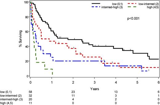
Because the techniques to determine the clonal relationship between the CLL and DLBCL are not routinely available at many centers, other prognostic approaches have been developed to predict outcome in patients with RS. In 2006, Tsimberidou and colleagues proposed an RS prognosis score based on the following 5 characteristics: (1) Eastern Cooperative Group (ECOG) performance status (>1); (2) serum LDH (≥1.5 × normal); (3) platelets (<100 × 109/L); (4) tumor size (>5 cm); and (5) number of prior therapies for CLL (>1). Patients were assigned 1 point for each adverse feature and then grouped into 1 of 4 risk categories based on their total score: low risk (score 0-1, median survival: 1.1 years); low-intermediate risk (score 2, median survival: 0.9 years); high-intermediate risk (score 3, median survival: 0.3 years); or high risk (score 4-5, median survival: 0.1 years).7 The RS prognosis score was validated in 120 RS patients seen at Mayo Clinic between 1995 and 2013 with median survivals in the low, low-intermediate, high-intermediate, and high-risk groups of 2.4 years, 0.7 years, 0.3 years, and 0.2 years, respectively (P < .001, Figure 3).2,33
Overall survival of RS patients seen at Mayo Clinic according to the RS prognosis score. Each of the following adverse characteristics is assigned one point to calculate the RS prognosis score: (1) ECOG performance score >1; (2) serum LDH >1.5 × normal; (3) platelets <100 × 109/L; (4) tumor size >5 cm; and (5) >1 prior therapy for CLL. Based on the total score, patients are grouped into: Low risk (score 0-1); Low-intermediate risk (score 2); High-intermediate risk (score 3); and High risk (score 4-5).
Overall survival of RS patients seen at Mayo Clinic according to the RS prognosis score. Each of the following adverse characteristics is assigned one point to calculate the RS prognosis score: (1) ECOG performance score >1; (2) serum LDH >1.5 × normal; (3) platelets <100 × 109/L; (4) tumor size >5 cm; and (5) >1 prior therapy for CLL. Based on the total score, patients are grouped into: Low risk (score 0-1); Low-intermediate risk (score 2); High-intermediate risk (score 3); and High risk (score 4-5).
Rossi and colleagues also devised a prognosis score for estimating overall survival in patients with RS based upon: (1) ECOG performance status; (2) presence of TP53 disruption (either by deletion or mutation); and (3) response to induction therapy. Patients were categorized as high risk if they had an ECOG performance score >1 (median survival: 0.7 years). Patients with an ECOG performance score of ≤1 but with either TP53 disruption or less than a complete response (CR) to induction were considered intermediate risk (median survival: 2 years). Patients with ECOG performance score ≤1 and no TP53 disruption who achieved a CR after induction therapy were considered low risk (median survival: not reached; 5-year survival: 70%).5 Although the inclusion of response to treatment as a variable in this prognostic model limits its clinical relevance (because it does not allow risk stratification at the time of RS diagnosis), this study illustrated the importance of TP53 disruption on prognosis in RS patients. Future efforts to incorporate clinical features (eg, ECOG performance score, LDH, number of prior CLL therapies, lymph node size), the clonal relationship of the DLBCL to the underlying CLL, and genetic factors (eg, TP53 disruption, loss of CDKN2A, NOTCH1 mutations) into a single prognostic model are likely to further improve the accuracy of risk stratification.
What are the treatment options for patients with RS?
Conventional chemotherapy approaches
Unfortunately, there are no randomized clinical trials to guide therapy for patients with RS, and thus the vast majority of evidence to guide practice is derived from single-arm phase 2 studies (Table 2). Traditionally, treatment regimens commonly used to treat other B-cell non-Hodgkin lymphomas have been used to treat CLL patients with RS.48,62,63 Anthracycline-based therapy (most commonly rituximab, cyclophosphamide, doxorubicin, vincristine, and prednisone [R-CHOP ]) has been the historical standard treatment of DLBCL,64 and has also been the traditional approach in patients with follicular lymphoma who experience histologic transformation to a more aggressive lymphoma.65,-67 Based in large part on extrapolation of this data, CHOP (and more recently R-CHOP) has been the initial treatment approach for CLL patients who experience RS. The efficacy of R-CHOP in CLL patients with RS was first prospectively evaluated in the CLL2G trial of the German CLL study group that enrolled 15 patients with RS. Although only 7% of patients achieved a CR, the overall response rate (ORR) was 67% and the median overall survival was 27 months. Treatment-related mortality was low (3%).68 In a retrospective series of 12 RS patients treated with R-CHOP at MD Anderson Cancer Center, the ORR was 50% and the median overall survival was 15 months.69 Other trials have explored alternative anthracycline-based regimens to try to improve efficacy. In a phase 2 study of 29 RS patients treated with hyperCVXD regimen (fractionated cyclophosphamide, vincristine, liposomal daunorubicin, and dexamethasone), the ORR was 41%, with 38% CRs, and a partial response rate of 3%. Although the reported CR rate was higher than that observed in the studies of R-CHOP, the median survival with hyperCVXD was only 10 months.70 A subsequent study of 30 patients added rituximab and granulocyte macrophage-colony stimulating factor to the hyperCVXD platform and incorporated high-dose methotrexate and cytarabine every other cycle. Unfortunately, the ORR was similar to the hyperCVXD regimen and median survival was only 8.5 months.71 Treatment-related mortality with both these hyperCVXD trials exceeded 10%.
Platinum-containing regimens have also been evaluated as initial therapy in CLL patients with RS. Investigators at the MD Anderson Cancer Center tested the combination of oxaliplatin, fludarabine, cytarabine and rituximab (OFAR) using 2 different dosing schedules. Among the 55 RS patients treated on both trials, the ORR ranged from 43% to 50% and median survival was 6 to 8 months. Unfortunately, the OFAR platform was associated with grade 3/4 neutropenia and thrombocytopenia occurring in approximately 80% patients under both schedules.69,72 Given the efficacy of purine analogs in patients with CLL, a pilot study tested the backbone of fludarabine and cytarabine in combination with either cyclophosphamide (PFA) or cisplatin (CFA) in 12 RS patients. The ORR was 45% with 18% CRs and a median survival of 17 months.73 Based on these results, all 4 agents (cisplatin, fludarabine, cyclophosphamide, cytarabine) were combined in a follow-up study of 15 RS patients. The 4-drug combination caused profound toxicity (20% treatment-related mortality in the first 4 weeks) and only 1 patient (5%) achieved a response.74 Radioimmunotherapy with 90Y ibritumomab tiuxetan was also tested in a pilot trial of 7 RS patients; however, no patient responded.75
Novel therapeutic approaches
Ibrutinib has demonstrated single-agent activity in a series of patients with relapsed/refractory de novo DLBCL (n = 70) with an ORR of 23%. Notably, ibrutinib appeared to be more active in patients with the ABC subtype (41%) relative to those with the germinal center B-cell subtype (5%, P = .007).76 Although ibrutinib has also shown remarkable efficacy in CLL patients, 7 (8%) of the 85 patients with relapsed/refractory CLL treated in the initial ibrutinib trial developed RS.46 Although this information does not directly provide insight into whether ibrutinib may have clinical activity in patients with RS, it suggests ibrutinib monotherapy is not an adequate approach for many RS patients. Recent studies of ibrutinib in combination with R-CHOP in patients with de novo DLBCL (n = 33) reported an ORR of 100%.77 Phase 3 testing of this combination in de novo DLBCL is now under way, and this may be an approach worth exploring in RS. Other novel approaches that may have promise in the management of RS as single agents or in combination with traditional chemotherapy approaches include ABT-199 (BCL-2 antagonist), idelalisib, and NOTCH1 inhibitors.
Role of stem cell transplantation
Given the poor response rates and survival with chemotherapy alone in RS, both autologous and allogeneic stem cell transplantation (SCT) have been explored as postremission therapy and/or salvage therapy. Unfortunately, most patients with RS do not achieve adequate response to induction to proceed to transplant. Of the 120 patients with RS seen at Mayo Clinic over a 19-year interval (1995-2013), only 13 (11%) patients were able to proceed to SCT (10 autologous SCT, 2 allogeneic SCT, and 1 autologous followed by allogeneic SCT). The median survival of patients who received SCT was better than those who did not receive SCT (5.2 years vs 0.7 years; P = .005).33 Tsimberidou et al described the outcomes of 20 RS patients (of a cohort of 148 [14%]) who underwent SCT (17 allogeneic SCT and 3 autologous SCT). As a group, patients who underwent SCT had an improved outcome compared with those that did not. Among those who underwent SCT, the estimated cumulative 3-year survival probability was 75% for those who were transplanted in CR or PR, compared with 21% for patients who underwent SCT as salvage therapy for relapsed/refractory RS. Furthermore, the estimated 3-year survival probability was 27% for those patients who responded to initial chemotherapy for RS, but did not undergo subsequent SCT, suggesting that postremission transplant increases the durability of remission in patients who respond to induction.7
The European Group for Blood and Marrow Transplantation recently evaluated its experience in 59 RS patients who underwent SCT (34 autologous SCT and 25 allogeneic SCT).78 The estimated 3-year survival was 36% for those who underwent allogeneic SCT compared with 59% in those who underwent an autologous SCT. This comparison should be interpreted with caution given the retrospective nature of the study. Among patients who underwent an allogeneic SCT, disease status at the time of SCT (CR/PR vs progressive disease) was the only parameter associated with survival. In a multivariate analysis of relapse-free survival among allogeneic SCT recipients, age <60 years, reduced intensity conditioning, and CR/PR at the time of transplantation were associated with superior relapse-free survival. Although there was no clear plateau in OS or relapse-free survival among the 34 patients who underwent autologous SCT, only 11 of 17 relapses were related to RS (the remainder were due to CLL), suggesting autologous SCT may eradicate the RS component in many patients even though the underlying CLL may persist.78 Several other studies exploring the role of SCT in the treatment of transformed low-grade B-cell lymphoproliferative neoplasms (most commonly follicular lymphoma), have included a small proportion of patients with CLL. These studies suggest that SCT is feasible in patients with RS and may be associated with improved outcomes.65,67 Although all of these studies are subject to potential selection bias and are limited by the fact that the clonal relationship of the CLL and DLBCL was not determined, collectively they suggest that “fit” RS patients with limited comorbidities who have a donor available and demonstrate chemosensitive disease benefit from postremission SCT.
How we treat RS
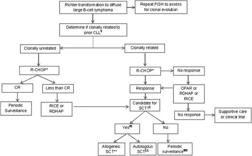
The experience with intense combination chemotherapy-based regimens (Table 2) suggests that traditional chemotherapy approaches provide inadequate disease control for most patients. It is hoped that novel, targeted therapies will improve the efficacy of therapy for RS and appropriate patients should be treated on well-designed clinical trials. A summary of our approach to CLL patients who develop RS and who are not treated on clinical trials is provided in Figure 4.
Management of CLL patients with Richter transformation to diffuse large B-cell lymphoma in which no clinical trial is available.¶The simplest way to determine clonality is to compare the κ and λ light chain restriction between the CLL and the DLBCL. For patients in whom they are different (eg, κ light chain–restricted CLL and λ light chain–restricted DLBCL), DLBCL is clonally unrelated to the CLL. In those patients in whom the light chain restriction is the same, clonality is determined either by sequencing the IGHV V-D-J genes or by comparing immunoglobulin gene rearrangements between the CLL and DLBCL tissue samples. *Consider a platinum-containing regimen for patients with prior anthracycline exposure. We generally administer pegfilgrastim to all patients undergoing intensive chemotherapy for RS. §Age, functional status, comorbidities, and chemosensitive disease determine if patient is a suitable candidate for stem cell transplantation. ¶¶Both autologous and allogeneic SCT may play a role in improving durability of remission. Factors that influence whether autologous or allogeneic is preferred strategy include patient age/fitness, availability of a suitable donor, the presence or absence of del(17p13.1)/TP53 mutation, and whether the patient’s underlying CLL is purine analog refractory. **An allogeneic stem cell transplantation is preferred in patients with clonally related RS as well as those with clonally unrelated DLBCL whose underlying CLL is purine analog–refractory or harbors the del(17p13.1)/TP53 mutation. §§Autologous SCT is our preferred approach for patients with clonally unrelated DLBCL whose CLL does not harbor the del(17p13.1)/TP53 mutation. An autologous SCT may be considered in select patients with clonally related RS who do not have a suitable donor or who are not candidates for allogenic SCT based on age/comorbidity. The primary intent of autologous SCT in such cases is to eradicate the DLBCL component rather than simultaneously treat the underlying CLL. ¶¶¶At this time, outside of a clinical trial, there are no specific recommendations for a postremission strategy among patients with clonally related RS who demonstrate a response to induction chemotherapy and are not considered candidates for SCT.
Management of CLL patients with Richter transformation to diffuse large B-cell lymphoma in which no clinical trial is available.¶The simplest way to determine clonality is to compare the κ and λ light chain restriction between the CLL and the DLBCL. For patients in whom they are different (eg, κ light chain–restricted CLL and λ light chain–restricted DLBCL), DLBCL is clonally unrelated to the CLL. In those patients in whom the light chain restriction is the same, clonality is determined either by sequencing the IGHV V-D-J genes or by comparing immunoglobulin gene rearrangements between the CLL and DLBCL tissue samples. *Consider a platinum-containing regimen for patients with prior anthracycline exposure. We generally administer pegfilgrastim to all patients undergoing intensive chemotherapy for RS. §Age, functional status, comorbidities, and chemosensitive disease determine if patient is a suitable candidate for stem cell transplantation. ¶¶Both autologous and allogeneic SCT may play a role in improving durability of remission. Factors that influence whether autologous or allogeneic is preferred strategy include patient age/fitness, availability of a suitable donor, the presence or absence of del(17p13.1)/TP53 mutation, and whether the patient’s underlying CLL is purine analog refractory. **An allogeneic stem cell transplantation is preferred in patients with clonally related RS as well as those with clonally unrelated DLBCL whose underlying CLL is purine analog–refractory or harbors the del(17p13.1)/TP53 mutation. §§Autologous SCT is our preferred approach for patients with clonally unrelated DLBCL whose CLL does not harbor the del(17p13.1)/TP53 mutation. An autologous SCT may be considered in select patients with clonally related RS who do not have a suitable donor or who are not candidates for allogenic SCT based on age/comorbidity. The primary intent of autologous SCT in such cases is to eradicate the DLBCL component rather than simultaneously treat the underlying CLL. ¶¶¶At this time, outside of a clinical trial, there are no specific recommendations for a postremission strategy among patients with clonally related RS who demonstrate a response to induction chemotherapy and are not considered candidates for SCT.
The most important step in deciding the treatment plan for patients with RS is to determine if the DLBCL is clonally related to the underlying CLL. For the 20% of patients with clonally unrelated DLBCL, we recommend the use of standard therapy with R-CHOP (similar to patients with de novo DLBCL). For patients who achieve a CR, we do not pursue SCT, and recommend periodic surveillance as outlined by the National Comprehensive Cancer Network guidelines.58 For those patients with clonally unrelated DLBCL who do not achieve a CR, salvage therapy with rituximab, ifosfamide, and etoposide (RICE) or with rituximab, dexamethasone, cytarabine and cisplatin (RDHAP), followed by an autologous SCT should be considered, analogous to the approach used for patients with de novo DLBCL.79 We favor allogeneic transplant in suitable patients with clonally unrelated DLBCL who have purine analog refractory CLL or CLL with TP53 disruption (because these patients have an indication for allogeneic SCT for the treatment of the underlying CLL even if they did not have RS).
For the 80% of patients with clonally related DLBCL, our first choice of therapy is a clinical trial given the suboptimal efficacy of the standard treatment approaches. When a clinical trial is not available, we generally consider R-CHOP as the initial treatment option given its favorable toxicity profile, followed by consolidation with SCT. If patients have received prior anthracycline therapy, platinum-based induction regimens such as OFAR, RDHAP, or RICE may be considered instead of R-CHOP. Age, functional status, comorbidities, and chemosensitivity of the disease determine if the patient is a suitable candidate for SCT. Allogeneic SCT is the preferred approach for patients with clonally related RS if they have a suitable donor and are a candidate for this approach based on age and performance status. An autologous SCT may be considered in select patients with clonally related RS who do not have a suitable donor or who are not candidates for allogenic SCT based on age/comorbidity with a primary goal of eradicating the DLBCL (not necessarily the underlying CLL). For patients who do not respond to induction with anthracycline and/or platinum-based therapy, clinical trials of novel agents and/or supportive care should be considered depending on age, comorbidities, performance status, and patient preference.
Special considerations
Hodgkin transformation
Hodgkin transformation of CLL is a rare event and clonally unrelated to the underlying CLL in most cases.80 At our institution, in a cohort study of 1644 newly diagnosed CLL patients followed prospectively, the prevalence of HL after 10 years was 0.5% (∼0.05%/year).81 The median interval from CLL diagnosis to development of HL was 6.2 years (0-24.5 years), and the median survival after diagnosis of HL was 3.9 years (1.0-5.0 years). A systematic review of the literature between 1975 and 2011 identified 86 CLL patients who developed HL. The median interval between the diagnosis of CLL and subsequent development of HL was 4.3 years (0-26 years), and median survival after HL diagnosis was 1.7 years (0-14 years).80 In contrast to DLBCL, EBV infection in the HL tissue is present in approximately 70% of CLL patients who develop HL.48,80
In the Mayo series, the International Prognostic Score for HL82 proved useful for predicting survival in CLL patients who developed HL. Patients with HL who received previous treatment of CLL, particularly fludarabine-based treatment, appear to have a worse outcome after diagnosis of HL.80 Combination chemotherapy using doxorubicin, bleomycin, vinblastine, and dacarbazine is the most widely used treatment strategy,80 which is also our standard approach.
Summary
RS in a CLL patient is associated with very rapid disease progression, limited therapeutic options, and generally poor survival. Knowledge from recent studies demonstrates that approximately 20% of DLBCL cases arising in CLL patients are clonally unrelated to the underlying CLL and represent de novo DLBCL with a median survival of 5 years. In contrast, the median survival is approximately 1 year in the remaining 80% patients who have clonally related RS. The discovery of several key genetic aberrations that appear to contribute to transformation of CLL to clonally related RS, such as CDKN2A and TP53 loss and NOTCH1 mutations, has improved our understanding of the pathophysiology of RS. It is hoped that clinical trials incorporating drugs that target these molecular abnormalities will improve clinical outcome for CLL patients who experience RS.
Resolution of clinical vignette
The patient underwent a bone marrow biopsy, which demonstrated 80% involvement by CLL, trisomy 12 in 28% of nuclei by FISH testing, and no marrow involvement by DLBCL. His ECOG performance score was 1, serum LDH was 565 U/L (normal, 122-222 U/L), and platelet count was 75 × 109/L. The RS prognosis score was calculated to be 3, ie, high-intermediate risk (projected median survival = 4 months). His IGHV V-D-J rearrangement analysis showed that the DLBCL was clonally related to the underlying CLL. The patient received 6 cycles of R-CHOP with a CR. Consolidation with allogeneic SCT was pursued. Six months after his transplant, the patient continues to be in complete remission.
Acknowledgments
The authors thank Drs David Viswanatha, Curtis Hanson, and Grzegorz Nowakowski for their critical review and input on selected aspects of this manuscript.
This publication was supported by a grant from the National Cancer Institute (CA95241) (N.E.K.). Tait D. Shanafelt is a clinical scholar of the Leukemia and Lymphoma Society.
Authorship
Contribution: S.A.P., N.E.K., and T.D.S. wrote and edited the manuscript.
Conflict-of-interest disclosure: N.E.K. has received research support from Celgene, Gilead and Hospira. T.D.S. has received research support from Celgene, Cephalon, Genentech, Glaxo-Smith-Kline, Hospira, and Polyphenon E International. The remaining author declares no competing financial interests.
Correspondence: Tait Shanafelt, Division of Hematology, Department of Medicine, Mayo Clinic, 200 First St SW, Rochester, MN 55905; e-mail: shanafelt.tait@mayo.edu.

