Abstract
Imatinib mesylate is a small molecule drug that in vitro inhibits the Abelson (Abl), Arg (abl-related gene), stem cell factor receptor (Kit), and platelet-derived growth factor receptor A and B (PDGFRA and PDGFRB) tyrosine kinases. The drug has acquired therapeutic relevance because of similar inhibitory activity against certain activating mutations of these molecular targets. The archetypical disease in this regard is chronic myeloid leukemia, where abl is constitutively activated by fusion with the bcr gene (bcr/abl). Similarly, the drug has now been shown to display equally impressive therapeutic activity in eosinophilia-associated chronic myeloproliferative disorders that are characterized by activating mutations of either the PDGFRB or the PDGFRA gene. The former usually results from translocations involving chromosome 5q31-33, and the latter usually results from an interstitial deletion involving chromosome 4q12 (FIP1L1-PDGFRA). In contrast, imatinib is ineffective, in vitro and in vivo, against the mastocytosis-associated c-kit D816V mutation. However, wild-type and other c-kit mutations might be vulnerable to the drug, as has been the case in gastrointestinal stomal cell tumors. Imatinib is considered investigational for the treatment of hematologic malignancies without a defined molecular drug target, such as polycythemia vera, myelofibrosis with myeloid metaplasia, and acute myeloid leukemia.
Introduction
The classic bcr/abl-negative chronic myeloproliferative diseases (CMPDs) include polycythemia vera (PV), essential thrombocythemia (ET), and myelofibrosis with myeloid metaplasia (MMM).1 Several other bcr/abl-negative clinicopathologic entities bear a certain degree of ontogenic and phenotypic resemblance to these classic CMPDs and are operationally classified as atypical CMPDs (Figure 1).2 Atypical CMPDs include a spectrum of eosinophilic disorders,3,4 systemic mastocytosis (SM),5 chronic myelomonocytic leukemia (CMML),6 juvenile myelomonocytic leukemia,7 chronic neutrophilic leukemia,8 and unclassified (mixed or hybrid) myeloproliferative disorders.9 Recent advances in the molecular characterization of some of the atypical CMPDs have identified molecular lesions that reliably predict outcomes to treatment with imatinib mesylate (imatinib).10-20 This translates into a need for tools that will allow patients with atypical CMPD to be rapidly classified into discrete molecular subgroups that allow for specific treatment choices to be adopted or discarded. We review here the therapeutically relevant molecular targets of imatinib in the bcr/abl-negative myeloid disorders, and we propose a semimolecular classification scheme that incorporates molecularly defined entities and the imatinib treatment experience for individual disorders, including those without known drug targets.
Operational classification of bcr/abl–negative chronic myeloproliferative disorders.
Operational classification of bcr/abl–negative chronic myeloproliferative disorders.
Mechanism of oncogenic activation of imatinib-relevant molecular targets
Imatinib-sensitive mutations in bcr/abl-negative myeloid disorders have involved a subset of receptor tyrosine kinases (RTKs), including platelet-derived growth factor receptor B (PDGFRB),16 PDGFRA,11 and Kit.15 Structurally, RTKs consist of an extracellular ligand-binding domain, a transmembrane domain, and an intracellular tyrosine kinase domain.21 The kinase domain contains an activation loop whose specific conformation is controlled by phosphorylation of tyrosine residues that reside within it, and this occurs when the otherwise monomeric surface receptors dimerize. Receptor dimerization induces kinase activity and is physiologically accomplished by ligand binding. In contrast, gain-of-function mutations of RTKs result in ligand-independent (constitutive) receptor dimerization and subsequent oncogenic activation of the receptor. Mutation-mediated RTK activation might involve the introduction of oligomerization domains to the extracellular segment of the receptor by fusion proteins (eg, fusion products of ETV6-PDGFRB, HIP1-PDGFRB, or ZNF198-FGFR1),10,22-24 possible disruption of an autoinhibitory region within the juxtamembrane domain of the RTK (eg, FLT3 and c-kit juxtamembrane mutations),25,26 or activation loop mutations that could stabilize an active kinase conformation (eg, c-kit D816V).27,28
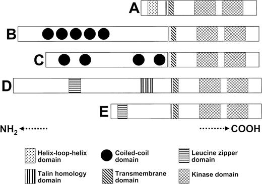
Oncogenic PDGFRB activation usually occurs as a result of a reciprocal chromosomal translocation. As illustrated in Table 1,29-32 the partner genes that are fused in-frame with the PDGFRB gene have been identified for 6 translocations seen in patients with myeloid disorders.10,18,23,33-36 Using this class of gene fusions as an example, it is possible to comment on several common themes in this regard (Figure 2). First, the oncogenic protein carries the entire PDGFRB tyrosine kinase catalytic domain fused to an N-terminal segment of the partner gene, such as ETV6-PDGFRB. Second, the reciprocal fusion transcript (eg, PDGFRB-ETV6) was not detected in any of the screened patients, and its pathogenetic contribution, if any, is unknown. Third, the 5′ fusion partner gene invariably encodes for one or more oligomerization domains that mediate oncoprotein dimerization. Fourth, the breakpoint in the PDGFRB gene is completely conserved for all 6 translocations. Fifth, for the 5′ partner fusion genes (eg, H4/D10S170 and Huntington interacting protein [HIP1]) in which detailed mutational analysis was performed, the data suggest that apart from the predicted critical role of the oligomerization and tyrosine kinase domains from either partner gene, other regions in the 5′ fusion partner gene may be essential for the transforming activity of the oncoprotein.
Representation of the oligomerization domain(s) within the 5′ partner genes that are fused with PDGFRB. The 5′ partner genes are (A) ETV6, (B) PDE4DIP or myomegalin, (C) rabaptin 5, (D) HIP1, and (E) H4/D10S170.
Representation of the oligomerization domain(s) within the 5′ partner genes that are fused with PDGFRB. The 5′ partner genes are (A) ETV6, (B) PDE4DIP or myomegalin, (C) rabaptin 5, (D) HIP1, and (E) H4/D10S170.
At least 2 distinct mechanisms have been demonstrated to operate on producing rearrangements of the PDGFRA gene. One is the reciprocal translocation, t(4;22)(q12;q11), associated with atypical chronic myeloid leukemia (CML), which produces the BCR/PDGFRA fusion37,38 and of which 3 cases have been reported. The other is the unique interstitial deletion of a small chromosomal fragment (approximately 800 bp) within the chromosome 4q12 region, which produces the FIP1L1/PDGFRA fusion (Table 1).11 Interestingly, for both mechanisms of PDFGRA activation, the breakpoint for the PDGFRA gene lies within exon 12 and involves the WW-like encoding region of the juxtamembrane domain in most patients.39 Unlike the other 5′ partner fusion genes involved in PDFGRB- or FGFR1-related fusions, FIP1L1 does not carry a homodimerization domain, at least based on homology to known oligomerization domains. Given the known autoinhibitory role of the juxtamembrane domain for the receptor tyrosine kinases EPHB240 and Kit,26 it is speculated that, for FIP1L1/PDGFRA, disruption of the WW domain serves as the primary mode of kinase activation, in lieu of any dimerization mediated by FIP1L1.
Mutations in c-kit have been classified into 2 groups, enzymatic-pocket–type mutations, exemplified by D816V, which alter the activation loop at the catalytic site, and regulatory-type mutations, which affect regions other than the enzymatic pocket to influence kinase activity.41 Regulatory-type mutations can involve the extracellular, juxtamembrane, and transmembrane Kit domains.15,28,42 Juxtamembrane mutations might alter protein residues in the juxtamembrane region of Kit that normally interact with the kinase domain and that autoinhibit ligand-independent kinase activation.26,43 The biochemical mechanism of oncogenic kinase activation in enzymatic pocket–type mutations might involve stabilization of the active conformation of the activation loop domain.44
Clinical phenotypes of imatinib-relevant atypical chronic myeloproliferative disorders
Patients with atypical CMPD display marked heterogeneity in clinical and bone marrow histologic features. In addition, a substantial degree of phenotypic mimicry exists among the relatively loosely defined disease entities. For example, a subset of predominantly male patients with the chromosome 5q31-33 (PDGFRB) rearrangement exhibits prominent blood eosinophilia that may be associated with monocytosis and, therefore, can be classified as CMML10,23,33 or chronic eosinophilic leukemia (CEL).16 Similarly, a subset of patients with the clinical phenotype of hypereosinophilic syndrome (HES) might be reclassified as having SM based on bone marrow histologic and mast cell immunophenotypic findings.20 This highlights the challenge entailed in identifying specific groups of patients who are likely to benefit from molecularly targeted therapy, including imatinib, and supports the identification and description of molecularly defined entities within the atypical CMPD category (Figure 1).
FIP1L1-PDGFRA–positive eosinophilic disorders
The seminal discovery11 of the association between FIP1L1-PDGFRA and clonal eosinophilia has allowed a unified molecular categorization of a clinicopathologic entity that has variously been described as HES,11 myeloproliferative variant of HES,45 SM associated with eosinophilia,14 and FIP1L1-PDGFRA–positive CEL.46 Most patients with FIP1L1-PDGFRA–positive eosinophilic disorder display hepatosplenomegaly, marked leukocytosis, elevated serum tryptase and vitamin B12 levels, and bone marrow pan-hyperplasia sometimes associated with myelofibrosis.20,45,46 In addition, some patients manifest typical clinical features of HES (eosinophilic cardiomyopathy, thrombosis)17,20,46 or SM (urticaria pigmentosa, mast cell mediator release symptoms, bone pain).20
In a single institutional cohort of 81 patients with primary blood eosinophilia (eosinophil count 1.5 × 10⁹/L [1500/μL] or higher), FIP1L1-PDGFRA was detected by fluorescence in situ hybridization (FISH) in 11 patients for an incidence rate of 14%.20 With the exception of a single patient,46 all other patients with FIP1L1-PDGFRA–positive eosinophilic disorders described so far in a combined series of 26 patients from 3 different studies have been men aged 26 to 72 years at diagnosis.17,20,46 The prognosis of FIP1L1-PDGFRA–positive eosinophilic disorder, in the absence of treatment with imatinib, is reported to be poor, and leukemic transformation has been documented in at least 2 patients, including 1 patient previously treated with imatinib.11,46
PDGFRB-rearranged CMPD
A recent comprehensive review of myeloid disorder–associated translocations involving chromosome 5q31-35 lists 34 reported cases of t(5;12)(q31-33;p12-13), including 11 with documented PDGFRB rearrangement, and 20 cases associated with other partner chromosomes (5 of the latter 20 were known to involve PDGFRB).47 Pathologic diagnosis in previously described 5q31-33–associated myeloid disorders have included CMML, CEL, atypical CML, atypical CMPD, and occasionally myeloid dysplastic syndromes (MDS) or acute myeloid leukemia (AML).16,32,44,47-49 Reported clinical and laboratory features have included splenomegaly, leukocytosis, and eosinophilia in most patients and hepatomegaly, monocytosis, and skin involvement in some.16,47
As with FIP1L1-PDGFRA–positive eosinophilic disorders, women are rarely affected with PDGFRB-rearranged CMPD.12,16,47 It is difficult to assign accurate incidence figures on PDGFRB-rearranged CMPD because of the heterogeneous clinical presentation. Nevertheless, in a retrospective review of 17 791 consecutive cytogenetic studies performed at the Mayo Clinic, only 2 patients t(5;12)(q31-33;p13) were identified, both of whom displayed prominent blood eosinophilia and monocytosis.50 In a separate cohort of 81 patients with primary eosinophilia higher than 1.5 × 10⁹/L (1500/μL), none were associated with a chromosome 5q31-33 translocation.20 In a series of 205 patients with CMML in whom cytogenetic studies were performed, not a single 5q translocation was reported despite the presence of cytogenetic abnormalities in 34% of the patients.6
c-kit–positive systemic mastocytosis
Among the myeloid disorders, activating c-kit mutations cluster primarily with mastocytosis, though such mutations have also been identified in patients with other myeloproliferative disorders and AML. Mutations in human c-kit at codons 560 (V560G) and 816 (D816V) that lead to its ligand-independent activation were first identified in the human mast cell leukemia cell line HMC-1.51 In mice, c-kit D814 is homologous to D816, and introducing murine mutant c-kit (D814Y) into the interleukin-3 (IL-3)–dependent mast cell line IC2 confirmed that such mutations mediate transformation in vitro and tumorigenicity in vivo.52 These data, and the cytotoxic effect of Kit inhibitors on cell lines that carry c-kit–activating mutations,53 indicate that such mutations are causally related to mastocytosis. Accordingly, the c-kit D816V mutation has been found to exhibit a strong association with adult and atypical pediatric mast cell disease.28
In an unselected series of 115 patients with a spectrum of chronic myeloid disorders, none of the 99 patients with a non–mast cell disease displayed a juxtamembrane or an activation loop c-kit mutation.54 In contrast, 5 (31%) of 16 patients with SM displayed the c-kit D816V mutation. In general, clinical and laboratory features of SM (urticaria pigmentosa, mast cell mediator release symptoms, bone pain, hepatosplenomegaly, blood eosinophilia) were not consistently influenced by the presence or absence of the c-kit D816V mutation.5,14,55
Diagnostic techniques to detect imatinib-relevant molecular markers in patients with bcr/abl-negative CMPD
In general, only a small minority of patients with bcr/abl-negative CMPD displays a microscopically visible karyotypic abnormality that offers any indication as to the presence or absence of an imatinib-relevant underlying molecular lesion.56 However, currently identified molecular translocations/rearrangements that are relevant to imatinib therapy produce characteristic breakpoint clusters, including those at 5q31-3310,16,18,23,33-35 and 4q12,11,14 which makes them amenable to detection by reverse transcription–polymerase chain reaction (RT-PCR) and FISH. However, neither of these diagnostic tests adequately substitutes for another. In some cases, cytogenetic abnormalities involving 5q31-33 that suggest a PDGFRB rearrangement cannot be confirmed by FISH.47 Whether this means that the PDGFR gene was not rearranged or was missed by the particular FISH strategy used is unclear.12 In contrast to PDGFRB mutations, except in rare instances,57 PDGFRA and c-kit mutations are usually not detected by karyotype and require either a FISH-based technique or RT-PCR, as previously described.11,14,20
Imatinib therapy for bcr/abl-negative myeloid disorders
Imatinib is a 2-phenylaminopyrimidine molecule that occupies the adenosine triphosphate–binding site and, hence, selectively inhibits the Abl (including Bcr/Abl)58,59 and a subset of the type 3 transmembrane RTKs. The latter category includes Kit (receptor for stem cell factor)60,61 and PDGFR.59,60 Imatinib has already been used in relatively large numbers of patients to demonstrate efficacy for the treatment of CML62-65 and the Kit-mediated gastrointestinal stromal tumor (GIST).66
As discussed and as illustrated in Table 1, imatinib-sensitive tyrosine kinases other than Abl might be constitutively activated in various bcr/abl-negative CMPDs through a variety of molecular mechanisms, including reciprocal chromosomal translocations (eg, ETV6/PDGFRB), complex translocations that fuse genes lying in opposite orientation in relation to the centromere (eg, ETV6/ABL), and submicroscopic interstitial chromosomal deletions (eg, FIP1L1-PDGFRA). Accordingly, several studies have explored the therapeutic value of imatinib in various myeloid disorders (Tables 2 and 367,68 ).
Eosinophilic disorders
Identifying the oncogenic FIP1L1-PDGFRA tyrosine kinase resulted from a reverse bedside-to-bench approach to the management of patients with eosinophilic disorders.11 The empiric use of imatinib in a relatively heterogenous group of patients with the common manifestation of blood eosinophilia led to the observation, in a number of studies (Table 2),69-73 that a subset rapidly achieved complete clinical and hematologic remissions at a relatively low-dose level (100 mg/d). These data, including the absence of activating mutations in other known imatinib-responsive genes (PDGFRB and c-kit),70,73 and response to a lower dose of imatinib (indicating a lower IC50 relative to known targets), suggested the presence of a novel imatinib-responsive target. Cloning of the FIP1L1-PDGFRA fusion gene was first reported in March 2003 from a patient with HES,11 and its presence in a subset of patients with eosinophilic disorders has since been confirmed by other studies.14,17,20,44,45
FIP1L1-PDGFRA–positive eosinophilic disorders. It is now well established that patients with FIP1L1-PDGFRA–positive eosinophilic disorders are likely to obtain clinical, hematologic, and even molecular remissions with standard-dose (400 mg/d)11,17 or low-dose (100 mg/d)14,20,46 imatinib therapy. The marked response at lower-than-standard drug doses is consistent with the observation that the IC50 for the growth of FIP1L1-PDGFRA–positive cell lines was substantially lower than that for bcr/abl-positive cell lines.11
In a combined series from 4 separate reports11,17,20,46 of 24 patients with FIP1L1-PDGFRA–positive eosinophilic disorders who were treated with imatinib starting at doses of 100 to 400 mg/d, all achieved complete hematologic remission and normalized blood eosinophil counts. It is important to note that this group of patients was variously labeled as having HES,11 myeloproliferative variant of HES,17 SM associated with eosinophilia,20 and FIP1L1-PDGFRA–positive CEL46 by different investigators. Time to complete hematologic response (defined as normalization of complete blood count, white blood cell differential, and eosinophil count) varied from 1 to 6 weeks. In addition, posttreatment bone marrow examinations, when performed, showed equally impressive improvements, including decreased cellularity, resolution of abnormal mast cell infiltration,74 and reversal of myelofibrosis.17,20,46 Furthermore, molecular remission was documented by FISH in 4 patients20 and by RT-PCR in 7 patients.17,46 Time to molecular remission ranged from 1 to 12 months. Minimal residual disease was documented by RT-PCR in 2 patients, including the only woman in the entire series.17,46
Numerous case studies have confirmed the favorable blood and bone marrow responses seen in imatinib-treated patients with FIP1L1-PDGFRA–positive eosinophilic disorders.75-77 Similarly, others have reported a similar experience in patients with a different mechanism of PDGFRA rearrangement that resulted from chromosomal translocation t(4;22)(q12;q11), involving the PDGFRA and bcr genes, respectively.33,57 In all these studies, the degree, rapidity, and duration of imatinib response was similar between the 100- and 400-mg daily dose schedules. In general, responses have been durable (up to more than 3 years), with only a single report of relapse that was associated with the development of an imatinib-resistant mutation.11
Hypereosinophilic syndrome (FIP1L1-PDGFRA–negative). In the first study in which molecular examination was performed, 4 of 5 patients with HES who had been shown to be negative for FIP1L1-PDGFRA responded completely to imatinib (400 mg/d), with response durations of 2 weeks to 16 months.11 However, in another study of 6 patients with HES, in whom the absence of FIP1L1-PDGFRA was documented by FISH, none achieved complete responses, and only 3 experienced partial responses with 400 mg/d imatinib.20 One patient from another study also did not respond to treatment with imatinib.46 More patients in this category of disease must be treated before any conclusions can be made.
Before the discovery of FIP1L1-PDGFRA, several pilot studies in patients with HES suggested the presence of a subgroup of patients with a dramatic response to low doses of imatinib therapy.69-73,78 From a combined series of 21 such patients from the 3 largest studies,70,72,73 10 (48%) were reported to have achieved complete hematologic responses, and 3 achieved partial responses. A more recent study consisting of 6 HES patients reports a complete remission rate of 100% with low-dose imatinib therapy.79 Whether these patients had true HES or FIP1L1-PDGFRA–positive eosinophilic disorder has not been ascertained, and the issue can only be resolved in a prospective treatment trial with the proper correlative laboratory studies.
PDGFRB-rearranged CMPD. Similar to what is seen with PDGFRA mutations, several activated PDGFRB fusion tyrosine kinases have been demonstrated to be imatinib sensitive (Table 3). In one study, all 4 patients with atypical CMPD characterized by eosinophilia and t(5;12)(q33;p13) (3 had the ETV6/PDGFRB fusion gene) achieved complete hematologic and cytogenetic remissions with imatinib given at 400 mg/d.16 In this report, the remissions were durable (at least 9 months long), and molecular remission was documented in one of the responders. Although in vitro imatinib toxicity to primary cells from these patients suggested an IC50 for ETV6/PDGFRB that was similar to that for bcr/abl,16 another study has shown higher imatinib sensitivity in RAB5/PDGFRB–expressing cell lines (IC50 = 0.03 μM) compared with those that expressed bcr/abl (IC50 = 0.3 μM).80 Similarly, FIP1L1-PDGFRA–transformed cell lines were substantially more sensitive to imatinib-induced growth inhibition (IC50 = 3.2 nM) compared with bcr/abl–transformed cell lines (IC50 = 582 nM).11 Regardless, because the IC50 of imatinib for all the PDGFRB fusion proteins is unknown, it is reasonable to start treatment at a dose of 400 mg/d. Several other case studies have confirmed the therapeutic value of imatinib in PDGFRB-rearranged CMPD regardless of the specific genes (chromosomes) that partner with PDGFRB.18,80-82
Systemic mastocytosis
The demonstration of imatinib-induced inhibition of Kit-associated signal transduction formed the rationale to use the drug in SM. In vitro, imatinib effectively inhibits normal mast cell growth and development.83 Additionally, in vitro, imatinib inhibits the growth of human mast cell lines that carry the V560G but not the D816V c-kit mutation.84 Similarly, the drug was lethal to clonal mast cells from patients who carry wild-type c-kit but not D816V c-kit mutations.84,85 The first study that evaluated imatinib therapy in SM was performed before the discovery of FIP1L1-PDGFRA and included 12 patients treated at a lower dose of the drug (100 mg/d).86 Overall, 5 patients (42%) experienced measurable responses. The most impressive response was seen in 3 patients with associated eosinophilia (SM-eos) later shown to carry the FIP1L1-PDGFRA mutation.14 In contrast, none of the 2 patients with SM-eos and the c-kit D816V mutation responded. In addition, 2 of the 7 patients with SM not associated with blood eosinophilia experienced partial remission.86
In vivo imatinib treatment results in SM are thus far consistent with in vitro data.87 Patients with enzymatic pocket–type c-kit mutations, exemplified by D816V, are refractory to imatinib therapy,14,20,86 whereas patients with regulatory-type mutations, such as the transmembrane F522C mutation, might be sensitive to such therapy.15 The F522C mutation appears to be associated with a unique variant of mastocytosis in that the mast cells have a well-differentiated phenotype with an absence of aberrant CD2 and CD25 expression typical for neoplastic mast cells associated with other activating c-kit mutations. A subset of SMCD patients without identifiable mutations, including FIP1L1-PDGFRA or c-kit juxtamembrane mutations, may derive clinical benefit that is generally partial or transient with imatinib therapy, presumably from the inhibition of wild-type c-kit signaling.86 Estimating clinical response a priori in these situations is not yet possible.
Acute myeloid leukemia
Although blasts in most patients (60%-90%) with de novo AML express Kit,88-90 its pathogenetic role and prognostic significance in AML has remained obscure. For a minority of AML patients, a constitutively activating c-kit mutation has been documented (eg, D816Y, in-frame deletions and insertion mutations involving exon 8, point mutations involving exon 10),91-94 but for the remainder, the role of unmutated Kit receptor expression on AML blasts, when detected, in promoting blast proliferation and survival remains unclear.
In preclinical studies, though imatinib was reported to inhibit SCF-induced Kit phosphorylation and cell proliferation in the human myeloid cell line M07e,61 it had little effect on inhibiting the proliferation of the Kit-positive, autonomously growing AML4 cell line or similarly positive blast cells obtained from AML patients.95 In the latter study, no autophosphorylation of Kit was observed in Kit-positive blasts before or after SCF stimulation, suggesting that the Kit receptor was expressed but might not have been functionally active.
The activity of imatinib in bcr/abl-negative AML has been recently evaluated in 2 pilot studies96,97 and in several case reports.98,99 In one study of Kit-positive refractory AML,96 5 of 21 patients receiving imatinib at a dose of 600 mg/d achieved hematologic responses. Genomic DNA sequencing of c-kit showed no mutations in exons 2, 8, 10, 11, 12, and 17. However, 6 of the 21 patients carried the alternatively spliced c-kit GNNK– isoform that is associated with increased transforming potential.100 In another study,97 none of the 10 patients with Kit-positive AML who received imatinib at 400 mg/d, a dose that is suboptimal for the treatment of CML blast crisis, achieved a sustained hematologic response. The c-kit mutational status of the patients in the latter study was not reported. Obviously, more work is needed, both in the laboratory and on the clinical front, before the role of imatinib in bcr/abl-negative AML is better defined.
Polycythemia vera
Imatinib has recently been reported to have clinical activity in the treatment of PV.97,101-103 Given that the precise molecular basis for sustenance of the autonomous erythropoiesis that characterizes PV is unknown, there is uncertainty regarding the molecular mechanism by which clinical benefit is derived for PV patients who respond to imatinib therapy. In preclinical studies, imatinib was shown to dramatically decrease erythroid burst-forming units (BFU-Es) from peripheral blood and bone marrow mononuclear cells from PV patients in the absence, but not in the presence, of exogenous growth-promoting cytokines.104 Whether this is linked to the presence of an imatinib-sensitive phosphoprotein in PV primary cells105 or represents a general effect on myeloid progenitor cells106 remains to be clarified.
In one pilot study,101 7 relatively young (median age, 53 years) PV patients who previously underwent phlebotomy with short disease durations (median, 12 months) were treated with imatinib at the dose of 400 mg/d (median treatment duration, 6 months). Six of 7 patients had modest decreases in phlebotomy requirements, and 2 patients experienced decreases in spleen size with treatment. Six patients needed increases in the imatinib dose for persistent thrombocytosis, splenomegaly, or phlebotomy frequency, and one patient was taken off study for grade 3 dermatitis. Another study reported the occurrence of fatal liver toxicity in a patient with spent-phase PV while on imatinib treatment.107 In our opinion, taking into consideration the relatively indolent nature of PV and the lack of a clearly defined drug target, it is unlikely for imatinib to play a major role in the treatment of PV.
Myelofibrosis with myeloid metaplasia
The rationale for imatinib use in MMM has centered around its inhibition of PDGF-mediated signaling, which, along with other cytokines such as transforming growth factor-β and basic fibroblast growth factor, is implicated in the pathogenesis of the bone marrow stromal reaction, including collagen fibrosis, neoangiogenesis, and osteosclerosis, that characterizes MMM.108 In patients with CML, the significant regression of bone marrow fibrosis109 and neoangiogenesis110 with imatinib therapy was associated with histologic normalization of the bone marrow, consistent with the widespread view that myelofibrosis in chronic myeloproliferative disorders is a reactive process that is mediated by cytokines, which are derived from one or more of the clonally involved myeloid cells. Reversal of myelofibrosis with imatinib therapy has similarly been reported for patients with FIP1L1-PDGFRA–positive eosinophilic disorder.17 To date, however, the results of imatinib therapy in MMM have not been impressive.
In one study,111 16 of 23 (70%) MMM patients treated at the 400-mg/d dose level had to have treatment held because of toxicity. Only 11 patients (48%), including those in whom treatment could be restarted at the 200-mg/d dose level, were able to complete 3 months of therapy. Although none experienced responses in anemia, 11 patients (all with baseline platelet counts greater than 105/μL) had a greater than 50% increase in the platelet count, and 2 patients had a greater than 50% decrease in spleen size. In another study,97 of 18 MMM patients treated at the 400-mg/d dose level, 4 experienced complete resolution of splenomegaly, and 3 experienced improvement in anemia or thrombocytopenia alone. Fourteen of the 18 patients discontinued treatment after a median of 15 weeks because of toxicity or of absent or transient response. Other studies have confirmed the limited activity of imatinib in reducing bone marrow fibrosis in MMM and the high frequency of drug toxicity, whereas the results on benefits were mixed.112-115 We do not recommend the use of imatinib in MMM outside a clinical trial.116
Imatinib treatment decisions in bcr/abl-negative CMPD
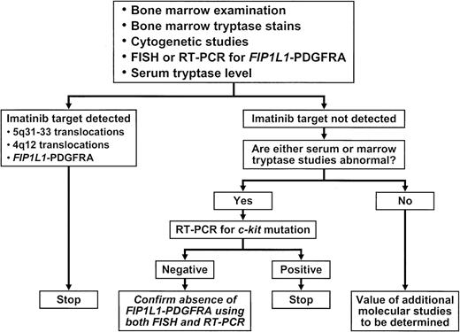
The diversity in the clinical phenotype and the underlying molecular lesion(s) in bcr/abl-negative CMPD continue to pose a significant challenge to the identification of patients who have imatinib-responsive disease. To date, most, if not all, bcr/abl-negative myeloid disorders that have displayed a marked response to imatinib therapy have been associated with prominent blood eosinophilia. Therefore, it is reasonable to routinely screen patients with eosinophilia-associated myeloid disorder for imatinib-sensitive and imatinib-resistant mutations. At present, this entails looking for the FIP1L1-PDFRA fusion by RT-PCR11,17,46 or FISH,14 c-kit mutations by RT-PCR,54 and chromosomal translocations that involve 5q31-33 by cytogenetic analysis.47 Figure 3 provides an algorithm in this regard.
Algorithm for detecting imatinib-sensitive or imatinib-resistant molecular targets in primary eosinophilic disorder.
Algorithm for detecting imatinib-sensitive or imatinib-resistant molecular targets in primary eosinophilic disorder.
PDGFRB-rearranged lesions are usually detected by standard cytogenetics.47 On the other hand, FISH may not be an adequate substitute for karyotype in this instance.47 RT-PCR and FISH are used to detect FIP1L1-PDGFRA. We encourage confirmation of the absence of FIP1L1-PDGFRA using FISH and RT-PCR, in c-kit mutation–negative primary eosinophilia, if the serum tryptase level or the bone marrow tryptase stain is abnormal (Figure 3). This is because of a previously documented association between the presence of FIP1L1-PDGFRA and an elevated serum tryptase level and because of abnormal mast cell infiltration of the bone marrow.45
In the presence of an imatinib-sensitive molecular target, treatment with imatinib is indicated. In addition, it might be prudent to consider treatment even in the absence of symptoms in the hope of preventing serious cardiovascular complications that might not be amenable to imatinib therapy once they occur.17,46 On the other hand, it is reasonable to withhold imatinib therapy in the presence of known, imatinib-resistant markers (eg, c-kit D816V mutation).20 Imatinib treatment decisions in the absence of drug-sensitive and drug-resistant molecular markers are not as straight-forward. It is possible that such patients may ultimately prove to have imatinib-responsive disease if given a therapeutic trial because of the activation of known targets by alternative molecular mechanisms or the presence of hitherto unidentified molecular targets. This point is amply illustrated in the paper by Cools et al,11 in which the molecular target for imatinib remained cryptic in 4 of 9 patients with HES who experienced durable remission with such therapy. This issue awaits resolution by a prospective study.
Finally, the treating physician should be aware of an infrequent but life-threatening complication of imatinib therapy in eosinophilic disorders—acute cardiogenic shock.14 To date, this complication has been reported in 3 patients within 1 week of the institution of imatinib treatment.117 All 3 patients displayed elevated serum troponin levels before or during the first few days of the start of therapy, but this was not documented in patients who did not develop the specific complication. Fortunately, the specific complication was reversed in all 3 patients with systemic corticosteroid therapy. Therefore, we recommend its prophylactic use for patients who display echocardiographic evidence of eosinophilic cardiac disease or elevated serum troponin levels.117
Prepublished online as Blood First Edition Paper, May 27, 2004; DOI 10.1182/blood-2004-01-0246.


