Abstract
The pathogenesis of acquired pulmonary alveolar proteinosis (PAP), a rare lung disease characterized by excessive surfactant accumulation within the alveolar space, remains obscure. Gene-targeted mice lacking the hematopoietic growth factor granulocyte-macrophage colony-stimulating factor (GM-CSF) or the signal-transducing β-common chain of the GM-CSF receptor have impaired surfactant clearance and pulmonary pathology resembling human PAP. We therefore investigated the hematopoietic effects of GM-CSF in patients with PAP. The hematologic response of 5 infants with congenital PAP to 5 μg/kg/d was of normal magnitude. By contrast, despite normal expression of GM-CSF receptor - and β-common chains on peripheral blood myelomonocytic cells (n = 6) and normal binding affinity of bone marrow mononuclear cells for GM-CSF (n = 3), each of the 12 patients with acquired PAP treated displayed impaired responses to GM-CSF; 5 μg/kg/d produced only minor eosinophilia, and doses of 7.5 to 20 μg/kg were required to induce ≥1.5-fold neutrophil increments in the 3 patients who underwent dose-escalation. However, neutrophilic responses to 5 μg/kg granulocyte colony-stimulating factor (G-CSF) were normal (n = 4). In vitro, the proportion of hematopoietic progenitors responsive to GM-CSF (16.1% ± 8.9%; P = .042) or interleukin-3 (IL-3; 19.3% ± 7.7%; P = .063), both of which utilize the β-common chain of the GM-CSF receptor complex, were reduced among patients with acquired PAP (n = 4) compared with normal bone marrow donor controls (47.2% ± 25.9% and 40.9% ± 18.6%, respectively). In the one individual who had complete resolution of lung disease during the period of study, this was temporally associated with correction of this defective in vitro response to GM-CSF and IL-3 on serial assessment. These data establish that patients with acquired PAP have an associated impaired responsiveness to GM-CSF that is potentially pathogenic in the development of their lung disease. Based on these observations, we propose a model of the pathogenesis of acquired PAP that suggests the disease arises as a consequence of an acquired clonal disorder within the hematopoietic progenitor cell compartment.
© 1998 by The American Society of Hematology.
THE STRIKING CAPACITY of granulocyte-macrophage colony-stimulating factor (GM-CSF) to stimulate the proliferation and differentiation of myeloid hematopoietic progenitor cells in vitro facilitated its initial purification and cloning1,2 and provides the basis for its clinical application in vivo.3 The pharmacologic administration of recombinant GM-CSF consistently leads to a dose-dependent stimulation of myeloid hematopoiesis resulting in peripheral blood neutrophilia, monocytosis, and eosinophilia.3 Each of these actions requires engagement of GM-CSF with the high-affinity receptor complex comprising a GM-CSF–specific α-chain and a common β-chain (βc) that is also a component of the receptor complexes for interleukin-3 (IL-3) and IL-5.4 5
Despite a detailed understanding of its actions on hematopoietic cells both in vitro and in vivo,1,3,5 the innate physiologic role of GM-CSF remained obscure until the recent generation of gene-targeted mice lacking either GM-CSF6,7 or βc.8,9 Surprisingly, these animals had no detectable impairment in steady-state hematopoiesis, but displayed lung abnormalities resembling human pulmonary alveolar proteinosis (PAP), an uncommon lung disease characterized by excessive intra-alveolar accumulation of surfactant phospholipid and protein.10,11PAP occurs in two distinct forms: congenital and acquired. The rare congenital form is transmitted in an autosomal recessive manner and is attributable to a deficiency of surfactant protein B in some kindreds.12 By contrast, 90% of cases of PAP are acquired, usually idiopathic, have no familial basis, and affect predominantly adults.10,11 Because the pathogenic basis of acquired PAP is unknown, treatment remains empirical, consisting of the periodic physical removal of accumulated surfactant by whole lung lavage under general anesthesia.11 Previous attempts to develop effective alternative therapies have been hampered by a poor understanding of the pathogenesis of the disease and the lack of a relevant animal model system.
Although GM-CSF is usually considered exclusively as a hematopoietic regulator, these recent animals studies have established that its activity is essential for normal pulmonary homeostasis.6-9In the absence of GM-CSF, the clearance of both surfactant protein and phospholipid from the alveolar space are dramatically reduced,13 but can be corrected by the localized expression of GM-CSF in airway epithelial cells.14 Similarly, in animals lacking βc, hematopoietic reconstitution with normal myeloid progenitors leads to reversal of the lung pathology.15 Collectively, these observations establish that GM-CSF has a unique local action upon myeloid-lineage hematopoietic-derived cells, which results in the enhancement of surfactant clearance. It is likely that alveolar macrophages are the effector cells involved; these cells are derived from hematopoietic progenitors,16 contribute significantly to the clearance and degradation of both surfactant protein and phospholipid,17,18 and are responsive to physiologic concentrations of GM-CSF.19-21 Because alveolar macrophages from patients with active acquired PAP are known to be functionally impaired22,23 and the lung abnormalities may recur following bilateral lung transplantation,24 the underlying pathogenic defect appears to be extrinsic to the pulmonary parenchyma and may lie within the hematopoietic compartment. This provides the rationale for the investigation of two facets of GM-CSF in PAP: as a novel therapeutic strategy and as a hematopoietic regulatory factor that may be causally involved in disease development. On this basis, we initiated a trial of recombinant GM-CSF in PAP.25 The markedly attenuated hematopoietic response to GM-CSF observed in patients with acquired PAP suggests that a defect in the GM-CSF receptor complex, or postreceptor signaling pathways, may contribute to the pulmonary disease in such patients.
MATERIALS AND METHODS
Details of clinical study and schedule of GM-CSF administration.
All patients enrolled before April 1998 in an ongoing open label phase-II study of GM-CSF in the therapy of PAP were analyzed.25 To be eligible, patients had to have a pathologically confirmed diagnosis of PAP and not have been exposed to cytotoxic agents, corticosteroids, cytokines, or growth factors in the preceding 4 weeks. The study was conducted in accordance with the Declaration of Helsinki, approved by the Institutional Ethics Committee at each participating institution, and all patients or their guardians provided written informed consent. Bacterially synthesized recombinant GM-CSF (Leucomax; Schering-Plough, Baulkham Hills, Australia; specific activity, 0.66 to 1.66 × 10⁷IU/mg protein; ≤25 Eu endotoxin per vial by the Limulusamoebocyte assay; data on file, Schering-Plough) was used for patients treated in Australia and Europe (n = 12). Because this preparation of GM-CSF is not approved for use in the United States, the 5 patients treated there received yeast-derived recombinant GM-CSF (Leukine; Immunex, Seattle, WA; specific activity, 5.6 × 10⁶ IU/mg protein; ≤25 Eu endotoxin per vial; data on file, Immunex).
GM-CSF treatment was initiated at 3.0 μg/kg subcutaneously daily for 5 days and increased to 5.0 μg/kg from day 6 onwards, provided that the total peripheral blood leukocyte count was less than 30 × 109/L. In one instance, for patient convenience, the dose of GM-CSF was capped at a single vial size (400 μg; 4.0 μg/kg). Patients with acquired PAP did not require hospitalization but were monitored with continuous pulse oximetry for 4 hours after the first dose and, provided that the arterial oxygen saturation did not decrease to less than 92%, all subsequent doses were self-administered without monitoring. Compliance with therapy was assessed by counting used vials and inspection of injection sites. The level of respiratory support required by patients with congenital PAP necessitated hospitalization through the period of study, and all doses of GM-CSF were administered by hospital staff. Toxicity was assessed by history and physical examination at least weekly and graded according to WHO criteria.26
To minimize possible detrimental effects of excessive leukocytosis, dose reductions were specified according to the total leukocyte count; if greater than 50 × 109/L at any time, GM-CSF was withheld until the total leukocyte count decreased less than 30 × 109/L and then resumed at 50% of the previous dose. If the total leukocyte count was greater than 30 × 109/L, but less than 50 × 109/L, the dose of GM-CSF was reduced by 30%, but daily administration continued. Peripheral blood counts were determined by automated full blood examinations (Sysmex SE-9000; Toa Medical Electronics Co Ltd, Kobe, Japan; or similar) performed at least 3 times per week until stable and then weekly for the duration of treatment. Confirmatory manual differential counts were performed intermittently without showing any significant discrepancies from automated counts. Baseline values were from peripheral blood samples collected on the first day of treatment, before GM-CSF administration. Where feasible, peripheral blood samples during treatment were obtained at least 8 hours after GM-CSF dosing to avoid sampling during the expected period of transient leukopenia after each dose.3,5 27
After analysis of the first 4 patients, the protocol was modified to allow exploratory dose-escalation of GM-CSF to define a dose able to induce a hematologic response, prospectively defined as a ≥1.5-fold increase in neutrophil count. Single doses of GM-CSF 7.5, 10.0, 15.0, 20.0, and 30.0 μg/kg were administered in a sequential fashion, with a minimum wash-out period of 3 days between doses and with blood counts taken at baseline and 4, 24, and 48 hours after each dose. Dose-escalation ceased once a hematologic response was attained, any toxicity of ≥grade 3 was seen, or the 30.0 μg/kg dose level was reached without response. Where possible, patients also received a single 5.0 μg/kg dose of recombinant granulocyte colony-stimulating factor (G-CSF; Filgrastim; Amgen, North Ryde, Australia) with peripheral blood counts monitored at baseline and 4, 24, and 48 hours after administration.
Definition of normal peripheral blood hematopoietic response to GM-CSF.
The peripheral blood changes anticipated in normal adults with the above dosing schedule were determined from published studies of daily, single-agent recombinant GM-CSF that fulfilled the following criteria: subcutaneous administration (either by bolus or brief infusion), or continuous intravenous infusion, no chemotherapy immediately preceding GM-CSF treatment, no pre-existing intrinsic abnormality of hematopoiesis or current malignant marrow infiltration, GM-CSF doses between 3.0 and 6.0 μg/kg (equivalent to ∼120 to 250 μg/m2), and the provision of either group mean, or individual patient data, for baseline and peak neutrophil and/or total leukocyte counts. Nine such studies were identified,27-35 only 4 of which presented individual patient data allowing optimal statistical comparisons27,28,30,34 (G.J. Lieschke, unpublished data, September 1997). The single study of GM-CSF in neonates without primary hematologic disorders used a dose of GM-CSF above this range (10 μg/kg), but demonstrated hematologic responses broadly comparable in magnitude to those observed in adults.36
Clonal culture methods.
Iliac crest bone marrow was aspirated using sterile technique either before or at least 2 weeks after GM-CSF administration, and colony-forming units (CFUs) were assayed in agar cultures using standard methods.37 Depending on separate institutional protocols, colony-forming unit–granulocyte-macrophage (CFU-GM) were enumerated from cultures stimulated with either (1) GM-CSF, G-CSF, and stem cell factor (SCF) or (2) G-CSF and SCF and burst-forming unit-erythroid (BFU-E) with either (1) GM-CSF, G-CSF, IL-3, IL-6, and erythropoietin or (2) SCF and erythropoietin (all recombinant human proteins; Amgen, Thousand Oaks, CA). Cultures were also established either at supramaximal concentrations or with titration of a single growth-factor (GM-CSF, IL-3, G-CSF, or SCF). Control marrow cells were obtained with informed consent from normal allogeneic marrow donors undergoing scheduled marrow harvest on the same day and at the same institution as the patient under evaluation. Colonies were defined as clones containing more than 50 cells and enumerated after 14 days of culture in a fully humidified atmosphere of 5% CO2 in air, with the exception that colonies from G-CSF–stimulated cultures were counted after 7 days.
Flow cytometric assessment of GM-CSF receptor expression.
Surface expression of growth factor receptor components was assessed in whole blood samples. For indirect flow cytometry, murine monoclonal antibodies recognizing the extracellular domains of βc(CDw131; S-16), the GM-CSF receptor α-chain (CD116; S-20), and the IL-3 receptor α-chain (CDw123; S-12) (Santa Cruz Biotechnology, Santa Cruz, CA and Pharmingen, Hamburg, Germany) were used and detected with fluorochrome-conjugated secondary goat-antimouse antibodies (Coulter, Krefeld, Germany). Direct staining was also performed with monoclonal antibodies against CD14, CD15, CD3, and CD20 (Immunotech, Hamburg, Germany). Analysis was performed using FACScan (Becton Dickinson, Heidelberg, Germany) according to published procedures.38Briefly, for indirect staining, cells were incubated with the primary antibody for 15 to 30 minutes at room temperature, washed twice with phosphate-buffered saline (PBS), and stained for 30 minutes with secondary antibody. Control samples were incubated with primary isotype-matched nonspecific mouse Ig (Dianova, Hamburg, Germany). For direct staining, cells were incubated with conjugated antibodies for 10 minutes at room temperature. All samples were preincubated with pooled Ig to minimize nonspecific staining. Results were recorded as the number of cells within the myeloid gate defined by physical light-scatter properties that were positive for the respective receptor components expressed as a percentage of total leukocytes and then corrected for the interpatient variation in myeloid cell content as determined by the percentage of cells expressing CD15. All samples were assayed in the same laboratory (Düsseldorf) and samples from normal volunteers were shipped overnight at ambient temperature, together with patient samples from collecting centers to control for specimen handling.
Radioligand studies of GM-CSF receptor quantification and binding affinity.
Statistics.
Numeric data are presented as the mean ± 1 SD, unless stated otherwise. The protocol prospectively specified separate analysis of congenital and acquired patient groups. Because the raw data comprised serial assessments, appropriate summary measures were used for some analyses,41 including maximum cell count and maximum change in cell counts from baseline. Hematologic parameters were analyzed witht-tests or one-way ANOVA, with logarithmic transformation applied where required by the non-normal distribution of data. After ANOVA, Dunnett’s pairwise comparison tests were performed to evaluate differences between the current study and previous reports. In practice, analyses with and without log-transformation gave similar results. Where data were expressed as proportions, comparisons were performed after appropriate logistic transformation.42Correlations were assessed using Spearman’s rank correlation coefficient. All analyses were performed using Minitab 9.2 (Minitab Inc, State College, PA; 1993) or SYSTAT 7.0 (SPSS Inc, Chicago, IL; 1997) and presented as two-sided comparisons, with P < .05 regarded as significant.
RESULTS
GM-CSF treatment.
Seventeen patients with PAP received 18 courses of GM-CSF treatment. Twelve patients had acquired disease (age of onset, 13 to 45 years; median, 30 years), and 5 infants had congenital PAP (onset, <1 month of age), 1 of whom (patient no. 15) was confirmed by immunohistochemical and molecular methods to be surfactant protein-B deficient.12 The remaining 4 infants had normal patterns of surfactant protein-B expression by immunohistochemistry. No patient had a previous malignancy, primary hematologic disorder, or any known exposure to mutagens, radiation, or cytotoxic chemotherapy, and pretreatment peripheral blood morphology was normal in all cases (Tables 1 and2). GM-CSF treatment was well tolerated; no subject showed any acute deterioration in oxygenation with the first dose or withdrew from treatment due to subjective intolerance. The observed adverse effects were limited to grade 2 nausea, vomiting, fever, and chills after the first dose (patient no. 1, first course of treatment only); 2 episodes of grade 2 fever and chills on days 20 and 137 (patient no. 3, described in Table 1); persistent grade 1 fevers (patient no. 14); and grade 1 erythema at injection sites in a total of 7 patients.
Patient Characteristics and Peripheral Blood Responses to GM-CSF (Acquired PAP)
| Patient No. . | Duration GM-CSF (d) . | Mean Daily Dose (day 1-21) (μg/kg)/(μg/m2) . | Baseline Cell Counts (×109/L) . | Peak Cell Counts Before Day 21 (×109/L) . | ||||
|---|---|---|---|---|---|---|---|---|
| Total WBC . | Neutrophils . | Eosinophils . | Total WBC . | Neutrophils . | Eosinophils . | |||
| 1 (course no. 1)* | 70 | 3.8/166 | 6.5 | 3.9 | 0.20 | 8.7 | 6.0 | 0.57 |
| 1 (course no. 2)* | 264 | 3.8/168 | 5.9 | 3.3 | 0.18 | 8.3 | 5.4 | 0.76 |
| 2 | 42 | 4.6/164 | 7.2 | 3.8 | 0.12 | 5.5 | 3.3 | 0.20 |
| 3-151 | 170 | 4.3/180-152 | 6.8 | 4.3 | 0.05 | 6.7 | 4.3 | 0.08 |
| 4 | 13 | 4.1/147-153 | 4.8 | 2.2 | 0.10 | 6.0 | 2.6 | 0.24 |
| 5 | 42 | 4.7/210 | 4.6 | 2.4 | 0.06 | 6.1 | 3.4 | 0.20 |
| 6¶ | 37 | 4.5/167 | 6.7 | 4.3 | 0.17 | 6.5 | 4.5 | 0.35 |
| 7 | 42 | 4.6/185 | 7.4 | 4.5 | 0.09 | 8.2 | 5.4 | 0.13 |
| 8¶ | 84 | 5.8/229 | 5.4 | 2.9 | 0.09 | 5.4 | 3.0 | 0.59 |
| 9 | 84 | 4.2/168 | 5.1 | 3.2 | 0.03 | 6.8 | 5.2 | 0.41 |
| 10 | 84 | 4.4/169 | 8.6 | 4.8 | 0.13 | 8.4 | 5.9 | 0.41 |
| 11 | 84 | 4.7/168 | 5.5 | 3.8 | 0.01 | 5.9 | 4.4 | 0.12 |
| 12¶ | 21+ | 4.5/204 | 6.8 | 3.9 | 0.07 | 7.4 | 4.3 | 0.28 |
| Mean (SD) | 71 (51) | 4.5 (0.5)/180 (23) | 6.3 (1.2) | 3.6 (0.8) | 0.09 (0.05) | 6.8 (1.1) | 4.3 (1.1) | 0.31 (0.18) |
*Results from the two treatment courses in patient no. 1 are averaged for the overall analysis.
Patient developed a grade 2 febrile reaction to GM-CSF on day 20 leading to the withdrawal of GM-CSF for 8 days. This reaction was treated with a single dose of intravenous hydrocortisone (100 mg) resulting in a leukocytosis of 11.1 × 10⁹/L, with a neutrophil count of 10.0 × 10⁹/L 48 hours later. Because these changes were most likely attributable to the corticosteroid, peripheral blood counts from days 21 and 22 were excluded from the above-noted analyses.
Data from the first 20 days of treatment only (see * note above).
Data from 13 days of treatment only.
¶These patients were treated with yeast-derived recombinant GM-CSF (Leukine; Immunex); all other patients received bacterially synthesized recombinant GM-CSF (Leucomax; Schering-Plough).
Patient Characteristics and Peripheral Blood Responses to GM-CSF (Congenital PAP)
| Patient No. . | Duration GM-CSF (d) . | Mean Daily Dose (d 1-21) (μg/kg) . | Baseline Cell Counts (×109/L) . | Peak Cell Counts Before Day 21 (×109/L) . | ||||
|---|---|---|---|---|---|---|---|---|
| Total WBC . | Neutrophils . | Eosinophils . | Total WBC . | Neutrophils . | Eosinophils . | |||
| 13 | 141 | 3.1 | 15.2 | 11.6 | 0.61 | 52.2 | 39.8 | 7.74 |
| 14 | 42 | 2.0 | 19.5 | 11.0 | 0.70 | 47.1 | 24.3 | 3.50 |
| 15* | 50 | 4.9 | 16.2 | 10.4 | 0.97 | 26.7 | 16.3 | 2.67 |
| 16 | 23 | 4.0 | 20.2 | 9.1 | 2.63 | 57.0 | 32.4 | 25.35 |
| 17* | 67 | 2.8 | 18.4 | 8.8 | 0.92 | 41.5 | 26.2 | 5.12 |
| Mean (SD) | 65 (46) | 3.4 (1.1) | 17.9 (2.1) | 10.2 (1.2) | 1.17 (0.83) | 44.9 (11.7) | 27.8 (8.9) | 8.88 (9.41) |
*These patients were treated with yeast-derived recombinant GM-CSF (Leukine; Immunex); all other patients received bacterially synthesized recombinant GM-CSF (Leucomax; Schering-Plough).
Definition of normal hematopoietic response.
GM-CSF elicits a predictable dose-dependent leukocytosis over the dose range 0.3 to 30 μg/kg/d when administered to adult patients with normal hematopoiesis.3,5 To define the expected magnitude of this response with the schedule used in this study, suitable published reports were sought (see Materials and Methods). Nine studies in adults (4 with individual patient data) dealing with a total of 48 patients were identified in which GM-CSF was administered in a fixed daily dose of 120 to 300 μg/m2 (equivalent to approximately 3 to 8 μg/kg/d) for a maximum of 14 days (Table 3).27-35 Within this dose range, there was no correlation between the GM-CSF dose and the peak leukocyte count (data not shown). Because the PAP patients on study had an increment in the daily dose of GM-CSF from day 6 of treatment, the peak counts attained at or before day 21 (allowing 15 days of treatment with a constant daily dose of 5.0 μg/kg GM-CSF) were used for comparative purposes (Tables 1-3).
Hematologic Response to GM-CSF in Current Patients With Acquired PAP and Adults With Normal Hematopoiesis
| First Author (reference no.) . | No. of Patients . | Daily Dose of GM-CSF . | Duration (d) . | Leukocyte Count (×109/L)* . | Neutrophil Count (×109/L)* . | ||
|---|---|---|---|---|---|---|---|
| Mean Peak . | Mean Increment† . | Mean Peak . | Mean Increment† . | ||||
| Individual patient data | |||||||
| Current study | 12‡ | 3.8-5.8 μg/kg [147-229 μg/m2] | 21 | 6.8 ± 1.1 | 0.5 ± 1.1 | 4.3 ± 1.1 | 0.7 ± 0.8 |
| Vadhan-Raj28 | 3 | 120-250 (mean 163) μg/m2 | 14 | 34.1 ± 12.12-153 | 25.0 ± 13.32-153 | 26.4 ± 6.52-153 | 19.8 ± 5.42-153 |
| Lieschke27 | 3 | 3 μg/kg | 10 | 22.6 ± 4.92-153 | 15.3 ± 3.5¶ | 15.4 ± 5.22-153 | 10.1 ± 3.9¶ |
| Haas30 | 7 | 250 μg/m2 | Mean 11.52-155 | 36.6 ± 8.42-153 | 29.7 ± 12.42-153 | 21.2 ± 6.22-153 | 15.8 ± 7.22-153 |
| Berthaud34 | 3 | 250 μg/m2 | 10 | 23.9 ± 5.92-153 | 19.5 ± 5.82-153 | 18.5 ± 5.72-153 | — |
| P value# | <.00001 | <.00001 | <.00001 | <.00001 | |||
| Grouped mean data | |||||||
| Steward29 | 6 | 3 μg/kg | 10 | 41.3 | 31.4 | 31.9 | 24.2 |
| Lord31 | 4 | 150-300 (mean 263) μg/m2 | 10 | 38.0 | 31.0 | 17.1 | 12.1 |
| Vadhan-Raj32 | 3 | 120 μg/m2 | 14 | 19.2 ± 2.1 | — | — | — |
| 3 | 250 μg/m2 | 14 | 42.7 ± 13.0 | — | — | — | |
| Bukowski33 | 4 | 125 μg/m2 | 14 | 21.2 | 14.7 | 16.9 | 11.4 |
| 5 | 250 μg/m2 | 14 | 65.9 | 56.5 | 36.8 | 27.9 | |
| Dale36 | 7 | 250 μg/m2 | 14 | — | — | 14.1 ± 4.5 | 11.9 |
*All data are given as the mean ± 1 SD. In some cases, values have been converted from the SEM provided (SD = SEM × √n).
Peak minus baseline count.
Thirteen courses of GM-CSF therapy were administered to 12 patients with acquired PAP; results from the two course of treatment in patient no. 1 are averaged for the overall analysis.
P < .004 for comparison between individual study and current patients using Dunnett’s pairwise comparison test.
¶P ≤ .026 for comparison between individual study and current patients using Dunnett’s pairwise comparison test.
Range of 5 to 22 days GM-CSF administration; patients with marrow infiltrate were excluded from analysis.
#Using ANOVA across all five studies.
Hematopoietic response—Congenital PAP.
Although none of the infants with congenital PAP had evidence of active infection, baseline leukocyte and neutrophil levels were mildly elevated. With GM-CSF treatment, there was a prompt and sustained neutrophil leukocytosis and marked eosinophilia (Table 2). In 4 cases, GM-CSF doses were reduced according to protocol guidelines due to excessive leukocytosis, resulting in the lower than projected mean daily dose of GM-CSF. Patient no. 16 received corticosteroids from day 14 onwards, but no other infants received any medications that may have influenced the magnitude of these hematopoietic changes. The magnitude of the hematopoietic response observed, whether assessed as increment in total leukocyte or neutrophil counts, did not differ from that previously reported for adults treated with similar doses of GM-CSF (Table 3; both P > .4).
Hematopoietic response—Acquired PAP.
Despite a median total duration of GM-CSF treatment of 71 days (range, 13 to 264 days; with 1 patient ongoing at 21+ days) in the 12 patients with acquired disease, the mean peak leukocyte, neutrophil, and eosinophil counts attained at any time on therapy were only 7.1, 4.6, and 0.46 × 10⁹/L, respectively. These low peak counts were not attributable to any myelosuppressive effects of other agents; apart from intermittent acetaminophen, the only concomitant medications used during the period of GM-CSF treatment were amoxycillin-clavulanic acid, warafarin, and co-trimoxozole in 1 patient each and a single dose of hydrocortisone in patient no. 3 (Table 1).
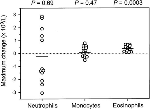
To investigate whether there was any detectable hematologic response to GM-CSF therapy, the maximum change in cell counts from baseline values at any time during treatment (whether an increase of decrease) were determined for each patient. Consistent with a biologic response to therapy, all patients displayed an increase in eosinophil counts (mean change, +0.37 × 10⁹/L; P = .0003). However, there was no discernible effect on either neutrophil (mean change, −0.25 × 10⁹/L; P = .69) or monocyte counts (mean change, +0.08 × 10⁹/L; P = .47; Fig 1).
Distribution of the maximum change from baseline levels (whether increase or decrease) in peripheral blood absolute neutrophil, monocyte, and eosinophil counts for the 12 patients with acquired PAP treated with GM-CSF. The horizontal bar represents the mean value, and the P values shown are for the comparison with a test mean of 0 (ie, no change with therapy) using a one-sample t-test.
Distribution of the maximum change from baseline levels (whether increase or decrease) in peripheral blood absolute neutrophil, monocyte, and eosinophil counts for the 12 patients with acquired PAP treated with GM-CSF. The horizontal bar represents the mean value, and the P values shown are for the comparison with a test mean of 0 (ie, no change with therapy) using a one-sample t-test.
Although overall there was no significant change in neutrophil counts with therapy, in 7 patients the greatest variation from baseline observed was a decline, which in 1 instance was particularly noteworthy. From a baseline count of 2.16 × 10⁹/L, patient no. 4 became progressively neutropenic while receiving GM-CSF in the absence of any other identifiable cause (nadir neutrophil count of 0.84 × 10⁹/L on day 12). This neutropenia was not artefactual, because all samples were obtained via venepuncture at least 12 hours after GM-CSF dosing, automated counts were confirmed by manual differential counts, and no leuko-agglutination was evident. The neutrophil count normalized after cessation of GM-CSF on day 13 (2.24 × 10⁹/L by day 22), and a bone marrow aspirate and biopsy, obtained on day 24 to investigate any possible underlying intrinsic hematopoietic disorder, were of normal cellularity without morphologic features of myelodysplasia. Myeloid maturation appeared normal, although mature neutrophils were somewhat prominent (13.0%; reference range, 3.8% to 11.0%), as were mature eosinophils (5.0%; reference range, 0% to 1.1%). Four other patients (no. 1, 2, 6, and 7) also underwent bone marrow examinations either before or at least 2 weeks after completing GM-CSF treatment; in each instance, granulopoiesis was normal without any morphologic abnormality, maturation defect, or features of myelodysplasia (data not shown).
The observed hematopoietic responses among patients with acquired PAP were markedly lower than in persons with normal hematopoiesis from previously published studies whether measured as absolute peak or maximum increment from baseline for both total leukocyte and neutrophil counts (each P < .00001; Table 3).
In vitro hematopoietic responsiveness.
To investigate the basis of this attenuated hematopoietic responsiveness in vivo among patients with acquired PAP, the response of hematopoietic progenitors was assessed in vitro using colony-forming assays with bone marrow cells obtained from patients no. 1, 2, 6, and 7. In patient no. 2, two separate experiments were performed with comparable results, and the mean of these was used for statistical purposes.
As has been previously observed in normal individuals using these assays, there was large biologic variation between individuals for the total number of both CFU-GM and BFU-E formed.43 However, overall, there was no consistent difference between patients with PAP and controls for either total CFU-GM (317 ± 262 and 166 ± 72 per 5 × 10⁴ mononuclear cells, respectively) or BFU-E (375 ± 255 and 253 ± 143 per 5 × 10⁴mononuclear cells, respectively).
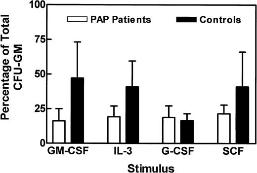
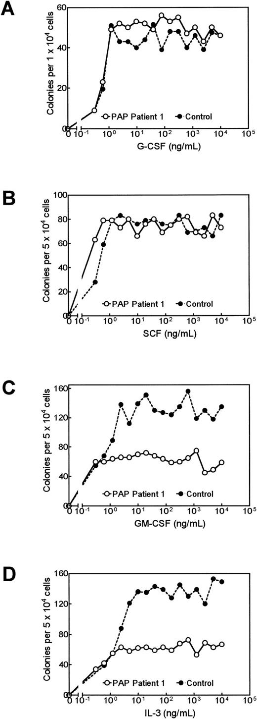
Whereas total CFU-GM in marrow samples can vary as much as 100-fold among normal persons, the proportion of these progenitors responsive to individual growth factors is more consistent.5 43 Thus, to compare responses to single growth factors, the number of colonies formed was expressed as a percentage of the total CFU-GM for that individual (Fig 2). Although moderate variability was still evident, the percentage of progenitors responsive to G-CSF or SCF among patients with acquired PAP were not consistently different from controls (P values of .16 and .81, respectively). However, among patients with PAP, the percentage of progenitors responsive to GM-CSF (16.1% ± 8.9%; P = .042) or IL-3 (19.3% ± 7.7%; P = .063) was lower than observed in controls (47.2% ± 25.9% and 40.9% ± 18.6%, respectively; Fig 2). This selective and concordant reduction in colony formation in response to GM-CSF and IL-3 is exemplified by the findings in patient no. 1 (Fig 3).
Summary of bone marrow colony formation in semisolid agar from patients with acquired PAP (n = 4) and concurrent normal controls (n = 5). Duplicate cultures of 5 × 10⁴ bone marrow mononuclear cells were stimulated with supramaximal concentrations of the individual growth factors shown, and the mean of the resulting colonies expressed as a percentage of the total CFU-GM from that individual formed in response to combined growth factor stimulation (see Materials and Methods). Data are expressed as the mean ± SD.
Summary of bone marrow colony formation in semisolid agar from patients with acquired PAP (n = 4) and concurrent normal controls (n = 5). Duplicate cultures of 5 × 10⁴ bone marrow mononuclear cells were stimulated with supramaximal concentrations of the individual growth factors shown, and the mean of the resulting colonies expressed as a percentage of the total CFU-GM from that individual formed in response to combined growth factor stimulation (see Materials and Methods). Data are expressed as the mean ± SD.
Bone marrow colony formation in semisolid agar from patient no. 1 with acquired PAP and a concurrent normal control in response varying concentrations of (A) G-CSF day-7 colonies, (B) SCF day-14 colonies, (C) GM-CSF day-14 colonies, and (D) IL-3 day-14 colonies. Data are shown as the mean of duplicate cultures.
Bone marrow colony formation in semisolid agar from patient no. 1 with acquired PAP and a concurrent normal control in response varying concentrations of (A) G-CSF day-7 colonies, (B) SCF day-14 colonies, (C) GM-CSF day-14 colonies, and (D) IL-3 day-14 colonies. Data are shown as the mean of duplicate cultures.
As previously described,25 GM-CSF treatment in patient no. 1 resulted in prolonged normalization of respiratory parameters and complete clearance of lung infiltrates on computerized tomographic (CT) scanning (data not shown). In this patient, a second bone marrow sample for colony assays was obtained 12 months after cessation of GM-CSF, while he remained free of any evidence of his alveolar proteinosis. The marrow was morphologically unchanged from his prior sample, and total CFU-GM and BFU-E were comparable to controls. The proportions of total progenitors responsive to GM-CSF and IL-3 were 48.3% and 58.1%, respectively. These results contrast to those obtained during the period of active lung disease (9.2% and 9.3%, respectively). These data demonstrate that the observed selective impairment of response to GM-CSF and IL-3 in vitro can, in some cases, be transient. In this one individual, the restoration of normal in vitro responsiveness to GM-CSF was coincident with the resolution of his lung disease.
Flow cytometric assessment of GM-CSF and IL-3 receptor expression.
To investigate the possible role of reduced receptor expression in the impaired response to GM-CSF, whole peripheral blood from 9 patients (6 with acquired and 3 congenital PAP) was evaluated by flow cytometry (Table 4). These samples were either obtained before or at least 12 weeks after cessation of GM-CSF treatment. There was no consistent reduction evident in the expression of components of the GM-CSF or IL-3 receptor complexes on peripheral blood cells from patients with acquired PAP. Similarly, there was no apparent difference in receptor expression between patients with congenital and acquired disease that may explain the observed differences in hematopoietic response.
Flow Cytometric Assessment of GM-CSF and IL-3 Receptor Component Expression on Peripheral Blood Cells
| Patient No. . | Myelomonocytic Cells* (% of total WBC) . | Expression of Specific Receptor Components (% relative to myelomonocytic cell content) . | ||
|---|---|---|---|---|
| βc . | GM-CSF α-chain . | IL-3 α-chain . | ||
| Acquired PAP | ||||
| 1 | 54 | 94 | 30 | 102 |
| 2 | 47 | 66 | 79 | 72 |
| 7 | 27 | 93 | 107 | 96 |
| 9 | 57 | 67 | 91 | 88 |
| 10 | 50 | 66 | 166 | 130 |
| 11 | 58 | 95 | 138 | 112 |
| Mean (SD) | 49 (11) | 80 (15) | 102 (47) | 100 (20) |
| Congenital PAP | ||||
| 12 | 70 | 76 | 80 | Not tested |
| 13 | 54 | 122 | 139 | 93 |
| 15 | 70 | 90 | 134 | 127 |
| Mean (SD) | 65 (9) | 96 (24) | 118 (33) | 110 (17) |
| Normal controls | ||||
| A | 45 | 93 | 69 | 93 |
| B | 70 | 86 | 101 | 84 |
| C | 47 | 64 | 87 | 77 |
| Mean (SD) | 54 (14) | 81 (15) | 86 (16) | 85 (8) |
*Myelomonocytic cells defined as those expressing CD15 by flow cytometry.
Radioligand assessment of receptor number and affinity.
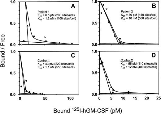
Binding studies using radiolabeled GM-CSF were performed on bone marrow mononuclear cells. Patients no. 1, 2, and 7 with acquired PAP were studied, each in conjunction with a normal allogeneic marrow donor control. All studies yielded comparable results in patients and controls (Fig 4). Each of the 3 patients with acquired PAP expressed high-affinity binding sites for GM-CSF, demonstrating the presence of functional extracellular portions of both βc, and GM-CSF receptor α-chains; the mean Kd value for patients was 81 pmol/L with a mean of 766 sites/cell, compared with 56 pmol/L and 134 sites/cell for the controls. In all controls and 2 of the 3 patients studied, low-affinity GM-CSF binding sites, consistent with excess free α-chains, were also detected; the mean Kd value for patients was 5.6 nmol/L with 800 sites/cell, compared with 4.8 nmol/L and 449 sites/cell for controls. In patient no. 7, no distinct low-affinity binding sites were detected, consistent with the majority of expressed α-chains contributing to high-affinity α/βc dimer formation without sufficient excess free α-chains remaining for reliable detection using this method.5
Representative Scatchard plots of the binding of125I human GM-CSF specific activity to bone marrow cells from patients no. 1 (A) and 2 (B) with acquired PAP and control individuals (C and D). The calculated dissociation equilibrium binding constants for the high-affinity (Kd1) and low-affinity (Kd2) sites are given along with the mean number of sites per cell (in parentheses).
Representative Scatchard plots of the binding of125I human GM-CSF specific activity to bone marrow cells from patients no. 1 (A) and 2 (B) with acquired PAP and control individuals (C and D). The calculated dissociation equilibrium binding constants for the high-affinity (Kd1) and low-affinity (Kd2) sites are given along with the mean number of sites per cell (in parentheses).
In vivo dose-escalation of GM-CSF.
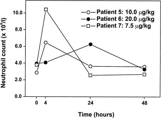
Three patients who had failed to demonstrate a significant neutrophilia despite prolonged administration of GM-CSF at 5.0 μg/kg/d received incrementally increasing doses of GM-CSF to determine a threshold for hematologic responsiveness in vivo. The highest neutrophil counts recorded during previous prolonged GM-CSF therapy at 5.0 μg/kg/d were 1.4-, 1.1-, and 1.2-fold baseline levels for patients no. 5, 6, and 7, respectively. Dose-escalation began at least 2 weeks after cessation of previous GM-CSF therapy, and all patients had significant persisting lung disease at that time. In each instance, the higher doses of GM-CSF produced a significant hematologic response (prospectively defined as ≥1.5-fold increase in neutrophil count), although the dose level necessary to attain such a response varied from 7.5 to 20.0 μg/kg (Fig 5). Consistent with the in vitro colony formation data, this demonstrates that responsiveness to GM-CSF is impaired, but not absent.
Hematologic response to GM-CSF dose-escalation in vivo. Three patients with acquired PAP who had failed to manifest any significant degree of neutrophilia in response to prolonged administration of 5.0 μg/kg/d of GM-CSF received incrementally increasing single doses of GM-CSF: 7.5, 10.0, 15.0, 20.0, and 30.0 μg/kg. Dose-escalation ceased once the patient had manifest a hematologic response (prospectively defined as ≥1.5-fold increase in neutrophil counts over baseline), and peripheral blood counts were assessed at the time points shown. The kinetics of peripheral blood neutrophil counts are plotted for the first dose, which induced such a hematologic response. Patient no. 6 received yeast-derived GM-CSF, whereas patients no. 5 and 7 received bacterially synthesized GM-CSF.
Hematologic response to GM-CSF dose-escalation in vivo. Three patients with acquired PAP who had failed to manifest any significant degree of neutrophilia in response to prolonged administration of 5.0 μg/kg/d of GM-CSF received incrementally increasing single doses of GM-CSF: 7.5, 10.0, 15.0, 20.0, and 30.0 μg/kg. Dose-escalation ceased once the patient had manifest a hematologic response (prospectively defined as ≥1.5-fold increase in neutrophil counts over baseline), and peripheral blood counts were assessed at the time points shown. The kinetics of peripheral blood neutrophil counts are plotted for the first dose, which induced such a hematologic response. Patient no. 6 received yeast-derived GM-CSF, whereas patients no. 5 and 7 received bacterially synthesized GM-CSF.
In vivo hematopoietic response to G-CSF.
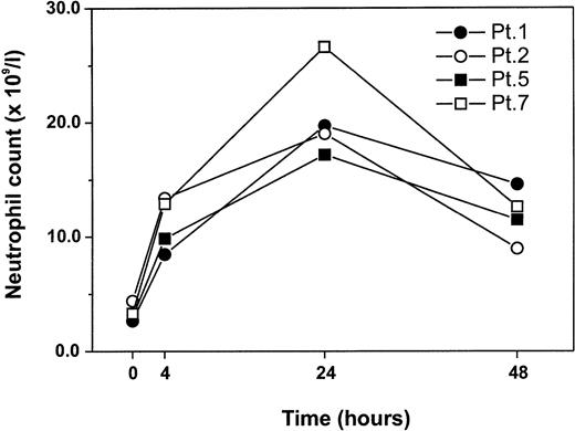
To explore the selectivity of the observed attenuated hematopoietic response, 4 patients received a single 5.0 μg/kg dose of recombinant G-CSF (Fig 6). Although each patient had previously demonstrated attenuated hematopoietic responsiveness to GM-CSF in vivo, all displayed a prompt and profound increase in peripheral blood neutrophil counts after a single dose of G-CSF; the mean peak attained was 20.6 × 10⁹/L (mean, 6.4-fold baseline counts). This is considerably greater that the previous peak neutrophil count seen during prolonged GM-CSF treatment at 5.0 μg/kg in these patients (mean of 4.7 ± 1.7 × 10⁹/L). Consistent with the in vitro colony formation data, this confirms that the observed hematopoietic defect is not attributable to an impairment of intrinsic proliferative capacity of the hematopoietic progenitors, but is restricted in a stimulus-specific manner.
Hematologic response to G-CSF in vivo. Four patients with acquired PAP received a single dose of 5.0 μg/kg recombinant G-CSF with hematologic assessment at the time points shown.
Hematologic response to G-CSF in vivo. Four patients with acquired PAP received a single dose of 5.0 μg/kg recombinant G-CSF with hematologic assessment at the time points shown.
DISCUSSION
The major finding of this study was the consistent impairment of hematopoietic response to 5 μg/kg/d of GM-CSF among all patients with acquired PAP evaluated. Although there are few reports of the administration of GM-CSF at similar doses to adult patients with normal marrow function, the hematopoietic responses in such studies have been quite consistent27-35; after 10 to 14 days, peak leukocyte counts have ranged from mean values of 19.2 to 65.9 × 10⁹/L, predominantly due to a marked neutrophilia (Table3). In contrast, the mean peak leukocyte and neutrophil counts of the 12 patients with acquired PAP attained during GM-CSF treatment were only 7.1 and 4.6 × 10⁹/L, respectively; significantly lower than in each published study using comparable doses of GM-CSF (Table 3). Similarly, although a mild eosinophilia was noted during GM-CSF treatment in the patients with acquired PAP (mean, 0.46 ± 0.28 × 10⁹/L), this was of a lesser degree than previously reported in patients with normal hematopoiesis (means of 2.9 ± 2.5,27 9.8 ± 11.1,30 and 5.3 ± 3.534). Incremental dose escalation of GM-CSF in 3 cases was able to induce a significant neutrophil increment, thus demonstrating that the observed impairment in responsiveness is relative, rather than absolute.
In contrast, the degree of leukocytosis, neutrophilia, and eosinophilia in the infants with congenital PAP treated with GM-CSF was comparable to that of similarly treated control subjects, demonstrating that impaired responsiveness to GM-CSF is not attributable to the severity of the underlying lung disease per se. These differences are not explained by differences in sampling frequency between patients or any variation in specific activity or cellular source between the batches of GM-CSF used, but suggest distinct pathogenic mechanisms in patients with idiopathic congenital disease and acquired PAP.
Acquired PAP can rarely occur as a consequence of primary hematopoietic stem cell disorders, including the myelodysplastic syndromes and myeloid leukemias, which may themselves lead to altered responses to GM-CSF.44 However, there was no evidence of any such underlying abnormalities in the peripheral blood of any patient (or bone marrow in 5 instances) at the time of GM-CSF administration, and no patient has developed such disorders during subsequent observation up to 33 months. Furthermore, this impaired hematopoietic response cannot be attributed to any intrinsic proliferative defect of hematopoietic cells, because patients responded normally to G-CSF administration in vivo,45 and bone marrow progenitors were able to proliferate and differentiate normally in vitro in response to combination growth factor stimulation, and SCF, or G-CSF in isolation (Figs 2 and 3A and B). Notably, there was a marked tendency for a selective reduction in the proportion of progenitor cells responsive to stimuli known to use the βc component of the GM-CSF receptor complex (GM-CSF and IL-3; Figs 2 and 3C and D). Such a reduction in maximal colony numbers, without an apparent shift in the dose-response relationship (IC50 values for GM-CSF were approximately 0.5 ng/mL in both patients with acquired PAP and controls, data not shown), is consistent with the presence of a significant population of progenitor cells selectively incapable of a proliferative response to these growth factors.
Such data suggest the possible involvement of βc, or a more distal shared component of the GM-CSF and IL-3 signaling pathways in the observed defects. Although 4 infants with congenital PAP who lacked βc expression in hematopoietic cells, a situation functionally analogous to the gene-targeted mice lacking βc,8,9 have recently been described,38 such a defect is inconsistent with our results. Flow cytometric assessment of the expression of GM-CSF receptor components did not demonstrate any reduction in the proportion of peripheral blood myelomonocytic cells expressing either the GM-CSF receptor α-chain or βc (Table 4). Furthermore, no consistent differences in receptor expression were found between patients with acquired and congenital PAP despite the clear discrepancy in hematopoietic responsiveness between patients in these disease categories. Radioligand binding studies of bone marrow cells in 3 patients also demonstrated the presence of functional high-affinity GM-CSF receptors with values for both binding affinity and receptor density equivalent to both concurrent controls and previous studies.5,46 47
Although we have demonstrated markedly attenuated responsiveness to GM-CSF in all 12 patients with acquired PAP studied, only the evaluation of many more patients can establish if this association is invariant. The demonstration of such an association does not conclusively prove that this impaired responsiveness to GM-CSF has a pathogenic role in the development of the lung disease. However, such a cause and effect relationship is biologically plausible, because some cases of congenital PAP are associated with the lack of GM-CSF receptor expression,38 and in the murine system, the action of GM-CSF is absolutely required for the maintenance of normal surfactant clearance.6-9 13 Furthermore, the restoration of normal in vitro responsiveness to GM-CSF after durable resolution of his lung disease in patient no. 1 supports such a causative relationship.
After the repeated administration of recombinant bacterially synthesized GM-CSF, immunocompetent patients have been reported to develop antibodies capable of binding and, in some cases, neutralizing the activity of recombinant GM-CSF activity in vitro.48 49There is no evidence that such antibodies impair the function of endogenous native GM-CSF. Although we did not assay for the development of GM-CSF antibodies during therapy, their induction could not account for the impaired responsiveness seen, which was evident from the initiation of therapy in all cases of acquired PAP. Similarly, the induction of such antibodies would not explain the bone marrow colony data obtained using isolated cell preparations.
Although the precise molecular nature of the underlying defect in patients with acquired PAP is yet to be determined and may indeed prove to be heterogeneous, any proposed model must explain the adult onset of disease manifestations, the impaired hematopoietic proliferation of isolated marrow cells in response to GM-CSF and IL-3 (both of which use βc) and yet allow for both the expression of a high-affinity GM-CSF receptor complex in normal numbers and some degree of biologic responsiveness to GM-CSF.25 At least two hypotheses are consistent with these data: an acquired mutation in the hematopoietic stem cell pool, which results in either (1) the functional impairment of an essential signaling intermediary or (2) involves a critical cytoplasmic component of the βc.
Although possible, an acquired defect in the GM-CSF intracellular signaling cascade appears the less likely explanation. Firstly, recent gene deletion experiments have generated mice lacking a number of the molecules involved in GM-CSF signaling, without reproducing the lung abnormalities noted in GM-CSF–deficient animals or leading to the observed degree of impairment of responsiveness to GM-CSF.50-56 Secondly, the signaling pathways for a number of cytokines use many of these molecules, and their functional loss, even if restricted to the hematopoietic lineage, would be expected to lead to impaired responsiveness to a broader range of growth factors than has been observed.4,5,57 58
Recent structural mapping studies of βc have established the absolute requirement for the small membrane-proximal region known as box 1 for transduction of the proliferative signal in response to GM-CSF.59-61 An acquired mutation disrupting the box 1 region of βc in a single hematopoietic stem cell that subsequently gave rise to a proportion of the active hematopoietic progenitors would be consistent with the observed impairment of proliferative response to GM-CSF in vivo and to GM-CSF and IL-3 in vitro. However, because the action of GM-CSF is not required for the physiologic proliferation, differentiation, and maturation of hematopoietic cells and their progeny under steady-state conditions in vivo,6-9 the presence of such a mutation would still allow these cells to contribute to the pool of alveolar macrophages.
The proposed model of an acquired mutation within the hematopoietic progenitor cell compartment predicts that (1) the phenotype (PAP-lung disease) attributable to the acquired defect in GM-CSF responsiveness would only be manifest if the progeny of the defective progenitors contributed significantly to the population of alveolar macrophages and (2) the duration of this lung disease would be determined by the period of activity of these progenitors. The lung disease would be expected to resolve fully once the defective stem cell population had ceased its contribution to mature hematopoiesis and the progeny of residual normal progenitors had repopulated the effector pool of alveolar macrophages. Such a process of clonal extinction provides a possible explanation for the phenomenon of spontaneous resolution of the lung disease observed after many years in some patient with acquired PAP.12 13
Such a model is adequate to explain the observed hyporesponsiveness to GM-CSF in vivo and in vitro and, importantly, has a number of experimentally verifiable implications. Regardless of possible future refinement, this model clarifies the previously undefined pathogenesis of acquired PAP and provides a basis for the rational development of future therapies to accelerate normal hematopoietic reconstitution and augment alveolar macrophage function through GM-CSF–independent pathways in afflicted individuals. Although the functional consequences are predominantly manifest within the pulmonary system, the proposed model suggests that the pathophysiologic basis of acquired PAP may originate, at least in some instances, within the hematopoietic compartment.
ACKNOWLEDGMENT
The authors thank Drs H. Dubach, M. Ho, P. Holmes, N. Nierhoff, W. Musk, P. Trembath, and Prof E. Russi for the kind referral of patients for study; Drs G. Downie, A. Hamvas, and S. Mukherjee for the clinical care of patients; Dr A. Grigg for assistance with hematologic analyses; R. Mansfield for her excellent technical assistance; the nursing staff of the 5W Day Care Centre, Royal Melbourne Hospital, for their high standard of patient care; Dr C. Sieff for performing colony assays on patient 6; Dr G. Lieschke for both provision of unpublished data and review of the manuscript; and Prof A. Burgess for his critical review of the manuscript. The recombinant human GM-CSF for in vivo administration was provided by the Schering-Plough Corp, ESSEX Chemie AG (Lucerne, Switzerland), and Immunex.
Supported in part by a National Health and Medical Research Council Medical Postgraduate Research Scholarship (J.F.S.) and the Cooperative Research Centre for Cellular Growth Factors (N.A.N.).
Presented at the American Society of Hematology Meeting, San Diego, CA, December 5-9, 1997.
Address reprint requests to John F. Seymour, MB, BS, FRACP, Peter MacCallum Cancer Institute, Locked Bag 1, A’Beckett Street, Victoria 3000, Australia; e-mail: jseymour@petermac.unimelb.edu.au.
The publication costs of this article were defrayed in part by page charge payment. This article must therefore be hereby marked "advertisement" is accordance with 18 U.S.C. section 1734 solely to indicate this fact.

This feature is available to Subscribers Only
Sign In or Create an Account Close Modal