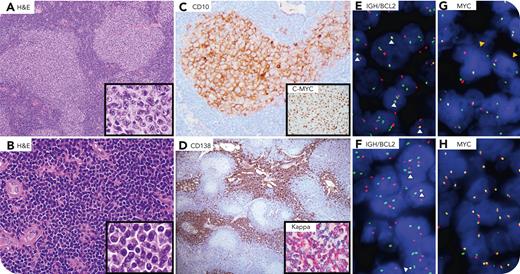
A 75-year-old man presented with anasarca, and imaging revealed generalized lymphadenopathy. An axillary lymph node biopsy demonstrated a high-grade follicular lymphoma (FL, grade 3A) and multifocal, interfollicular clusters of mature plasma cells (panel A: hematoxylin and eosin [H&E] stain, 10× lens objective, original magnification ×100; panel A inset: H&E stain, 40× lens objective, original magnification ×400; panel B: H&E stain, 40× lens objective, original magnification ×400; panel B inset: H&E stain, 40× lens objective, original magnification ×400). Immunophenotypic analysis revealed κ-restricted follicular B cells with a germinal center phenotype (BCL6+, HGAL+, CD10+ (panel C: CD10 stain, 40× lens objective, original magnification ×400) and BCL2 and CMYC expression (panel C inset: CMYC stain, 20× lens objective, original magnification ×200). The plasma cells (CD19+, CD79a+, CD56−, cyclin D1−, CD138+) were also κ restricted (panel D: CD138 stain, 4× lens objective, original magnification ×40; panel D inset: κ ISH stain, 40× lens objective, original magnification ×400). Fluorescence in situ hybridization analysis showed IGH::BCL2 translocations in both the FL and plasma cell components (panels E and F, fused signals, white arrowheads), but the MYC translocation was confined to FL cells (panels G and H, break-apart signals, orange arrowheads). Because a clonally related low-grade FL with plasmacytic differentiation was observed in a staging marrow biopsy, plasma cell differentiation occurred earlier in FL evolution, with the MYC translocation arising later, likely in a shared FL clone.
Plasmacytic differentiation of FL is uncommon (≈3% of FLs), but well recognized. Two patterns have been described, intrafollicular and interfollicular, the latter associated with BCL2 translocation and a mutation profile typical of FL. The presence of double-hit FL with a clonally related plasmacytic component, however, is an unusual finding.
For additional images, visit the ASH Image Bank, a reference and teaching tool that is continually updated with new atlas and case study images. For more information, visit https://imagebank.hematology.org.


This feature is available to Subscribers Only
Sign In or Create an Account Close Modal