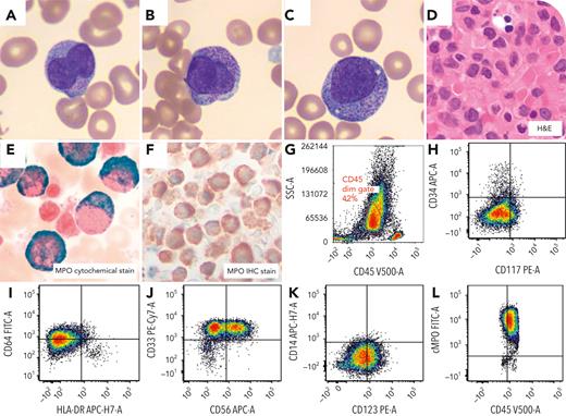
A 29-year-old woman with pancytopenia, worsening gum bleeding, bruising, and fatigue was referred for evidence of circulating blasts and concert for disseminated intravascular coagulation. Blood smear revealed atypical promyelocytes with bilobed nuclei and granules (panels A-C: Wright-Giemsa stain, 100× objective, original magnification ×1000). Bone marrow biopsy demonstrated increased promyelocytes (panel D: hematoxylin and eosin stain, 50× objective, original magnification ×500). Myeloperoxidase (MPO) cytochemical, and immunohistochemical stains were strongly positive (panels E-F: MPO cytochemistry, 100× objective, original magnification ×1000 [E]; immunohistochemistry, 50× objective, original magnification ×500 [F]). Flow-cytometry analysis showed a blast population with high side scatter merging with granulocytes and was CD34−, HLA-DR−, and MPO (bright+) (panels G-L). The findings were suggestive of acute promyelocytic leukemia (APL). The patient began to receive therapy with all-trans retinoic acid (ATRA). Fluorescence in situ hybridization (FISH) analysis demonstrated 3 copies of the RUNX1T1 locus (8q22) without translocation involving PML::RARA. The patient continued receiving ATRA therapy despite negative fusion. A FISH RARA break-apart probe revealed no rearrangement. Subsequent chromosome analysis showed an abnormal clone with trisomy 8 without t(15;17): 47,XX,+8[10]/46,XX[10]. A next-generation sequencing heme fusion panel detected PML::RARA fusion. The breakpoints in this case occur in the bcr3 region of PML and the common intron 2 region of RARA. Diagnosis of APL was confirmed, and the patient began receiving arsenic trioxide therapy.
The mechanism resulting in the fusion event is likely a cryptic insertion. Such submicroscopic insertion events are a rare, but recurrent, mechanism of PML::RARA fusion formation with no reported associated morphologic or clinical differences.
For additional images, visit the ASH Image Bank, a reference and teaching tool that is continually updated with new atlas and case study images. For more information, visit https://imagebank.hematology.org.


This feature is available to Subscribers Only
Sign In or Create an Account Close Modal