

In this issue of Blood, Grob et al1 integrate mutational analysis with clinical outcomes and propose merging TP53-mutated myelodysplastic syndrome with excess blasts (MDS-EB; bone marrow blasts 5% to 19%) and TP53-mutated acute myeloid leukemia (AML) into a unified diagnosis. They evaluate mutations in 2200 patients with AML or MDS-EB (International Prognostic Scoring System [IPSS] ≥1.5 or IPSS-revised (IPSS-R) >4.5) who were treated in clinical trials in the Haemato-Oncology Foundation for Adults in the Netherlands and the Swiss Group for Clinical Research (HOVON-SAKK) from 2001 through 2017. They identify 283 TP53 mutations in 230 patients (186 patients with AML and 44 patients with MDS-EB). Across multiple clinical parameters, they found no significant difference between the patients with TP53-mutated AML or TP53-mutated MDS-EB (age, sex, white blood cell counts, remission rates, consolidation treatment, allelic status, number of TP53 mutations, TP53 clone size, concurrent mutation patterns, or survival outcomes).
As a junior Hematology/Oncology fellow, I was told there were 2 types of physicians: splitters and mergers. That is, clinicians either seek to diagnose increasingly homogenously narrow groups of patients based on increasingly refined, shared characteristics, or they seek to find broad, overarching patterns that unite diagnostic classifications. Hematologic malignancies have been fertile ground for the diagnostic splitters of the world. On the other hand, there have been some noteworthy exceptions. Sometimes it is a technological advance that allows for the synthesis of disparate diagnoses. Paul Ehrlich’s cell staining techniques combined with improved microscopy allowed 19th-century clinicians to recognize shared features in splenic and myelogenous leukemias, unifying these diagnoses. Sometimes it is thoughtful speculation. William Damashek surveyed diverse clinical presentations and proposed a shared structure to myeloproliferative neoplasms. Grob and colleagues propose an interesting union of diagnoses (see figure).
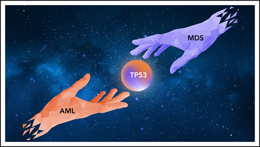
AML and MDS represent related diagnoses of clonal myeloid disorders. Grob and colleagues report that in patients with AML or MDS with TP53 mutations clinical characteristics and outcomes are overlapping and propose that these 2 diseases be considered as a single diagnosis.
AML and MDS represent related diagnoses of clonal myeloid disorders. Grob and colleagues report that in patients with AML or MDS with TP53 mutations clinical characteristics and outcomes are overlapping and propose that these 2 diseases be considered as a single diagnosis.
The diagnostic separation of MDS and AML has been the subject of changing guidelines. In the French-American-British schema, MDS and AML were separated by an arbitrary threshold of 30% bone marrow blasts, later revised in the World Health Organization classification to 20%, and MDS has been variably subcategorized further based on cytopenias, bone marrow blast counts, dysplastic morphology, and recurrent cytogenetics.
Genome sequencing has revealed striking shared features between MDS and AML. MDS and AML share a similar degree of disease burden in the bone marrow, with mutation variant allele frequencies which often suggest that most of the bone marrow cells are involved in the malignant clone, even in cases of MDS with low or absent blasts.2 MDS and AML also share overlapping mutational and cytogenetic patterns, even if some mutations may be more biased toward one diagnosis or the other (MDS: spliceosome and cohesin mutations; AML: NPM1, 11q23 rearrangements, core-binding factor mutations). Based on clinical progression and survival patterns, researchers have proposed merging low blast count MDS (blasts <2%) and clonal cytopenias of undetermined significance (CCUS) into a unified category of clonal cytopenias, whereas higher blast count MDS (2% to 19%) could be merged with AML into a disorder of oligoblastic leukemia.3,4
Diagnostic separation of MDS-EB and AML can be technically challenging. Variance in bone marrow collection quality and hematopathology analysis leads to discrepancies between repeat analysis of prepared slides and between repeat aspirates, and a second bone marrow evaluation has been associated with disease upstaging in 10% to 15% of MDS.5 A clear diagnostic separation within TP53-mutant diseases may be even more challenging. Consistent with other datasets, Grob and colleagues observed that TP53-mutated AML tends to present with lower white blood cell counts and lower bone marrow blast counts than TP53 wild-type AML. In MDS, the opposite has been observed,6 and patients with the TP53 mutation tend to have higher blast counts than their wild-type counterparts. Thus, TP53-mutant MDS and TP53-mutant AML may represent a technical diagnostic continuum.
Despite overlapping features, TP53 mutations may not be sufficient to independently form a unifying diagnosis of cases across all myeloid disorders (clonal hematopoiesis of indeterminate prognosis [CHIP], CCUS, myeloproliferative neoplasm, chronic myelomonocytic leukemia [CMML], MDS, and AML). TP53 mutations are associated with more rapid progression in patients with CHIP and in myeloproliferative diseases compared with patients with wild-type TP53, but those with CHIP or with TP53 mutations appear to have longer survival than their counterparts with MDS or AML.7,8 Unlike patients with MDS or AML, those with CMML with TP53 mutations do not commonly have co-occurring complex karyotypes.9 Also, not all TP53 mutations are created equally; most are missense variants that occur at a series of hotspot nucleotides and may be associated with second mutations, loss of heterozygosity, or deletion of the alternate allele. Some have suggested that more deleterious mutations may be associated with dominant-negative or gain-of-function effects, which induce more rapid clinical progression than isolated deletion or nonsense variants.10
Why does a name matter? Juliet famously pines for unity with her Romeo, “What is in a name? … Doff thy name, and for that name which is no part of thee, take all myself.” But Romeo cannot shed his Montague heritage, even when bound to her gentleness. Outcomes for TP53-MDS and -AML are poor, and treatment options are limited. Bringing these diseases together may increase the number of related cases that can be analyzed in individual datasets and may enable more consistent enrollment onto TP53-mutant–focused clinical trials. Also, as a name, “dysplastic syndrome” does not convey the perilous journey that lies ahead for a patient with TP53-mutated MDS. In the end, Romeo and Juliet find themselves sealed together in a tomb of tragedy, and their families are admonished to “know their true descent … meantime forebear and let mischance be slave to patience.” Perhaps it is time to bring these star-crossed lovers together in their own house, and patiently seek the instruments, “fit to open these dead men’s tombs.” “O brother Montague, give me thy hand.”
Conflict-of-interest disclosure: J.S.W. receives research support from Janssen Pharmaceuticals and Notable Labs.

This feature is available to Subscribers Only
Sign In or Create an Account Close Modal