Key Points
The diagnosis of recurrent ipsilateral DVT is challenging because of persistent intravascular abnormalities after previous DVT.
The incidence of VTE recurrence after negative MRDTI was low, and MRDTI proved to be a feasible and reproducible diagnostic test.
Abstract
The diagnosis of recurrent ipsilateral deep vein thrombosis (DVT) is challenging, because persistent intravascular abnormalities after previous DVT often hinder a diagnosis by compression ultrasonography. Magnetic resonance direct thrombus imaging (MRDTI), a technique without intravenous contrast and with a 10-minute acquisition time, has been shown to accurately distinguish acute recurrent DVT from chronic thrombotic remains. We have evaluated the safety of MRDTI as the sole test for excluding recurrent ipsilateral DVT. The Theia Study was a prospective, international, multicenter, diagnostic management study involving patients with clinically suspected acute recurrent ipsilateral DVT. Treatment of the patients was managed according to the result of the MRDTI, performed within 24 hours of study inclusion. The primary outcome was the 3-month incidence of venous thromboembolism (VTE) after a MRDTI negative for DVT. The secondary outcome was the interobserver agreement on the MRDTI readings. An independent committee adjudicated all end points. Three hundred five patients were included. The baseline prevalence of recurrent DVT was 38%; superficial thrombophlebitis was diagnosed in 4.6%. The primary outcome occurred in 2 of 119 (1.7%; 95% confidence interval [CI], 0.20-5.9) patients with MRDTI negative for DVT and thrombophlebitis, who were not treated with any anticoagulant during follow-up; neither of these recurrences was fatal. The incidence of recurrent VTE in all patients with MRDTI negative for DVT was 1.1% (95% CI, 0.13%-3.8%). The agreement between initial local and post hoc central reading of the MRDTI images was excellent (κ statistic, 0.91). The incidence of VTE recurrence after negative MRDTI was low, and MRDTI proved to be a feasible and reproducible diagnostic test. This trial was registered at www.clinicaltrials.gov as #NCT02262052.
Introduction
Despite major technical advances in recent years, critical limitations in currently available diagnostic techniques for venous thromboembolism (VTE) have been found in specific settings. The failure to provide an accurate diagnosis may lead to misdiagnosis and subsequent mistreatment, affecting both morbidity and mortality.1,2 One of these settings is suspected recurrent ipsilateral deep vein thrombosis (DVT) of the leg, in which the safety of ruling out recurrent DVT by applying clinical decision scores and D-dimer testing has not been established.2 Moreover, the diagnosis of recurrent DVT using compression ultrasonography (CUS) is complicated by residual vascular abnormalities after a first DVT episode in up to 50% of patients after 1 year, despite adequate anticoagulant treatment.3-5 CUS has been proposed to be diagnostic of recurrent DVT in cases of a new, noncompressible venous segment or a 2- to 4-mm increase in vein diameter of a previously noncompressible vein, in comparison with a prior CUS.6-9 However, in clinical practice, a prior CUS is often unavailable, and comparisons with previous CUS examinations are subject to major interobserver variability.10 Similarly, these residual vascular abnormalities complicate the interpretation of all other diagnostic modalities, including contrast venography. As a consequence, recurrent ipsilateral DVT cannot be ruled out in up to 30% of patients in daily practice, resulting in overtreatment.3
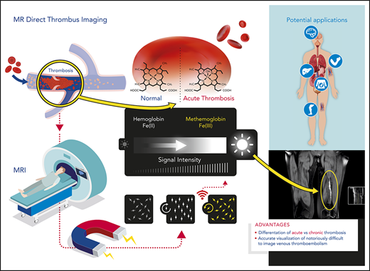
Magnetic resonance direct thrombus imaging (MRDTI) is a technique with a short (10-minute) acquisition time that is based on the formation of methemoglobin in a fresh thrombus that appears as a high signal when imaged on a T1-weighted magnetic resonance imaging (MRI) sequence by measurement of the shortening T1 signal (supplemental Appendix A; available on the Blood web site).11 This technique does not require intravenous gadolinium contrast. MRDTI can accurately diagnose a first DVT and distinguish acute recurrent DVT from chronic residual thrombotic abnormalities with a sensitivity and specificity of at least 95%.12,13 MRDTI therefore has potential to be used as a single test to diagnose or rule out recurrent ipsilateral DVT, but a formal outcome study has not been performed.14 We have conducted a prospective management study to evaluate the safety of ruling out acute recurrent ipsilateral DVT of the leg by a MRDTI negative for DVT.
Methods
Study design and patients
The Theia Study was a prospective, international, multicenter, diagnostic management study conducted at 5 academic and 7 nonacademic teaching hospitals across 5 countries. From March 2015 through March 2019, we included patients aged 18 years or older with clinically suspected acute recurrent ipsilateral DVT of the leg. Exclusion criteria were DVT diagnosed by CUS within 6 months before presentation (to prevent false-positive MRDTI findings because of a previous recent DVT episode15 ), symptom duration of more than 10 days, suspected concurrent acute pulmonary embolism (PE), hemodynamic instability at presentation (as a consequence of concurrent PE or other clinical conditions), medical or psychological condition preventing completion of the study or of signing informed consent (including life expectancy less than 3 months), and general contraindications for MRI. Furthermore, patients treated with full-dose anticoagulation that had been initiated ≥48 hours before the eligibility assessment were excluded. Notably, from August 2015 onward, patients with suspected recurrent DVT while receiving therapeutic anticoagulant treatment ≥48 hours were also enrolled in the study, as they were found to represent a high proportion of the screened study population (30%) in the first year after study initiation and thus formed a clinically relevant patient group.
The study protocol and its amendments were approved by the Institutional Review Board of the Leiden University Medical Center (LUMC) (Leiden, The Netherlands; for all participating hospitals in The Netherlands) and by the institutional review boards at the Danderyd Hospital (Stockholm, Sweden), Østfold Hospital (Østfold, Norway), Ottawa Hospital (Ottawa, ON, Canada), and Rambam Health Care Campus (Haifa, Israel). All patients provided written informed consent. All participating centers were provided with a training set of MRDTI images and performed a test MRDTI before the study started. The study was initiated only if the quality of the scan was judged adequate by the LUMC team of expert radiologists.
Procedures
Consecutive patients who fulfilled all inclusion criteria and met none of the exclusion criteria were eligible for enrollment, and their treatment was managed according to the study algorithm (Figure 1). The diagnosis and treatment decisions were based solely on the result of the MRDTI of the affected leg, which was performed within 24 hours of inclusion. MRDTI was performed with a 1.5- or 3.0-Tesla (T) unit with maximum gradient amplitude of 45 mT/m, slew rate of 200 T/m per second, using an integrated 16-channel posterior coil and a 16-channel anterior body coil for signal reception.15-17 The complete MRDTI sequence is provided in supplemental Appendix B.
Study flowchart of patients with clinically suspected acute recurrent ipsilateral DVT. The reference CUS in patients with MRDTI negative for DVT was performed within 48 hours and did not influence the treatment decision.
Study flowchart of patients with clinically suspected acute recurrent ipsilateral DVT. The reference CUS in patients with MRDTI negative for DVT was performed within 48 hours and did not influence the treatment decision.
In case MRDTI was not instantly available at the time of presentation and in the absence of absolute contraindications, patients received a single dose of therapeutic anticoagulation per local treatment guidelines. Acute recurrent DVT, as diagnosed by the MRDTI protocol, was defined as a high signal in the location of a deep vein segment against the suppressed background greater than that observed in the corresponding or contiguous segments of the ipsilateral vein, as judged by the attending radiologist.12,13
Patients with a MRDTI negative for DVT were left untreated, or treatment remained unadjusted if they had already received anticoagulants for a previous indication. In these patients a standardized CUS examination within 48 hours after the MRDTI was performed. This examination served as a reference test in case a patient returned with symptoms of DVT recurrence during the follow-up period, but it was not used for management decisions at baseline. In case of a MRDTI positive for DVT, anticoagulant treatment was initiated in accordance with international and local guidelines, or modified in patients with recurrent DVT who were on anticoagulant therapy.
All patients were followed up for recurrence of symptomatic VTE, anticoagulation-associated major bleeding, and all-cause mortality over a period of 3 months after inclusion. Patients were instructed to return to the hospital before the 3-month appointment if symptoms of recurrent VTE occurred, at which time objective tests were performed.18-20
Outcomes
The primary outcome was the 3-month incidence of recurrent symptomatic VTE in patients with MRDTI negative for DVT. The diagnosis of recurrent DVT during follow-up was defined as incompressibility of a new venous segment or a ≥2- to 4-mm increase in vein diameter of a previous noncompressible venous segment upon CUS.9 In cases of suspected recurrence during the follow-up period, investigators were also encouraged to perform a repeat MRDTI. PE was considered to be present if computed tomography pulmonary angiography (CTPA) showed at least 1 filling defect in the pulmonary artery tree and if PE was judged to be a probable cause of unexplained death unless proven otherwise by autopsy. An independent committee, blinded for all diagnostic procedures and treatment decisions at baseline, assessed and adjudicated all suspected cases of VTE and deaths that occurred during follow-up.
After study initiation, we observed a relevant prevalence of patients with a MRDTI negative for DVT but positive for superficial thrombophlebitis. These patients were not anticipated in the protocol and were mostly treated with a half-therapeutic dose of anticoagulants for 6 weeks, per local guidelines. Because patients who are treated with anticoagulants have a lower risk of development of recurrent DVT during follow-up, the primary outcome was modified by adding another subgroup: patients with MRDTI negative for both DVT and thrombophlebitis and off anticoagulant treatment at inclusion.
The main secondary outcome was the interobserver agreement of MRDTI in daily clinical practice and was assessed post hoc: the first 10 scans of each study site were reassessed by the expert team at LUMC, who were blinded to the clinical presentation and follow-up of the study patients. Their ruling was compared to the ruling of the attending local radiologist at the moment of clinical presentation. Also, we assessed the feasibility of MRDTI: the number of patients who could not be included because of MRDTI unavailability, as well as the median time between study inclusion and MRDTI scanning.
Statistical analysis
We aimed to mirror the risk of a false-negative test ruling by MRDTI to that of a ruling by CUS. In the 2012 American College of Chest Physicians guidelines, the upper limit of the 95% confidence interval (95% CI) of the risk of a false-negative serial CUS result in suspected recurrent ipsilateral DVT was estimated to be 6.5% in the setting of a 15% DVT prevalence.9 In the largest relevant published study, the overall diagnostic failure rate of normal ultrasonographic findings, compared to a reference CUS, was 3.3% (5 of 153; 95% CI, 1.2-7.6).7 Accordingly, assuming a 3.3% incidence of our primary outcome and considering a maximum recurrent VTE failure rate of 6.5% as the upper limit of a safe test, we determined that a sample of 246 patients who had a MRDTI negative for DVT and who completed follow-up would provide 80% power to reject the null hypothesis that the incidence of recurrent symptomatic VTE would be greater than 6.5%, at an overall 1-sided significance level of .05. Assuming a 15% prevalence of DVT at baseline and anticipating a 5% incidence of loss to follow-up, we aimed to include 305 patients.
Baseline characteristics are expressed as the mean with standard deviation or the median with interquartile range (IQR). The primary outcome was calculated with corresponding exact 95% CI. For the secondary outcome, in which we assessed interobserver agreement of MRDTI reading, the κ-statistic was calculated. The κ value for agreement was interpreted as follows: poor (≤0.20), fair (0.21–0.40), moderate (0.41–0.60), good (0.61–0.80), or excellent (0.81–1.00).21 Analyses were performed with the use of SPSS software, version 25.0.
Results
Patients
From March 2015 through March 2019, 444 consecutive patients with clinically suspected acute recurrent ipsilateral DVT of the leg were screened; 139 patients (31%) were excluded for various reasons, per the predefined exclusion criteria (Figure 2). The baseline characteristics of the 305 study patients are summarized in Table 1.
Flowchart of study patients. *From August 2015 onward, patients with suspected acute recurrent ipsilateral DVT on anticoagulant treatment were enrolled in the study, because they were found to represent a high proportion (30%) of the screened study population. §The patient with a venous iliac stent in whom the stent could not be visualized and the patient in whom MRDTI could not be performed because of extreme pain were both receiving anticoagulant treatment at inclusion. Hence, a total of 68 patients were on anticoagulant treatment at inclusion, including 12 patients with a MRDTI scan positive for DVT, 1 patient with inconclusive MRDTI scan, 1 patient in whom MRDTI could not be performed, 53 patients with MRDTI negative for DVT, and 1 patient with MRDTI negative for DVT but diagnostic for superficial thrombophlebitis.
Flowchart of study patients. *From August 2015 onward, patients with suspected acute recurrent ipsilateral DVT on anticoagulant treatment were enrolled in the study, because they were found to represent a high proportion (30%) of the screened study population. §The patient with a venous iliac stent in whom the stent could not be visualized and the patient in whom MRDTI could not be performed because of extreme pain were both receiving anticoagulant treatment at inclusion. Hence, a total of 68 patients were on anticoagulant treatment at inclusion, including 12 patients with a MRDTI scan positive for DVT, 1 patient with inconclusive MRDTI scan, 1 patient in whom MRDTI could not be performed, 53 patients with MRDTI negative for DVT, and 1 patient with MRDTI negative for DVT but diagnostic for superficial thrombophlebitis.
Baseline characteristics of 305 patients with suspected recurrent ipsilateral DVT of the leg
| Characteristics . | Data . |
|---|---|
| Mean age (±SD), y | 58 (16) |
| Male, n (%) | 152 (50) |
| Median duration of complaints (IQR), d | 4 (2-7) |
| More than 1 prior VTE episode, n (%) | 98 (32) |
| Mean time since the last DVT episode (±SD), y | 7 (9) |
| Active malignancy, n (%) | 18 (5.9) |
| Immobility for >3 d or recent long travel >6 h in the past 4 wk, n (%) | 21 (6.9) |
| Trauma/surgery during the past 4 wk, n (%) | 11 (3.6) |
| Hormone (replacement) therapy, n (%) | 6 (2.0) |
| Known genetic thrombophilia, n (%) | 42 (14) |
SD, standard deviation.
MRDTI results
Of the 305 study patients, 189 (62%) had a MRDTI negative for DVT (Figure 2). Of the 189 patients, 122 patients (65%) had a MRDTI negative for both DVT and thrombophlebitis and were not receiving anticoagulant treatment at inclusion. These patients were left untreated.
The MRDTI was negative for DVT but positive for superficial thrombophlebitis in 14 patients (7.4%). Twelve of these were treated with a short course of half-therapeutically dosed anticoagulants, whereas 1 patient was treated with a short course of therapeutically dosed anticoagulants. One patient who was diagnosed with superficial thrombophlebitis was on anticoagulant treatment at the time of inclusion, and treatment was modified.
The remaining 53 patients (28%) were on anticoagulants at inclusion and continued with unmodified treatment, per previous indications.
Two of the 305 patients (0.66%) had an inconclusive MRDTI: 1 patient had imaging artifacts secondary to a knee prosthesis, and 1 patient had a venous iliac stent that could not be visualized. Both patients were considered to have recurrent DVT based on elevated D-dimer and ultrasonography results. MRDTI could not be performed in 2 additional patients, 1 of whom had extreme pain and 1 of whom had claustrophobia. These 2 patients were also judged to have recurrent DVT based on available diagnostic test results. One patient was incorrectly included and had both suspected recurrent DVT and acute PE at baseline. CTPA confirmed acute PE, and treatment was started before MRDTI of the leg could be performed (which was considered to be a protocol deviation).
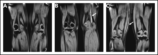
A total of 111 patients (36%) had a MRDTI positive for DVT, of whom 99 were not receiving anticoagulant treatment at the time of inclusion in the study and started anticoagulant treatment (Figure 2). Twelve patients were on anticoagulants at the time of study inclusion, and their treatment was modified after diagnosis. Thus, the overall prevalence of recurrent DVT at baseline, including 111 patients with MRDTI positive for DVT and the above-mentioned 5 patients with recurrent VTE diagnosed otherwise, was 38% (116 of 305). The baseline prevalence of recurrent DVT in patients on anticoagulants at inclusion was 21% (14 of 68; Figure 2). Figure 3 and supplemental Videos 1-6 show examples of MRDTI images and movies of 3 patients in which clear high signal intensities were seen in cases of acute thrombus and symmetrical low signal intensity in the absence of an acute thrombus.
Coronal MRDTI images from 3 study patients. (A) MRDTI negative for DVT with symmetric low signal intensity in both popliteal veins, despite an incompressible popliteal vein in the left leg upon CUS. (B) Asymmetrical high signal intensity in the left popliteal vein diagnostic of acute recurrent DVT of the left leg (arrow). (C) Asymmetrical high signal intensity in the right great saphenous vein diagnostic for acute thrombophlebitis, but not DVT, in the right leg (arrow).
Coronal MRDTI images from 3 study patients. (A) MRDTI negative for DVT with symmetric low signal intensity in both popliteal veins, despite an incompressible popliteal vein in the left leg upon CUS. (B) Asymmetrical high signal intensity in the left popliteal vein diagnostic of acute recurrent DVT of the left leg (arrow). (C) Asymmetrical high signal intensity in the right great saphenous vein diagnostic for acute thrombophlebitis, but not DVT, in the right leg (arrow).
Primary outcome
In total, 5 patients met the primary outcome (Table 2), including 2 of the 122 patients who had a MRDTI negative for both DVT and thrombophlebitis and were not receiving anticoagulant treatment at baseline. The first patient developed CUS-confirmed ipsilateral DVT 21 days after immobilization during a long-haul airplane flight. In addition to CUS, which showed new incompressible venous segments compared to the reference CUS, a repeat MRDTI showed a positive signal for acute recurrent DVT. The second patient was referred for a reference CUS 1 day after the MRDTI that was negative for DVT, but instead presented at the emergency department with sudden shortness of breath. CTPA showed segmental PE. Both patients were treated with anticoagulants in an outpatient setting and had an uncomplicated follow-up. Three of the 122 patients developed thrombophlebitis during follow-up and were treated with anticoagulants; recurrent DVT was ruled out in all 3 patients. The incidence of recurrent VTE in patients with MRDTI negative for both DVT and thrombophlebitis and who were not treated with any anticoagulant during follow-up was thus 1.7% (2 of 119; 95% CI, 0.20%-5.9%; Table 3).
Overview of confirmed venous thromboembolism events during follow-up
| Baseline . | Follow-up . | |||||||||
|---|---|---|---|---|---|---|---|---|---|---|
| Patient . | Sex . | Age, y . | Wells’ score, points . | D-dimer concentration, ng/mL . | Anticoagulant therapy at presentation . | MRDTI result . | Interval to event, d . | Outcome . | Clinical presentation . | Adjudication . |
| 1 | Female | 60 | 2 | 6200 | No | Negative | 1 | Pulmonary embolism | Patient was referred for a reference CUS 1 d after MRDTI negative for DVT, but presented at the emergency department with sudden shortness of breath. CTPA showed bilateral PE. | Nonfatal pulmonary embolism |
| 2 | Male | 75 | 1 | 3200 | No | Positive | 4 | Pulmonary embolism | Patient presented with acute dyspnea. CTPA showed PE in left pulmonary artery and bilaterally in lobar arteries. | Nonfatal pulmonary embolism |
| 3 | Female | 33 | 1 | <220 | No | Negative | 22 | Proximal DVT | Patient had recurrent ipsilateral proximal DVT after immobilization during a long-haul airplane flight as demonstrated by a D-dimer test 3291 ng/mL, a CUS showing new incompressible venous segments and MRDTI indicative of acute DVT. | Nonfatal recurrent DVT |
| 4 | Male | 27 | 2 | 860 | No | Positive | 26 | Pulmonary embolism | Patient presented at emergency department with 2 d of thoracic pain. CTPA showed PE in right segmental pulmonary artery. | Nonfatal pulmonary embolism |
| 5 | Male | 48 | 5 | 240 | Yes | Inconclusive | 77 | In-stent thrombosis | Patient was diagnosed on CUS with recurrent iliac in-stent thrombosis. | Nonfatal recurrent DVT |
Primary outcome of the study
| Category . | Patients, n . | Incidence of the primary outcome (95% CI), % . |
|---|---|---|
| Patients with MRDTI negative for both DVT and thrombophlebitis who were not treated with any anticoagulant during follow-up* | 119 | 1.7 (0.20-5.9) |
| All patients with MRDTI negative for DVT | 189 | 1.1 (0.13-3.8) |
Patients who developed thrombophlebitis during follow-up were not included in this cohort because they received a course of anticoagulant treatment.
Reference CUS
All 189 patients with MRDTI negative for DVT were subjected to a reference CUS examination after the treatment decision was made, showing incompressibility in 88 (47%). The report of these reference CUS examinations mentioned specifically that recurrent DVT was likely or could not be excluded in 57 patients (30%). Notably, prior CUS examinations for comparison were available in only 90 patients with MRDTI negative for DVT (48%). Of these 90 patients, recurrent DVT was likely or could not be excluded in 24 (27%).
Secondary outcomes
The agreement between the initial local reading and the post hoc central reading of the MRDTI images was excellent (κ statistic, 0.91). Among the 444 screened patients, only 16 (3.6%) could not be included, because the MRDTI was not available or could not be performed within 24 hours. The median time from study inclusion to performing the MRDTI was 4 hours (IQR, 2-22 hours).
Discussion
In our study, the incidence of VTE recurrence after negative MRDTI was low. The failure rate among patients with baseline MRDTI negative for DVT who remained without anticoagulant treatment during follow-up was 1.7%, with the upper limit of the 95% CI, well below the predefined 6.5% safety threshold, as was the failure rate and upper limit of the CI in all patients with a MRDTI negative for DVT.
MRDTI is a noninvasive technique that can visualize the metabolism of a fresh thrombus. When red bloods cells are trapped within a thrombus, hemoglobin within the red blood cells undergoes oxidative denaturation to methemoglobin, which causes shortening of the T1 signal and results in a high signal on a T1-weighted sequence.11 Before the DTI signal can become positive, methemoglobin must be formed reliably within an acute clot. Profuse acquired or congenital methemoglobinemia will therefore not result in a positive DTI signal.11 MRDTI was first described as diagnosing a first episode of DVT, an observation that was confirmed in several cohorts.11-13,15 Histological proof of the ability of MRDTI to detect acute thrombosis has been provided in the setting of chronic thromboembolic pulmonary hypertension: the location of a positive MRDTI signal in the pulmonary artery correlated 1:1 with fresh clots found in the surgical specimens of pulmonary artery endarterectomy performed 1 day after the MRDTI.22
The main advantage of the MRDTI technique in the setting of suspected recurrent ipsilateral DVT is the clear distinction between acute and chronic thrombosis, leading to a large reduction of inconclusive diagnoses from 30% in a previous cohort (mainly due to the poor interobserver agreement of the thrombus diameter measurement by CUS and the unavailability of reference CUS examinations)3 to less than 1% (2 of 305) in the present study. The interobserver agreement of the MRDTI in our study was excellent (κ statistic, 0.91). This finding is consistent with the interrater agreement observed in a prospective study that evaluated the diagnostic accuracy of MRDTI for distinguishing acute recurrent ipsilateral DVT from chronic thrombi in leg veins (κ statistic, 0.98).13 Moreover, MRDTI proved to be a feasible and reproducible diagnostic test across international academic and nonacademic study sites.
An important methodological aspect of our study requires comment. From August 2015 onward, patients with suspected acute recurrent ipsilateral DVT while on therapeutic anticoagulants were enrolled in the study, because they represented a high proportion of the screened study population. Canadian researchers have recently reported that 15% of VTE patients in a large management study were subjected to testing for suspected recurrence within the first year of treatment, underlining our experience.23 In the setting of our study, many of the clinical presentations of recurrent DVT during anticoagulant treatment were most likely attributable to the postthrombotic syndrome, considering overlapping symptoms as well as the established association between incomplete thrombus resolution for both postthrombotic syndrome and recurrent VTE.24,25 To date, no published study has focused on the optimal diagnostic management of suspected recurrent ipsilateral DVT in anticoagulated patients. Given the clinical relevance and considering this current “evidence-free zone,” we decided it was reasonable to include these patients in the study. The 21% baseline prevalence of confirmed DVT in this patient group reassured us of the importance and validity of that decision.
What are the clinical implications of our study? First, MRDTI can now be used for therapeutic management decisions in patients with suspected recurrent ipsilateral DVT. Considering the relatively limited availability of MRI and its associated costs, MRDTI cannot currently be suggested to be performed in all patients with suspected recurrent DVT. CUS is sufficient when there are no incompressible vein segments or if a thrombus is detected in a venous segment that was previously not affected by DVT or that was normalized on a reference CUS. Second and equally important, the application of MRDTI in several other settings of notoriously difficult to diagnose acute VTE is now worth evaluating, including upper extremity DVT,26 isolated pelvic vein thrombosis in pregnancy,27 cerebral vein thrombosis,28 and splanchnic vein thrombosis (supplemental Appendix A).
The strengths of our study include the prospective design, the large number of consecutive patients, the near complete follow-up, and the independent adjudication of suspected end points. Moreover, the study was performed across several countries and hospital settings, and both 1.5- and 3.0-T MRI machines of several manufacturers were used. Importantly, MRDTI had not been performed in two-thirds of the study sites before the start of the study, which supports the external validity of our study and the wide applicability of our method and its results.
The main limitation of our study is the absence of a control group. Because this was not a randomized study, we could not compare the safety of MRDTI to the current standard diagnostic approach with CUS, nor could we accurately determine the number of patients in whom anticoagulant treatment was prevented by MRDTI. Based on the reports of the reference CUS performed in patients with MRDTI negative for DVT, we estimate this latter number to be up to 19% (57 of 305) of the total study population, which is a considerable improvement in current practice. Second, although we do not expect a fast normalization of the MRDTI signal in patients with symptom duration exceeding 10 days, we excluded such patients from our study. Therefore, we cannot disregard the possibility of a lower sensitivity of MRDTI in patients with a longer or unknown duration of symptoms. Furthermore, 29% of patients with MRDTI negative for DVT were receiving anticoagulants at inclusion and continued treatment during the follow-up period and were thus largely protected from recurrent VTE. By analyzing the patients without any anticoagulant treatment during follow-up separately, we corrected for this potential bias. Moreover, the high number of patients receiving anticoagulant treatment presenting with suspected recurrent DVT and their high 21% baseline prevalence of recurrent DVT support the decision to include these patients, especially in regard to the lack of evidence of diagnostic and therapeutic management of this patient subgroup. Last, we had estimated that 246 patients with MRDTI negative for DVT would be necessary to reject the null hypothesis. Because the baseline prevalence of recurrent DVT was higher than anticipated and because of the inclusion of patients on anticoagulant treatment, this number was not met. The sample size was not adjusted, as this was not anticipated in the study protocol and because such an adjustment was not feasible after study initiation. Nevertheless, the upper limit of the 95% CI of the primary end point in patients with MRDTI negative for DVT left untreated remained well below the predetermined safety threshold. Furthermore, according to a recent statement of the Scientific and Standardization Committee of the International Society on Thrombosis and Haemostasis, our observed low rate of diagnostic failures despite the high baseline DVT prevalence underlines the safety of ruling out recurrent ipsilateral DVT by MRDTI.29
To summarize, the incidence of VTE recurrence after negative MRDTI was low. MRDTI proved to be a simple, feasible, and reproducible diagnostic test. We suggest, that MRDTI be considered for therapeutic management decisions in patients with suspected recurrent ipsilateral DVT and an inconclusive CUS result. Furthermore, MRDTI creates new opportunities for accurate diagnosis in other challenging settings of suspected acute venous thrombosis.
Complete deidentified participant data collected for this study will be made available after publication to researchers whose proposed use of the data has been approved with a signed data-access agreement. Requests for access to the clinical study data can be submitted via email to f.a.klok@lumc.nl.
The online version of this article contains a data supplement.
The publication costs of this article were defrayed in part by page charge payment. Therefore, and solely to indicate this fact, this article is hereby marked “advertisement” in accordance with 18 USC section 1734.
Acknowledgments
This work was supported by an unrestricted grant from BMS/Pfizer (CV185-390). F.A.K. and C.E.A.D. are supported by Dutch Thrombosis Association grant 2013-01. The International Network of Venous Thromboembolism Clinical Research Networks (INVENT) endorsed and supported this study.
The study was designed by the authors with no involvement of any commercial entity. The authors vouch for the accuracy and completeness of the data and analyses and for the fidelity of the study to the protocol. No one who is not an author contributed to the writing of the manuscript. All trial investigators had access to all clinical trial data in order for the results to be published.
Authorship
Contribution: C.E.A.D., M.V.H., L.J.M.K., and F.A.K. designed the study; L.F.v.D., C.E.A.D., L.J.M.K., G.G., E.W., and F.A.K. managed the study with support and input from Å.E., W.G., J.G., A.v.H., H.M.A.H., M.M.C.H., M.V.H., S.K., A.T.A.M., M.N., M.A.v.d.R., C.J.v.R., R.E.W., and J.W.; L.F.v.D., C.E.A.D., G.G., E.W., and F.A.K. analyzed the data, which were interpreted by Å.E., W.G., J.G., A.v.H., H.M.A.H., M.M.C.H., M.V.H., S.K., A.T.A.M., M.N., M.A.v.d.R., C.J.v.R., R.E.W., J.W., and L.J.M.K.; and L.F.v.D., M.V.H., and F.A.K. wrote the first draft of the manuscript, which was reviewed, modified and approved by C.E.A.D., G.G., Å.E., W.G., J.G., A.v.H., H.M.A.H., M.M.C.H., S.K., A.T.A.M., M.N., M.A.v.d.R., C.J.v.R., R.E.W., J.W., E.W., and L.J.M.K.
Conflict-of-interest disclosure: The authors declare no competing financial interests.
A complete list of the members of the Theia Study Group appears in “Appendix.”
Correspondence: Frederikus A. Klok, Department of Thrombosis and Hemostasis, Leiden University Medical Center, Albinusdreef 2, 2300 RC Leiden, The Netherlands; e-mail: f.a.klok@lumc.nl.
Appendix: study group members
The members of the Theia Study Group are listed below.
Writing group: The Netherlands: Frederikus A. Klok, Lisette F. van Dam, Charlotte E. A. Dronkers, Lucia J. M. Kroft, and Menno V. Huisman (Leiden University Medical Center, Leiden); Herman M. A. Hofstee (Haaglanden Medical Center, The Hague); Marcel A. van de Ree and Stan Kolman (Diakonessenhuis, Utrecht); Robin E. Westerbeek (Deventer Hospital, Deventer); Mathilde Nijkeuter and Jan Westerink (University Medical Center Utrecht, Utrecht); Albert T. A. Mairuhu and Cornelis J. van Rooden (Haga Teaching Hospital, The Hague); and Marcel M. C. Hovens (Rijnstate Hospital, Arnhem). Sweden: Gargi Gautam, Eli Westerlund, Åsa Eckerbom, and Anders von Heijne (Karolinska Institute, Danderyd Hospital, Stockholm). Norway: Jostein Gleditsch (Østfold Hospital Trust, Østfold) and Waleed Ghanima (Østfold Hospital Trust, Østfold, and Institute of Clinical Medicine, Oslo).
Contributing authors: The Netherlands: Guido van Haren, Albert de Roos, and Alexander Šrámek (Leiden University Medical Center Leiden, Leiden); Danny M. Cohn (Amsterdam University Medical Center, Amsterdam); Frank M. Zijta and Fleur I. de Korte (Haaglanden Medical Center, The Hague); and Kaspar J. van Everdingen (Diakonessenhuis, Utrecht). Canada: Grégoire Le Gal (Ottawa Hospital, Ottawa). Israel: Benjamin Brenner (Rambam Hospital, Haifa).
Adjudication committee: The Netherlands: Hugo ten Cate and Karly Hamulyák (Maastricht University Medical Center, Maastricht).
REFERENCES
Author notes
L.F.v.D., C.E.A.D., and G.G. contributed equally to this study.


This feature is available to Subscribers Only
Sign In or Create an Account Close Modal