Despite the success of chimeric antigen receptor T (CART) cell therapy, it is limited by 1) lower rates of durable responses related to inadequate CART cell expansion and trafficking to tumor sites and 2) development of life-threatening complication such as cytokine release syndrome (CRS). Development of a strategy to efficiently and robustly image and track CART cells in the clinic would allow the in vivo characterization of T cell expansion and trafficking to tumor sites as well as the development of strategies to potentially overcome these limitations. The sodium iodide symporter (NIS) is a characterized and sensitive reporter system that has been used for cell imaging in the clinic. We hypothesized that the incorporation of NIS into CART cells would be a sensitive and efficient way to assess CART cell expansion, trafficking, and toxicity.
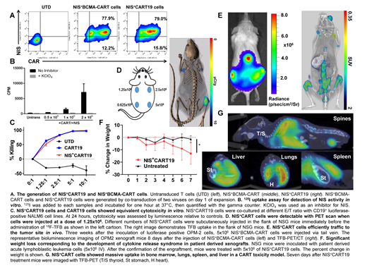
To test our hypothesis, we used two CART cell constructs that are characterized in preclinical models and studied extensively in the clinic: CART19 (41BB costimulated) and BCMA-CART cells (41BB costimulated). First, we generated NIS+CART19 and NIS+BCMA-CART cells through dual transduction of lentiviral vectors (Fig A) and revealed the exclusive 125I uptake by these NIS+CARTs and its inhibition by the NIS inhibitor KClO4in vitro (Fig B). We then analyzed T cell functions of NIS+CART cells. Here, NIS+CART19 or CART19 cells were cultured with the CD19+ acute lymphoblastic leukemia (ALL) cell line NALM6. There was no difference in CART cell cytotoxicity (Fig C), proliferation, or cytokine production (not shown) between NIS+CART19 and CART19. This indicates that the incorporation of NIS into CART cells does not impair their antitumor activity.
Next, we evaluated the sensitivity of NIS+CART19 cell detection by TFB-PET in vivo; imaging was performed using an Inveon TFB-PET/CT scanner. Mice received 250 μCi 18F-TFB 45 minutes prior to image acquisition. NIS+CART cells were detectable with TFB-PET when cells were subcutaneously injected at a dose of 1.25x10⁶ cells (Fig D).
Having demonstrated that the incorporation of NIS in CART cells provides a sensitive way of their detection by TFB-PET and does not interfere with their effector functions, we tested its efficiency to assess CART cell trafficking in vivo, using multiple myeloma (MM) xenografts. Here, immunocompromised NOD-SCID-ɣ-/- (NSG) mice were engrafted with the BCMA+OPM2 MM cell line (1x10⁶ IV). After engraftment, the tumor burden was assessed by bioluminescence imaging (BLI) and mice were randomized to receive 1) BCMA-CART or 2) NIS+BCMA-CART cells (5x10⁶ IV). Mice were then serially imaged for 1) bioluminescence as a measure of disease burden, and 2) with TFB-PET to assess CART cell expansion and trafficking. As expected, BLI demonstrated that MM predominantly engrafts in bones (Fig E). TFB-PET confirmed trafficking of the NIS+BCMA-CART cells to the bones, corresponding to the areas involved by MM based on BLI (Fig E, right). Both BCMA-CART and NIS+BCMA-CART cells exhibited similarly potent antitumor activity in this model (not shown).
Finally, we aimed to explore whether TFB-PET can detect CART massive expansion in vivo and predict the development of CRS. Here, we used an established CRS model in our laboratory. NSG mice were engrafted with patient derived relapsed ALL blasts (5x10⁶ IV). Engraftment was confirmed by peripheral blood sampling. When the leukemic burden is >10 CD19+ cell/µl, mice were treated with either high dose NIS+CART19 cells (5x10⁶ IV) or PBS control. One week after NIS+CART19 cell treatment, mice developed muscle weakness, hunched bodies, and weight loss (Fig F), which correlate with an extreme elevation of cytokines (Sterner et al. Blood 2018). TFB-PET revealed a significant uptake in the bone marrow, spleen, liver, and lungs (Fig G) of the diseased mice but not control mice. Mice were then euthanized, and tissues were harvested. Flow cytometric analysis confirmed an extensive infiltration of CART cells in the liver and spleen. This demonstrates the ability of TFB-PET to detect NIS+CART cell expansion in vivo, correlating with the development of CRS.
In summary, our results robustly show that the incorporation of NIS into CART cells provides a sensitive, clinically applicable platform to image CART cells using TFB-PET and to assess their expansion, trafficking to tumor sites, and the development of CRS. These studies illuminate a novel way to noninvasively assess CART cell functions in vivo.
Sakemura:Humanigen: Patents & Royalties. Suksanpaisan:Imanis: Employment. Cox:Humanigen: Patents & Royalties. Parikh:Janssen: Research Funding; AstraZeneca: Honoraria, Research Funding; Pharmacyclics: Honoraria, Research Funding; MorphoSys: Research Funding; AbbVie: Honoraria, Research Funding; Acerta Pharma: Research Funding; Ascentage Pharma: Research Funding; Genentech: Honoraria. Kay:Agios: Other: DSMB; Infinity Pharmaceuticals: Other: DSMB; Celgene: Other: Data Safety Monitoring Board; MorphoSys: Other: Data Safety Monitoring Board. Peng:Imanis: Equity Ownership. Russell:Imanis: Equity Ownership. Kenderian:Kite/Gilead: Research Funding; Lentigen: Research Funding; Morphosys: Research Funding; Tolero: Research Funding; Humanigen: Other: Scientific advisory board , Patents & Royalties, Research Funding; Novartis: Patents & Royalties, Research Funding.
Author notes
Asterisk with author names denotes non-ASH members.


This feature is available to Subscribers Only
Sign In or Create an Account Close Modal