

Background
Dasatinib potently inhibits the Src family kinase Lck at therapeutic concentrations. Lck plays a critical role in signalling from the T cell receptor with immediate downstream targets including ZAP70 and LAT. The cell surface scaffolding protein LAT has been previously shown to play a central role in normal Treg development. STAT5, the downstream target of IL-2, also plays a critical role in Treg differentiation and maintenance of FOXP3 expression through its binding of the promoter region of the FOXP3 gene.
The proportion of Tregs has been shown to inversely correlate with improved molecular response in patients on dasatinib and a subset of patients taking dasatinib will develop a large granular lymphocytosis (LGL) which is associated with inflammatory toxicity and improved outcome (Mustjoki et al. Leukaemia 2009).
We hypothesised that a reduction in Treg frequency and function would correlate with the expansion of a clonal LGL population in certain patients taking dasatinib. To investigate this, we performed ex-vivo analysis of the phosphorylation of key cell signalling proteins and intracellular cytokine production in Tregs and T effectors from CML patients and healthy controls.
Methods
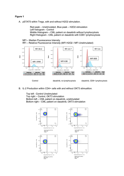
We first performed phosphoflow cytometry in Tregs and T effectors to assess the effect of dasatinib on signalling downstream from the TCR, including activated/phosphorylated ZAP70 (pZAP70), LAT (pLAT) and STAT5 (pSTAT5). 11-colour flow cytometry was performed after cells were activated with H2O2 for 15 minutes, due to its activity as a potent phosphatase inhibitor. A gating strategy of CD3+/CD4+/CD25+/FOXP3+/CD127lo cells was used for identification of Tregs.
We then performed 10-colour intracellular flow cytometry assessing the impact of dasatinib on cellular cytokine production including TNF, IFN gamma, IL-2, IL-4 and IL-10 after stimulation with OKT3.
Results
8 patients with CML and 2 healthy controls were recruited. Of the CML patients 5 were taking dasatinib, 100mg daily, two were taking imatinib and one nilotinib. Patients on dasatinib had lower proportion of Tregs compared with the non dasatinib group with a mean proportion of CD3+ cells of 1.4 vs 2.1.
Patients on dasatinib had significantly reduced phosphorylation of ZAP70 compared with the non dasatinib group in CD3+ cells, CD4+ cells and Tregs, following H2O2 stimulation. The mean increase in median fluorescence intensity (MFI) of pZAP70 was 1.7 vs 3.6 in CD3+ cells, 1.4 vs 3.5 in CD4+ cells and 1.5 vs 2.8 in Tregs (p=0.006, p=0.004, p=0.045). Similarly, pLAT showed a lower increase in phosphorylation in the dasatinib group in all T cell subsets evaluated, with mean increase in MFI of 4.3 vs 11.9 in CD3+ cells, 4 vs 12.3 in CD4+ cells and 4.3 vs 8.2 in Tregs. STAT5 also showed reduced phosphorylation in the dasatinib group with a mean increase in MFI of 6.4 vs 21.2 in CD3+ cells, 5.7 vs 20.3 in CD4+ cells and 5.9 vs 19.7 in Tregs (p=0.007, p=0.003, p=0.01).
Two patients on dasatinib had reversal of normal CD4:CD8 ratio, with one of these also having absolute lymphocytosis, in keeping with likely development of clonal LGL populations. These patients had lower increase in MFI of pZAP70, pLAT and pSTAT5 following stimulation within isolated Tregs compared to patients on dasatinib with normal CD4:CD8 (Figure 1A).
Within CD4+ cells the mean proportional increase in IL-2 production was lower in the dasatinib group at 0.4 vs 5 (p=0.003). Using an additional panel, a single patient was also evaluated and shown to have strikingly reduced production of TNF within isolated Tregs and Il-2 within CD4+cells compared to control (Figure 1B).
Conclusion
Dasatinib inhibits phosphorylation of ZAP70, LAT and STAT5 within T cell subsets including Tregs, with the strongest effect seen against STAT5. In addition, dasatinib causes a reduction in pro-inflammatory cytokine production within CD4+ cells, with the most significant inhibitory effect seen against IL-2. Tregs have abundant expression of the IL-2 receptor on the cell surface and binding leads to STAT5 signalling resulting in transcription of FOXP3.
A more pronounced inhibition of phosphorylation of ZAP70, LAT and STAT5 is seen in two patients on dasatinib with reversal of CD4:CD8 when compared with other patients on dasatinib, suggestive of a more potent reduction of Treg function. Reduction in number and function of Tregs might explain the immunostimulatory effects seen in patients who develop a monoclonal LGL population.
Harrington:Bristol-Myers Squibb: Research Funding. Dillon:TEVA: Consultancy, Honoraria; Pfizer: Consultancy, Honoraria; Novartis: Consultancy, Honoraria; Abbvie: Consultancy, Honoraria. Rousselot:Incyte: Research Funding; Pfizer: Research Funding. Radia:Blueprint Medicines: Consultancy; Novartis: Consultancy, Speakers Bureau. Harrison:Promedior: Honoraria; Gilead: Speakers Bureau; Sierra Oncology: Honoraria; Roche: Honoraria; Celgene: Honoraria, Speakers Bureau; CTI: Speakers Bureau; Shire: Speakers Bureau; AOP: Honoraria; Janssen: Speakers Bureau; Incyte: Speakers Bureau; Novartis: Honoraria, Research Funding, Speakers Bureau. Kordasti:Celgene: Research Funding; Novartis: Research Funding; Boston Biomed: Consultancy; API: Consultancy. De Lavallade:Bristol Myers Squibb: Research Funding; Bristol Myers Squibb: Research Funding.

This feature is available to Subscribers Only
Sign In or Create an Account Close Modal