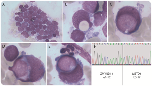
A 19-year-old man was admitted for fatigue without other clinical abnormality. Blood tests showed anemia (115 g/L), thrombocytopenia (81 × 109/L), and 17% blast cells among leukocytes (4.1 g/L). Bone marrow smears revealed infiltration by 90% blast cells mostly organized in small clusters (panel A; May-Grünwald Giemsa stain, original magnification ×500). Rare blast cells showed hemophagocytic activity of erythrocytes or platelets (panel B; May-Grünwald Giemsa stain, original magnification ×1000). More frequently, we have observed 1% to 5% of blast vs blast phagocytic activity, previously referred to as cannibalistic leukemia (panels C-E; May-Grünwald Giemsa stain, original magnification ×1000). Flow cytometry immunophenotyping revealed the following profile: CD45dim/SSClo, CD34+hi, CD38lo/−, CD123+, HLADR−, CD13+/−, CD33+, CD117+, CD7+/−, CD56++. A t(10;17)(p15;q21) translocation was identified. Reverse transcription polymerase chain reaction followed by sequencing confirmed at the messenger RNA level a fusion of ZMYND11 and MBTD1 (panel F). A diagnosis of acute myeloid leukemia (AML) with minimal differentiation was established. Complete remission was obtained with a 3 + 7 regimen.
Whereas hemophagocytic activity of AML blast cells has been well described, especially in cases with a t(16;21)(p11;q22) or t(8;16)(p11;p13) translocation, cannibalistic features are extremely rare in AML and have been described in another case with the same t(10;17) translocation. Further studies are required to understand the link between the ZMYND11-MBTD1 fusion, for which different breakpoints have been described, and the occurrence of “cannibalistic” phenomenon in AML.
For additional images, visit the ASH Image Bank, a reference and teaching tool that is continually updated with new atlas and case study images. For more information, visit http://imagebank.hematology.org.


This feature is available to Subscribers Only
Sign In or Create an Account Close Modal