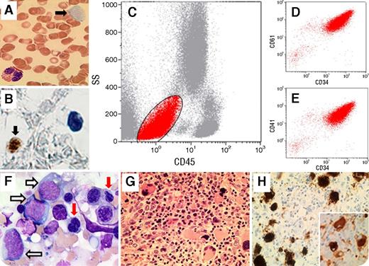
A 65-year-old woman presented with fatigue and exertional dyspnea. The peripheral blood showed: hemoglobin, 91 g/L; leukocytes, 4.8 × 10⁹/L with 1% blast cells; and platelets, 35 × 109/L with megakaryocytic fragments (panel A, filled black arrow; original magnification ×100; May-Grünwald-Giemsa stain). By flow cytometry on the peripheral blood specimen, there was a population of CD45− and low side scatter (18%) that was positive for CD34, CD41, and CD61 (panels C-E, red), and negative for all myeloid- and lymphoid-associated markers. Those were CD34+ megakaryocytic fragments confirmed by immunohistochemistry (panel B, filled black arrow; original magnification ×100; CD34 stain). The bone marrow aspiration and biopsy revealed hypercellularity with 12% blasts (panel F, unfilled arrows; original magnification ×100; May-Grünwald-Giemsa stain), dyserythropoiesis (red arrows), and megakaryocytic hyperplasia and dysplasia (panel G; original magnification ×40; hematoxylin and eosin stain). Immunohistochemistry showed coexpression of CD61 (panel H; original magnification ×40; CD61 stain) and CD34 (panel H, inset; original magnification ×40; CD34 stain) on megakaryocytes. Cytogenetics showed normal karyotype. The diagnosis of myelodysplastic syndrome (MDS) with excess blasts-2 was rendered.
CD34 can be expressed in a subset of megakaryocytes in patients with MDS. Megakaryocytic fragments are larger platelet-like fragments which detach earlier and closer to the megakaryocytic core as a rapid response to thrombocytopenia, then cross the sinusoidal barrier and enter the blood. Our case also highlights the importance of reviewing peripheral blood smear in MDS patients, and performing accurate morphological and immunophenotypic analysis in the presence of a cell population positive for CD34.
For additional images, visit the ASH IMAGE BANK, a reference and teaching tool that is continually updated with new atlas and case study images. For more information visit http://imagebank.hematology.org.


This feature is available to Subscribers Only
Sign In or Create an Account Close Modal