Key Points
SIRT6 is highly expressed in multiple myeloma cells and blocks expression of ERK-regulated genes.
Targeting SIRT6 enzymatic activity sensitizes multiple myeloma cells to DNA-damaging agents.
Abstract
Multiple myeloma (MM) is characterized by a highly unstable genome, with aneuploidy observed in nearly all patients. The mechanism causing this karyotypic instability is largely unknown, but recent observations have correlated these abnormalities with dysfunctional DNA damage response. Here, we show that the NAD+-dependent deacetylase SIRT6 is highly expressed in MM cells, as an adaptive response to genomic stability, and that high SIRT6 levels are associated with adverse prognosis. Mechanistically, SIRT6 interacts with the transcription factor ELK1 and with the ERK signaling-related gene. By binding to their promoters and deacetylating H3K9 at these sites, SIRT6 downregulates the expression of mitogen-activated protein kinase (MAPK) pathway genes, MAPK signaling, and proliferation. In addition, inactivation of ERK2/p90RSK signaling triggered by high SIRT6 levels increases DNA repair via Chk1 and confers resistance to DNA damage. Using genetic and biochemical studies in vitro and in human MM xenograft models, we show that SIRT6 depletion both enhances proliferation and confers sensitization to DNA-damaging agents. Our findings therefore provide insights into the functional interplay between SIRT6 and DNA repair mechanisms, with implications for both tumorigenesis and the treatment of MM.
Introduction
Genomic instability is a common feature of monoclonal gammopathies, resulting in complex genetic changes associated with disease progression from monoclonal gammopathy of undetermined significance to active multiple myeloma (MM) to plasma cell leukemia.1,2 Although alterations in DNA damage checkpoint proteins are less common (10% to 15%) in blood cancers compared with solid tumors,3-5 MM cells do manifest a dysfunctional DNA-damage response (DDR), a key determinant of their genomic instability.6-8 Identifying proteins and signaling pathways that protect MM cells from cumulative genomic instability may therefore lead to innovative therapeutic opportunities, as exemplified by the clinical efficacy of PARP inhibitors in the context of breast and ovarian tumors lacking functional BRCA1 or BRCA2.9,10 In MM cells, direct evidence of homozygous loss or mutations in BRCA1/2 or other DDR genes is lacking, but increased DNA repair activity has been reported.11,12 Thus, identification of adaptive pathways for coping with genomic instability in MM may similarly provide the framework for new therapeutic strategies.
Sirtuins (SIRTs) are NAD+-degrading enzymes involved in a variety of biological processes, ranging from metabolism to lifespan regulation.13,14 Of the 7 sirtuin family members, only SIRT6 clearly contributes to DNA repair.15-18 Consistently, murine SIRT6 knockout cells exhibit genomic instability and hypersensitivity to DNA-damaging agents.15,17,19 Moreover, solid cancers, including breast and prostate cancer, express high levels of SIRT6 and DDR to overcome chemotherapy-induced DNA damage.20,21 In contrast, SIRT6 appears to be downregulated in other types of solid tumors (ie, gastrointestinal cancers, including colorectal and pancreatic cancer), which results in increased glycolysis, glucose uptake, and MYC activity.18,22
To date, the role of SIRT6 in hematologic malignancies, including MM, is unknown. In this study, we have characterized the biological role, prognostic significance, and potential clinical relevance of SIRT6 expression in MM. Our data demonstrate a key role for SIRT6 in the DDR and sensitivity to DNA-damaging agents (DDAs) in MM cells and provide the rationale for targeting SIRT6 as novel strategy to enhance activity of standard anti-MM therapies.
Materials and methods
For a more detailed description of the methods used, see supplemental Methods (available on the Blood Web site).
Cell lines and primary tumor specimens
Cell lines were obtained from the ATCC or kindly provided by sources indicated in supplemental Experimental Procedures.
Reagents
Nicotinamide was obtained from Sigma-Aldrich (St. Louis, MO). Doxorubicin and melphalan were purchased from Selleck Chemicals (Houston, TX) and Sigma-Aldrich, respectively; SIRT6 chemical inhibitor (OSSL_128167) was obtained from MolPort (Riga, Latvia). MEK1/2 selective inhibitors (AS-703026 and UO126-EtOH) and RSK2 (FMK) inhibitor were purchased from Selleck Chemicals and Axon Medchem (Reston, VA), respectively.
Gene editing by lentiviral transgenesis
SIRT6 was knocked down using a pGIPz vector as well as a tetracycline-inducible pTRIPz-Turbo-RFP vector (Thermo Scientific, Pittsburg, PA) containing the target sequence (indicated in the list below) or scramble control, according to the manufacturer’s specifications. Based on preliminary analysis, we selected clone #134911 for the subsequent experiments. To obtain stable silenced cells, the following protocol was used. 293T cells were plated (300 000 cells on 6-cm plates) in Dulbecco’s modified Eagle medium, 5% fetal bovine serum, and 0.1% penicillin-streptomycin. After 24 hours, when cells were 60% to 70% confluent, 6000 ng of the vector of interest together with Trans-Lentiviral Packaging Mix was cotransfected with CaCl2 (Thermo Scientific). After 12 hours, the 293T medium was changed with Dulbecco’s modified Eagle medium, 20% fetal bovine serum, and 10% penicillin-streptomycin to promote viral production. The supernatant containing lentiviral particles was harvested 24 and 48 hours after transfection, filtered with a 0.45-μm-diameter filter, and used to infect 2 500 000 MM cells. MM cells were spinoculated at 750g for 45 min with 8 μg/mL polybrene (Santa Cruz Biotechnology), incubated with viral supernatant for 6 hours, and left in culturing medium. After the second cycle of infection, cells were selected with a suitable concentration of puromycin (2 μg/mL). The transduction efficiency was approximated 48 and 72 hours after selection by counting the proportion of cells expressing the fluorescent protein (green fluorescent protein [GFP] or red fluorescent protein [RFP]) using a fluorescence microscope (Nikon Eclipse 80i; Nikin, Melville, NY), and the knockdown efficiency was validated by detecting SIRT6 protein level by western blot analysis. For the inducible model, MM cells expressing short hairpin RNA (shRNA) were pretreated with 2μg/mL doxycycline for 3 days to achieve SIRT6 knockdown (SIRT6-KD). The efficacy of the induction was confirmed by examining the cells for the presence of Turbo-RFP and western blot analysis. Subsequently, such cells were treated with indicated stimuli.
Overexpression of SIRT6 was obtained in MM cells using precision LentiORF/SIRT6 (SIRT6+) with EV used as control (Thermo Scientific). Overexpression efficiency was assessed using western blot analysis.
Statistical analyses
All data are shown as means ± standard deviation (SD). The Student t test was used to compare 2 experimental groups using Graph-Pad Prism software. Correlation of SIRT6 expression with disease progression and overall survival was measured using the Kaplan-Meier method, with log-rank test for group comparison. Significance was P < .05.
Results
SIRT6 is overexpressed in human MM cells
We first investigated the relevance of SIRT6 in the biology of MM by characterizing its expression in bone marrow biopsies from 10 newly diagnosed MM patients and in 5 healthy donors by immunohistochemical staining. As shown in Figure 1A, SIRT6 staining was significantly greater in MM cells than in normal cells, with a predominant nuclear localization confirmed by immunofluorescence analysis of MM cell lines and patient MM cells (Figure 1B). Similar results were observed in MM cells ectopically overexpressing a GFP-tagged SIRT6 protein, as well as in protein extracts from MM cell lines (Figure 1C-D). Overall, these findings on SIRT6 localization were consistent with those from previous studies in other cell types.19,23,24 Notably, SIRT6 was virtually undetectable in peripheral blood mononuclear cells from healthy donors and from MM patients, suggesting its tumor selectivity.
SIRT6 expression and prognostic relevance in MM. (A) Immunohistochemical analysis of 3 representative BM specimens derived from normal and MM patients (ND #1, 2, and 5 and MM #1, 4, and 8) show SIRT6 expression (positive cells are brown). Original magnification ×20 (×100 in insets). (B) Immunofluorescence showing subcellular distribution of SIRT6 in MM cell lines and patient MM cells (n = 5). (C) MM.1S and U266 cells were transfected with SIRT6 GFP-tagged plasmid. SIRT6 protein was visualized with an antibody directed against GFP, and cells were counterstained with DAPI to visualize nuclei. (D) Western blot analysis (1 of 3 representative blots) confirms the nuclear localization of SIRT6 in MM cell lines, but not in peripheral blood mononuclear cells (PBMCs) collected from healthy donors and MM patients. Cell lysates from cytoplasm and nuclear fractions were analyzed for SIRT6 expression. GAPDH and nucleolin were used as loading controls for the cytoplasmic and nuclear fractions, respectively. GAPDH, glyceraldehyde 3-phosphate dehydrogenase. (E) SIRT6 locus (human chromosome 19p13.3) copy-number heatmap for 45 (left side) MM cell lines (Mayo Clinic database) and 254 (right side) MM patients (Multiple Myeloma Research Consortium [MMRC]) (http://www.broad.mit.edu/mmgp). Color indicates degree of copy-number loss (blue) or gain (red). (F) SIRT6 expression in plasma cells from patients with MM (data set GSE2658). Increased SIRT6 expression is observed in high-risk compared with low-risk MM (unpaired t test; ****P < .0001). (G) Kaplan-Meier plots showing prognostic relevance of SIRT6 expression on overall survival of MM patients (based on GSE4581; deposited by Dr J. D. Shaughnessy Jr, University of Arkansas for Medical Sciences). The patient group with higher SIRT6 expression (blue line) had shorter overall survival than the patient cohort with lower SIRT6 expression (red line) (log-rank test).
SIRT6 expression and prognostic relevance in MM. (A) Immunohistochemical analysis of 3 representative BM specimens derived from normal and MM patients (ND #1, 2, and 5 and MM #1, 4, and 8) show SIRT6 expression (positive cells are brown). Original magnification ×20 (×100 in insets). (B) Immunofluorescence showing subcellular distribution of SIRT6 in MM cell lines and patient MM cells (n = 5). (C) MM.1S and U266 cells were transfected with SIRT6 GFP-tagged plasmid. SIRT6 protein was visualized with an antibody directed against GFP, and cells were counterstained with DAPI to visualize nuclei. (D) Western blot analysis (1 of 3 representative blots) confirms the nuclear localization of SIRT6 in MM cell lines, but not in peripheral blood mononuclear cells (PBMCs) collected from healthy donors and MM patients. Cell lysates from cytoplasm and nuclear fractions were analyzed for SIRT6 expression. GAPDH and nucleolin were used as loading controls for the cytoplasmic and nuclear fractions, respectively. GAPDH, glyceraldehyde 3-phosphate dehydrogenase. (E) SIRT6 locus (human chromosome 19p13.3) copy-number heatmap for 45 (left side) MM cell lines (Mayo Clinic database) and 254 (right side) MM patients (Multiple Myeloma Research Consortium [MMRC]) (http://www.broad.mit.edu/mmgp). Color indicates degree of copy-number loss (blue) or gain (red). (F) SIRT6 expression in plasma cells from patients with MM (data set GSE2658). Increased SIRT6 expression is observed in high-risk compared with low-risk MM (unpaired t test; ****P < .0001). (G) Kaplan-Meier plots showing prognostic relevance of SIRT6 expression on overall survival of MM patients (based on GSE4581; deposited by Dr J. D. Shaughnessy Jr, University of Arkansas for Medical Sciences). The patient group with higher SIRT6 expression (blue line) had shorter overall survival than the patient cohort with lower SIRT6 expression (red line) (log-rank test).
As previously mentioned, SIRT6 was proposed to act as a tumor suppressor in at least some types of solid tumors (ie, gastrointestinal cancers), in which SIRT6 locus is typically lost.18,25 However, an array-based comparative genomic hybridization analysis revealed that the genomic region encompassing the SIRT6 gene (on chromosome19) is amplified in 62% of primary MM cells and in 85% of MM cell lines (Figure 1E). We subsequently evaluated SIRT6 messenger RNA (mRNA) expression in a publicly available data set26 and found that SIRT6 transcript levels are higher in monoclonal gammopathy of undetermined significance and MM than in normal plasma cells (supplemental Figure 1). In a second, independent data set of 414 newly diagnosed patients,27 SIRT6 expression was found to be higher in the genetically defined high-risk group than in the low-risk MM group (Figure 1F). Finally, high SIRT6 expression in MM was found to correlate with shorter overall survival (Figure 1G). Overall, these findings suggested a relevant role for SIRT6 in the biology of MM.
SIRT6 reduces MM cell proliferation by opposing the MAPK-signaling pathway
To define the biological role(s) played by SIRT6 in MM cells, we first silenced it by RNA interference, using lentiviral vectors carrying green fluorescent protein (GFP) as a reporter. Surprisingly, as compared with the control shRNA, SIRT6-KD appeared to confer a growth advantage to MM cells, as shown by the progressive accumulation of GFP+ cells (Figure 2A). Indeed, SIRT6-KD MM cells formed more colonies and exhibited increased proliferation, adhesion, and migration compared with controls (Figure 2B-C and supplemental Figure 2A-B). Altogether, these data indicated, as in solid tumors,18 that SIRT6 reduces MM cell proliferation. Notably, overexpressing SIRT6 wild-type (WT) in MM cells did not affect cell proliferation (Figure 2C), likely due to the constitutively high SIRT6 levels.
SIRT6 reduces MM cell proliferation by opposing MAPK-signaling pathway. (A) SIRT6 silencing using the lentiviral vector pGIPz (after 48 hours with 2 μg/ml puromycin selection) in MM.1S cells. Shown is a western blot for SIRT6 in SIRT6-silenced cells (with 3 different shRNAs) and scramble control. After 4 days of infection, cells were harvested and whole-cell lysates were subjected to immunoblot analysis. The indicated MM cell lines were infected with lentivirus coexpressing GFP and SIRT6 shRNA (clone #911) or scramble control (top). Western blot analyses were performed 4 days after transfection. Data are representative of 2 independent experiments (middle). The percentage of GFP+ cells was measured over time by flow cytometry and normalized to the percentage of scramble GFP+ cells. Data represent the mean values ± SD and represent a minimum of triplicates (bottom). (B) Colony formation of MM cells transduced with lentivirus expressing GFP-tagged scramble or shSIRT6. Light and florescence microscopy is shown. Numbers of colonies after 2 weeks (average of 3 independent experiments; unpaired t test; *0.05 < P < .04) (top). (C) Cell proliferation of MM.1S cells transduced with SIRT6-specific shRNA (clone #911), SIRT6 wild-type (WT) or specific controls was determined by [3H]- thymidine uptake. The results presented are a mean ± SD of triplicate samples (Student t test; *0.45 < P < .03; **0.006 < P < .001; ***0.009 < P < .006; ****P < .0001). (D) Representative western blots showing activation of MAPK-signaling pathway in MM cells depleted of SIRT6 compared with control. GAPDH was used as loading control. One representative blot of two is shown. (E) Representative western blots showing SIRT6 regulating ERK2 activity. MM.1S cells stably expressing shRNA-targeting SIRT6 were transduced with pLOC carrying SIRT6 open reading frame sequence lacking the 3′ UTR region (which is targeted by the siRNA), permitting rescue of the target-specific shRNA phenotype. (F) Western blots showing specific role of SIRT6 in MM.1S cells. One representative blot of two is shown.
SIRT6 reduces MM cell proliferation by opposing MAPK-signaling pathway. (A) SIRT6 silencing using the lentiviral vector pGIPz (after 48 hours with 2 μg/ml puromycin selection) in MM.1S cells. Shown is a western blot for SIRT6 in SIRT6-silenced cells (with 3 different shRNAs) and scramble control. After 4 days of infection, cells were harvested and whole-cell lysates were subjected to immunoblot analysis. The indicated MM cell lines were infected with lentivirus coexpressing GFP and SIRT6 shRNA (clone #911) or scramble control (top). Western blot analyses were performed 4 days after transfection. Data are representative of 2 independent experiments (middle). The percentage of GFP+ cells was measured over time by flow cytometry and normalized to the percentage of scramble GFP+ cells. Data represent the mean values ± SD and represent a minimum of triplicates (bottom). (B) Colony formation of MM cells transduced with lentivirus expressing GFP-tagged scramble or shSIRT6. Light and florescence microscopy is shown. Numbers of colonies after 2 weeks (average of 3 independent experiments; unpaired t test; *0.05 < P < .04) (top). (C) Cell proliferation of MM.1S cells transduced with SIRT6-specific shRNA (clone #911), SIRT6 wild-type (WT) or specific controls was determined by [3H]- thymidine uptake. The results presented are a mean ± SD of triplicate samples (Student t test; *0.45 < P < .03; **0.006 < P < .001; ***0.009 < P < .006; ****P < .0001). (D) Representative western blots showing activation of MAPK-signaling pathway in MM cells depleted of SIRT6 compared with control. GAPDH was used as loading control. One representative blot of two is shown. (E) Representative western blots showing SIRT6 regulating ERK2 activity. MM.1S cells stably expressing shRNA-targeting SIRT6 were transduced with pLOC carrying SIRT6 open reading frame sequence lacking the 3′ UTR region (which is targeted by the siRNA), permitting rescue of the target-specific shRNA phenotype. (F) Western blots showing specific role of SIRT6 in MM.1S cells. One representative blot of two is shown.
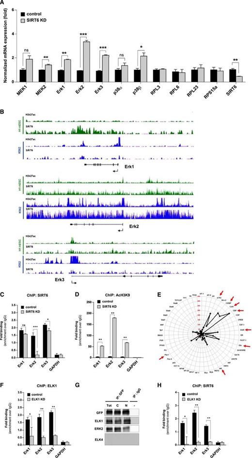
To define the mechanism underlying the increased proliferation of SIRT6-KD MM cells, we focused on the activity of the growth-promoting mitogen-activated protein kinase (MAPK) signaling pathway and measured their proliferation after treatment with 2 MEK1/2 inhibitors (U0126 and AS703026). As shown in supplemental Figure 3A, these agents significantly reduced proliferation in both control and SIRT6-KD cells, confirming that MAPK signaling mediates MM cell proliferation, including increased proliferation in SIRT6-KD versus control MM cells. Moreover, knockdown of SIRT6 increased ERK2 expression and p90RSK phosphorylation (but not p38 expression or phosphorylation) (Figure 2D and supplemental Figure 3B). Concomitant expression of the SIRT6-shRNA and of a wild-type SIRT6 allele in MMS.1 cells restored ERK2 protein to control levels, confirming the specificity of the effects observed with SIRT6 shRNA (Figure 2E). In addition, transiently transfecting MM.1S cells with synthetic SIRT6 small interfering RNAs (siRNAs), but not with SIRT1 siRNAs, also led to a marked increase in ERK2, confirming the role of SIRT6, but not SIRT1, in regulating this pathway (Figure 2F). Consistent with the western blot experiments, quantitative reverse-transcription polymerase chain reaction also showed increased Mek1, Mek2, Erk1, Erk2, and Erk3 mRNA levels in SIRT6-KD compared with control cells (Figure 3A), whereas expression of RPL3, RPL6, RPL23, and RPS15a, which are unrelated to this pathway, was unchanged. These findings suggest that MAPK-signaling regulation by SIRT6 may occur at the transcriptional level of key members of this cascade.
SIRT6 destabilizes MAPK pathway by deacetylating Histone H3 lysine9 and ELK1 targeting in MM cells. (A) Real-time PCR analysis of MEK/ERK-signaling–related genes in wild-type (WT) and SIRT6-silenced MM cells. *P = .02, ** 0.0062 < P < .0018, ***0.007 < P < .005 (Student t test). (B) ChIP-seq binding density for H3K27ac and SIRT6 at the promoter of indicated genes in K562 cells (blue) and human H1-ES cells (green). (C-D) ChIP analyses to detect SIRT6 binding (C) and H3K9 acetylation (D) at the promoter of indicated genes, performed with SIRT6 or AcH3K9-specific antibody and immunoglobulin G (IgG) control, in SIRT6-WT and SIRT6-KD MM.1S cells. The binding of SIRT6 and AcH3K9 at promoters is shown relative to background with IgG control antibody. Data are presented as mean values ± SD of triplicates. Student t test was applied to calculate the P value: ns, not significant; *P = .001; ** 0.0006 < P < .0004; *** P = .0005. (E) Radar chart displays fold-change values of the TF activation profile in SIRT6-KD and SIRT6-WT MM.1S cells. TFIID was used as loading control. (F) ChIP quantitative polymerase chain reaction analysis of control or ELK1-KD MM1S cells. ELK1 immunoprecipitates (IPs) were measured relative to control (IgG) (mean values ± SD of triplicate experiments). GAPDH, which is bound by neither SIRT6 nor ELK1, was a negative control. *P = .04; ** 0.007 < P < .001. (G) SIRT6 binding to ELK1 and ERK2, shown by western blots of GFP-tagged SIRT6 or control IgG antibody IPs from MM.1S cells. C, cytoplasmic fraction; N, nuclear fraction; Tot, total. (H) SIRT6 occupancy at promoters of target genes in ELK1-KD versus ELK1-WT MM.1S cells determined by ChIP (mean values ± SD of triplicates). *P = .04, **.007 < P < .002 (Student t test).
SIRT6 destabilizes MAPK pathway by deacetylating Histone H3 lysine9 and ELK1 targeting in MM cells. (A) Real-time PCR analysis of MEK/ERK-signaling–related genes in wild-type (WT) and SIRT6-silenced MM cells. *P = .02, ** 0.0062 < P < .0018, ***0.007 < P < .005 (Student t test). (B) ChIP-seq binding density for H3K27ac and SIRT6 at the promoter of indicated genes in K562 cells (blue) and human H1-ES cells (green). (C-D) ChIP analyses to detect SIRT6 binding (C) and H3K9 acetylation (D) at the promoter of indicated genes, performed with SIRT6 or AcH3K9-specific antibody and immunoglobulin G (IgG) control, in SIRT6-WT and SIRT6-KD MM.1S cells. The binding of SIRT6 and AcH3K9 at promoters is shown relative to background with IgG control antibody. Data are presented as mean values ± SD of triplicates. Student t test was applied to calculate the P value: ns, not significant; *P = .001; ** 0.0006 < P < .0004; *** P = .0005. (E) Radar chart displays fold-change values of the TF activation profile in SIRT6-KD and SIRT6-WT MM.1S cells. TFIID was used as loading control. (F) ChIP quantitative polymerase chain reaction analysis of control or ELK1-KD MM1S cells. ELK1 immunoprecipitates (IPs) were measured relative to control (IgG) (mean values ± SD of triplicate experiments). GAPDH, which is bound by neither SIRT6 nor ELK1, was a negative control. *P = .04; ** 0.007 < P < .001. (G) SIRT6 binding to ELK1 and ERK2, shown by western blots of GFP-tagged SIRT6 or control IgG antibody IPs from MM.1S cells. C, cytoplasmic fraction; N, nuclear fraction; Tot, total. (H) SIRT6 occupancy at promoters of target genes in ELK1-KD versus ELK1-WT MM.1S cells determined by ChIP (mean values ± SD of triplicates). *P = .04, **.007 < P < .002 (Student t test).
A previous study showed that SIRT6 counters IGF-1–driven signaling cascades via epigenetic regulation and transcription factor (TF) activity.28 To explore the possibility that a similar mechanism may occur in MM cells, we next interrogated a publicly available SIRT6 chromatin immunoprecipitation sequencing (ChIP-seq) data set from human embryonic stem cells and erythroleukemia cells (K562).29 We found that SIRT6 does bind to the MAPK genes promoter: a sharp peak of SIRT6 binding co-localized with H3K27Ac on ERK2 gene promoter in K562 cells, but not in human embryonic stem cells, indicating that ERK2 may be a specific target of SIRT6 in tumor cells (Figure 3B). Conversely, SIRT6-KD significantly decreased SIRT6 binding to these promoters, confirming the specificity of this binding (Figure 3C).
Based on its specific activity, we next asked whether SIRT6-KD would result in increased H3K9 acetylation at the promoters of these MAPK-signaling–related genes. Consistent with this hypothesis, ChIP experiments clearly showed increased H3K9 acetylation enrichment on ERK1/2/3 gene promoter regions in SIRT6-KD MM cells compared with controls (Figure 3D). Importantly, the most abundant levels of acetylated H3K9 in response to SIRT6-KD were observed on the ERK2 gene promoter, further confirming that SIRT6 regulates ERK2 expression. Overall, these data are consistent with those obtained by Sundaresan and colleagues in heart tissue28 indicating that SIRT6 suppresses the expression of key MAPK-pathway–related genes, such as ERK2, by binding to their promoters and deacetylating H3K9 at these sites.
Because SIRT6 also acts by modulating the activity of TFs,28,30,31 we next sought to identify specific TFs downstream of MAPK genes whose activity would be similarly downregulated by SIRT6 by screening the DNA-binding activities of different TFs in SIRT6-KD and control MMS.1 cells. As shown in Figure 3E, CBF, C/EBP, CAR, ATF-2, EGR, Pbx1, IRF, and ETS activities were significantly increased in SIRT6-KD cells. A subsequent computational analysis revealed enrichment of common TF binding sites in SIRT6-bound promoter sequences of MEK/ERK signaling related-genes (data not shown). Among the 8 TFs whose activity was increased in SIRT6-KD cells, the most frequent DNA motifs corresponded to consensus-binding sites for ETS family members,32 with ELK1 showing the highest matrix similarity score for SIRT6- and MAPK-signaling–regulated genes (supplemental Figure 4). These data led us to further investigate the interaction between this ETS family member and SIRT6. We first demonstrated that ELK1 binds several SIRT6 target promoters containing the ETS consensus motif (ERK1/2/3), but not promoters lacking this motif (glyceraldehyde-3-phosphate dehydrogenase [GAPDH]) (Figure 3F). Subsequently, in coimmunoprecipitation experiments, we found that SIRT6 directly interacts with ELK1, but not with other ETS proteins including ELK4 (Figure 3G and supplemental Figure 5A). Western blot analysis of immunoprecipitated ELK1 (both endogenous and ectopically overexpressed) confirmed this interaction (supplemental Figure 5B). The functional sequelae were further assessed by measuring the effect of ELK1 knockdown on SIRT6 occupancy at specific target promoters. As shown in Figure 3H, depletion of ELK1 led to a significant decrease in SIRT6 occupancy at ERK1/2/3 promoters, but not at the GAPDH promoter. Likewise, ELK1 knockdown increased H3K9 acetylation at ERK genes promoters, but not at promoters lacking the ETS motif (supplemental Figure 6A). Finally, we tested the effects of SIRT6 on transcriptional activity of ELK1 using a luciferase reporter assay. SIRT6-KD, as well as treatment with SIRT6 chemical inhibitor, enhanced transcriptional activity of endogenous ELK1 (supplemental Figure 6B). ChIP analysis with an ELK1-specific antibody revealed that ELK1 binds promoters of ERK signaling-related genes, and SIRT6-KD was found to enhance this binding (supplemental Figure 6C). Therefore, ELK1 and SIRT6 mediate the functional interaction between SIRT6 and MEK/ERK-driven transcription in MM cells.
SIRT6 plays multiple roles in the DDR of MM cells
We next examined whether SIRT6 overexpression in MM cells has additional effects besides conferring reduced cell proliferation. The MAPK-signaling pathway, in addition to promoting cell cycle progression and cell proliferation, also regulates DDR; specifically, ERK/p90RSK phosphorylates the checkpoint kinase Chk1 at inhibitory site Ser 280, thereby inhibiting ATR/CHK1 signaling and preventing activation of the G2 DNA damage checkpoint.33,34 Thus we reasoned SIRT6 overexpression in MM, by reducing ERK/p90RSK activity, promotes DDR, genomic stability, and resistance to anticancer agents, and conversely, that reducing or inhibiting SIRT6 may sensitize MM cells to DNA-damaging agents.
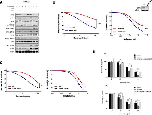
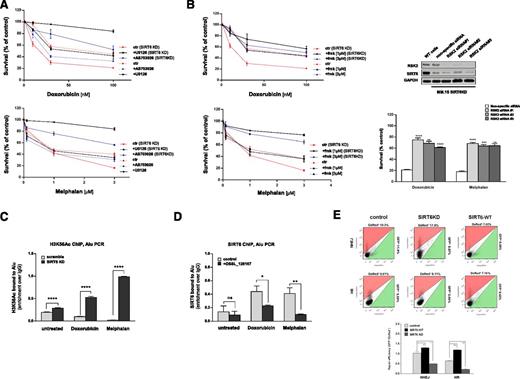
We monitored DNA damage signaling activity following conditional SIRT6 silencing by engineering MM.1S and NCI-H929 cells to express doxycycline-inducible SIRT6 shRNA. Although SIRT6 depletion did not affect ATM, Chk1, or RPA protein levels after genotoxic stress, it markedly decreased their function. In the absence of doxycycline, treatment with melphalan or doxorubicin triggered RPA phosphorylation on Ser4 and Ser8, as well as increased ATM, Chk1(Ser317), and p53 phosphorylation, associated with accumulation of the lower-molecular-weight protein γH2AX. In contrast, DDA treatment of doxycycline-induced SIRT6-KD MM.1S and NCI-H929 cells decreased phosphorylation of Chk1(Ser317), RPA32, p53, and ATM as well as increased γH2AX (Figure 4A and supplemental Figure 7A-C). SIRT6-KD was also associated with p90RSK activation and phosphorylation of Chk1 at Ser280, which was retained after genotoxic stress. Importantly, SIRT6-silenced cells exhibited increased sensitivity to melphalan and doxorubicin (Figure 4B and supplemental Figure 8C). Treatment with the pan-sirtuin inhibitor nicotinamide (NAM) (supplemental Figure 8A-B) and with the specific SIRT6 inhibitor OSS_12816735 (Figure 4C) triggered similar DDA sensitivity. Indeed, OSS_128167 induced chemosensitization in primary MM cells, as well as in melphalan-resistant (LR-5) and doxorubicin-resistant (Dox40) MM cell lines (supplemental Figure 8D-E). Conversely, OSSL_128167 did not enhance cytotoxic activity DDAs in SIRT6KD cells, confirming that its activity as a chemosensitizer reflects on-target effects. This specificity of the chemosensitizing effect achieved by SIRT6 silencing was further confirmed by overexpressing WT-SIRT6 lacking the 3′ UTR sequence in SIRT6KD cells, which abolished the DDA sensitization due to SIRT6 shRNA (Figure 4D) or by ectopically overexpressing SIRT6 (supplemental Figure 9A). Finally, to confirm that increased ERK/p90RSK signaling mediated DDA hypersensitivity in SIRT6-KD cells, we treated these cells with MEK1/2 inhibitors and then assessed their sensitivity to DDAs. These inhibitors (Figure 5A), as well as RSK2 suppression via a specific small-molecule inhibitor (fmk) or specific siRNAs, abrogated hypersensitivity to genotoxic stress in SIRT6-depleted cells (Figure 5B and supplemental Figure 10A-B).
SIRT6 depletion/inhibition sensitizes MM cells to genotoxic agents. (A) MM.1S cells inducibly expressing shRNA targeting SIRT6 were grown with or without doxycycline, and then treated with vehicle, doxorubicin (Doxo; 1 μM) or melphalan (mel; 100 μM) for 1 hour prior to the preparation of lysates and analyzed by western blotting. Representative immunoblots (n = 3) for the indicated proteins in MM cells are shown. (B) Survival curves of NCI-H929 cells transfected with scramble or SIRT6-targeting shRNA and then treated with doxorubicin or melphalan for 48 hours. Western blot shows SIRT6-KD 4 days after viral transduction (insert). (C) Survival curves of the NCI-H929 cell line treated with DMSO or OSSL_126167 (200 μM) and increasing concentration of doxorubicin (10-100 nM) or melphalan (0.3-3 μM). (D) MM.1S cells stably expressing shRNA-targeting SIRT6 were transduced with pLOC carrying SIRT6 open reading frame sequence to rescue shRNA phenotype. Next, doxorubicin (10-100 nM) and melphalan (0.3-3 μM) activities were measured. (B-D) All data are shown as the mean values ± SD of triplicates (1 representative experiment performed in triplicate). ns, not significant, **0.009 < P < .001, ****P < .0001 (Student t test). ctr, control.
SIRT6 depletion/inhibition sensitizes MM cells to genotoxic agents. (A) MM.1S cells inducibly expressing shRNA targeting SIRT6 were grown with or without doxycycline, and then treated with vehicle, doxorubicin (Doxo; 1 μM) or melphalan (mel; 100 μM) for 1 hour prior to the preparation of lysates and analyzed by western blotting. Representative immunoblots (n = 3) for the indicated proteins in MM cells are shown. (B) Survival curves of NCI-H929 cells transfected with scramble or SIRT6-targeting shRNA and then treated with doxorubicin or melphalan for 48 hours. Western blot shows SIRT6-KD 4 days after viral transduction (insert). (C) Survival curves of the NCI-H929 cell line treated with DMSO or OSSL_126167 (200 μM) and increasing concentration of doxorubicin (10-100 nM) or melphalan (0.3-3 μM). (D) MM.1S cells stably expressing shRNA-targeting SIRT6 were transduced with pLOC carrying SIRT6 open reading frame sequence to rescue shRNA phenotype. Next, doxorubicin (10-100 nM) and melphalan (0.3-3 μM) activities were measured. (B-D) All data are shown as the mean values ± SD of triplicates (1 representative experiment performed in triplicate). ns, not significant, **0.009 < P < .001, ****P < .0001 (Student t test). ctr, control.
SIRT6 plays multiple roles in the DDR of MM cells. (A) Cell viability assays of control and SIRT6KD MM cells following DDA treatment, with or without U0126 (10µM) or AS703026 (100 µM). Viable fraction is expressed as a percentage of the viability values obtained for the respective untreated conditions. (B) Survival curves of control and SIRT6-KD MM.1S cells upon DDAs treatment in the presence or absence of the specific RSK inhibitor fmk (1-3 μM). Results represent 3 experiments; error bars denote SD. Survival histogram of MM.1S SIRT6KD cells transfected with nontargeting (ctr) or RSK2-targeting siRNAs (3 individual clones) and treated for 48 hours with doxorubicin (100 nM) or melphalan (3 μM) (right). Survival fraction is expressed as percentage of the viability values obtained for the respective untreated conditions (mean ± SD). **0.003 < P < .001, ***P = .0009, ****0.0008 < P < .0001; ns, not significant (Student t test). Representative western blot (n = 3) shows RSK2 silencing 72 hours after transfection of MM.1S SIRT6-KD cells with 3 individual siRNAs targeting RSK2 (top). (C) ChIP analysis using a SIRT6-specific antibody to detect SIRT6 recruitment at Alu sequences after DNA damage triggered by doxorubicin (1 μM) or melphalan (100 μM), with or without pretreatment with OSS_128167 (200 μM). Occupancy at Alu sites is shown relative to background signal with IgG control antibody. n = 3 independent experiments. Data are presented as the mean ± SD. *P = .01, **P = .003 (Student t test) (D) ChIP analysis to detect H3K56 acetylation at DNA damage sites in SIRT6-WT and SIRT6-KD MM cells upon genotoxic stress. Antibody to acetylated H3K56 was used, and levels are shown relative to the background signal with IgG control antibody. n = 4 independent experiments. Data are presented as the mean ± SD. ****P < .0001 (unpaired t test). (E) Effect of SIRT6 depletion and overexpression on NHEJ and HR mechanisms. MM1S-overexpressing SIRT6-WT or SIRT6-depleted cells, as well as control cells, were cotransfected with NHEJ or HR reporter constructs and DsRed-encoding plasmids. The number of GFP+ and DsRed+ cells was determined by flow cytometry 72 hours later. The ratio of GFP+ to DsRed+ cells was used as a measure of repair efficiency. Representative fluorescence-activated cell sorter traces are shown (top). Data are shown as the mean values ± SD of triplicates. *0.03 < P < .02, **0.004 < P < .001 (Student t test). PCR, polymerase chain reaction.
SIRT6 plays multiple roles in the DDR of MM cells. (A) Cell viability assays of control and SIRT6KD MM cells following DDA treatment, with or without U0126 (10µM) or AS703026 (100 µM). Viable fraction is expressed as a percentage of the viability values obtained for the respective untreated conditions. (B) Survival curves of control and SIRT6-KD MM.1S cells upon DDAs treatment in the presence or absence of the specific RSK inhibitor fmk (1-3 μM). Results represent 3 experiments; error bars denote SD. Survival histogram of MM.1S SIRT6KD cells transfected with nontargeting (ctr) or RSK2-targeting siRNAs (3 individual clones) and treated for 48 hours with doxorubicin (100 nM) or melphalan (3 μM) (right). Survival fraction is expressed as percentage of the viability values obtained for the respective untreated conditions (mean ± SD). **0.003 < P < .001, ***P = .0009, ****0.0008 < P < .0001; ns, not significant (Student t test). Representative western blot (n = 3) shows RSK2 silencing 72 hours after transfection of MM.1S SIRT6-KD cells with 3 individual siRNAs targeting RSK2 (top). (C) ChIP analysis using a SIRT6-specific antibody to detect SIRT6 recruitment at Alu sequences after DNA damage triggered by doxorubicin (1 μM) or melphalan (100 μM), with or without pretreatment with OSS_128167 (200 μM). Occupancy at Alu sites is shown relative to background signal with IgG control antibody. n = 3 independent experiments. Data are presented as the mean ± SD. *P = .01, **P = .003 (Student t test) (D) ChIP analysis to detect H3K56 acetylation at DNA damage sites in SIRT6-WT and SIRT6-KD MM cells upon genotoxic stress. Antibody to acetylated H3K56 was used, and levels are shown relative to the background signal with IgG control antibody. n = 4 independent experiments. Data are presented as the mean ± SD. ****P < .0001 (unpaired t test). (E) Effect of SIRT6 depletion and overexpression on NHEJ and HR mechanisms. MM1S-overexpressing SIRT6-WT or SIRT6-depleted cells, as well as control cells, were cotransfected with NHEJ or HR reporter constructs and DsRed-encoding plasmids. The number of GFP+ and DsRed+ cells was determined by flow cytometry 72 hours later. The ratio of GFP+ to DsRed+ cells was used as a measure of repair efficiency. Representative fluorescence-activated cell sorter traces are shown (top). Data are shown as the mean values ± SD of triplicates. *0.03 < P < .02, **0.004 < P < .001 (Student t test). PCR, polymerase chain reaction.
Having shown that SIRT6 modulates MM cell response to genotoxic agents by inhibiting MAPK/ERK/p90RSK signaling, we next assessed whether SIRT6 plays a role in other DDR repair mechanisms19,36,37 by assessing SIRT6 recruitment to DNA break sites after genotoxic stress using ChIP assays. As shown in Figure 5C, treatment with DDAs resulted in SIRT6 enrichment at double-strand break sites; conversely, treatment with OSS_128167 abolished such binding. Given that acetylated H3K56 is a direct substrate of SIRT638,39 and that H3 is frequently deacetylated at DNA break sites,16 we next asked whether SIRT6 acts as an H3K56 deacetylase at DNA damage sites in MM cells by evaluating the effect of SIRT6 depletion on H3K56 acetylation at Alu sites after genotoxic stress. SIRT6-KD cells treated with DDAs showed higher H3K56 acetylation at Alu sites than control MM cells, suggesting that DNA damage in MM cells triggers SIRT6 recruitment to DNA damage sites, resulting in local histone H3K56 deacetylation (Figure 5D). We also examined the specific role of SIRT6 in DNA repair mechanisms in MM cells using a transient direct-repeat GFP/I-Scel system, which allows for independent measurement of both homologous recombination (HR) and nonhomologous end joining (NHEJ).36 As shown in Figure 5E, SIRT6-depleted cells displayed defects in both mechanisms of repair, whereas increased efficiency of HR and NHEJ was observed in SIRT6-overexpressing cells. Thus, SIRT6-overexpressing cells exhibited an enhanced recruitment of key repair factors, including 53BP1, Rad51, RPA, and γH2AX, to sites of DNA damage following DDA treatment (supplemental Figure 9B). Finally, HR and NHEJ in a panel of MM cell lines were strongest in drug-resistant cells and correlated with SIRT6 protein levels (r = 0.76 and P = .02 and r = 0.86 and P = .005 for NHEJ and HR, respectively) (supplemental Figure 11). These studies therefore delineate mechanisms whereby SIRT6 preserves DNA stability in MM cells and, conversely, mechanisms of DDA hypersensitivity in SIRT6-KD MM cells.
Inhibiting SIRT6 activity enhances anti-MM activity of doxorubicin in vivo
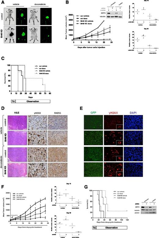
To demonstrate the in vivo relevance of our findings, we used 2 murine xenograft models of human MM. First, GFP-expressing NCI-H929 scramble or SIRT6-KD stably transduced MM cells were injected subcutaneously into the right flank of CB17-SCID mice. Fourteen tumor-bearing mice in each group were randomly assigned to receive either 3 mg/kg doxorubicin administered intraperitoneally (at days 1 and 5) or vehicle control. On day 30, 3 mice in each group were sacrificed and evaluated for tumor bulk by fluorescence imaging and histologic analysis. SIRT6-depletion significantly increased tumor size due to enhanced tumor growth. Importantly, doxorubicin-treatment induced greater anti-MM activity in SIRT6-depleted tumors than scramble-control tumors (Figure 6A-B); the mean relative tumor volume (RTV) in mice bearing SIRT6-KD cells treated with doxorubicin was 83% (P = .004) and 93.2% (P = .0003) lower than the RTV in treated mice harboring SIRT6-WT cells at days 16 and 30, respectively. Moreover, doxorubicin treatment significantly prolonged overall survival compared with vehicle-treated animals; the median overall survival of mice treated with doxorubicin was significantly longer in the absence of SIRT6 than in its presence (45 vs 29 days; P = .04), whereas SIRT6 silencing in vehicle-treated mice resulted in shorter survival than scramble control tumors (14 and 24 days, respectively) (Figure 6C). Finally, we assessed the effects of SIRT6-depletion in tumors from doxorubicin-treated mice. Consistent with our in vitro results, doxorubicin treatment increased γH2AX cellular foci and reduced Rad51 positive cells in SIRT6-KD harvested tumors compared with controls, indicating a higher degree of DNA damage due to impaired DDR (Figure 6D-E).
SIRT6 inhibition sensitizes MM cells to doxorubicin treatment in vivo. (A) Whole-body imaging of CB17-SCID mice bearing GFP-expressing NCI-H929 scramble (scr) or SIRT6-KD stably transduced cells treated with vehicle or doxorubicin. (B) Growth of NCI-H929 control and SIRT6-depleted xenografts in mice treated with vehicle or doxorubicin (doxo) (P = .011 and P = .013, respectively). Data are mean tumor volume ± SD. SIRT6 was measured by western blot in representative tumors harvested from mice on day 30. Mean RTV ± SEM (n = 4) for mice treated in panel A on days 16 and 30 is expressed compared with tumor volumes on day 1 (insert). (C) Kaplan-Meier analysis showing median survival times of mice bearing tumors with or without SIRT6, before and after treatment with vehicle or doxorubicin. (D) Immunohistochemical analysis for hematoxylin and eosin (H&E), γH2AX, and RAD51 focus formation in control and SIRT6-KD NCI-H929 xenografts harvested from mice treated with either vehicle or doxorubicin. Black arrows indicate γH2AX nuclear foci. (E) Tumor sections from treated and untreated mice were subjected to immunostaining with anti-GFP and anti-γH2AX. Nuclear staining was performed with DAPI. For panels, Original magnification ×20 (D-E) (×40 in insets). (F) Mean tumor growth assessments showed smaller tumor sizes in mice with SIRT6-KD xenografts than in other groups (left). RTV for mice treated on days 16 and 30 (right). (G) Kaplan-Meier survival plot showing median survival of mice bearing conditional SIRT6-KD xenografts with doxycycline diet and treated with vehicle or doxorubicin (left). Western blot of harvested tumors confirms SIRT6 silencing in doxycycline-fed mice, as well as increased γH2AX after treatment (right). Tx, therapy.
SIRT6 inhibition sensitizes MM cells to doxorubicin treatment in vivo. (A) Whole-body imaging of CB17-SCID mice bearing GFP-expressing NCI-H929 scramble (scr) or SIRT6-KD stably transduced cells treated with vehicle or doxorubicin. (B) Growth of NCI-H929 control and SIRT6-depleted xenografts in mice treated with vehicle or doxorubicin (doxo) (P = .011 and P = .013, respectively). Data are mean tumor volume ± SD. SIRT6 was measured by western blot in representative tumors harvested from mice on day 30. Mean RTV ± SEM (n = 4) for mice treated in panel A on days 16 and 30 is expressed compared with tumor volumes on day 1 (insert). (C) Kaplan-Meier analysis showing median survival times of mice bearing tumors with or without SIRT6, before and after treatment with vehicle or doxorubicin. (D) Immunohistochemical analysis for hematoxylin and eosin (H&E), γH2AX, and RAD51 focus formation in control and SIRT6-KD NCI-H929 xenografts harvested from mice treated with either vehicle or doxorubicin. Black arrows indicate γH2AX nuclear foci. (E) Tumor sections from treated and untreated mice were subjected to immunostaining with anti-GFP and anti-γH2AX. Nuclear staining was performed with DAPI. For panels, Original magnification ×20 (D-E) (×40 in insets). (F) Mean tumor growth assessments showed smaller tumor sizes in mice with SIRT6-KD xenografts than in other groups (left). RTV for mice treated on days 16 and 30 (right). (G) Kaplan-Meier survival plot showing median survival of mice bearing conditional SIRT6-KD xenografts with doxycycline diet and treated with vehicle or doxorubicin (left). Western blot of harvested tumors confirms SIRT6 silencing in doxycycline-fed mice, as well as increased γH2AX after treatment (right). Tx, therapy.
In a second in vivo model, we subcutaneously injected MM.1S cells expressing a doxycycline-inducible SIRT6-shRNA (or a control shRNA). Mice were fed with doxycycline-containing diets to induce either SIRT6 or scramble silencing and then treated as described above. As in the previous model, tumor cells from control mice continued to grow normally, whereas SIRT6-KD resulted in more rapid tumor growth. Treatment with doxorubicin inhibited tumor growth of both control and SIRT6-KD MM xenografts. However, the anticancer effect of doxorubicin was more pronounced in SIRT6-KD tumors (Figure 6F); the mean RTV of mice bearing SIRT6-KD MM cells and treated with doxorubicin was 70.4% (P = .017) and 87.3% (P = .007) lower than RTV of mice bearing tumors with high levels of SIRT6 treated with doxorubicin at days 16 and 30, respectively. Western blot analysis of tumors from doxorubicin-treated mice confirmed SIRT6 downregulation, as well as increased γH2AX. Finally, Kaplan-Meier analyses indicated that median survival after treatment with doxorubicin was significantly longer in mice bearing SIRT6-KD than in control tumors (50 days vs 23 days; P = .001) (Figure 6G).
Discussion
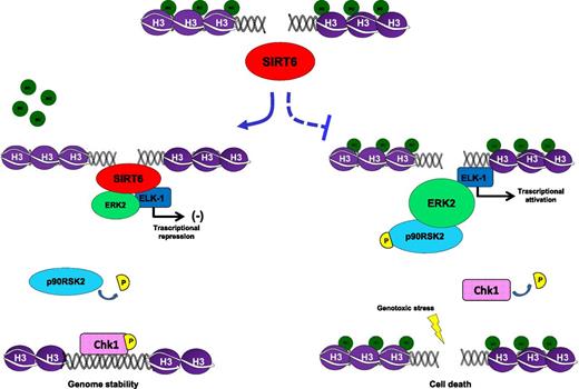
In this study, we identify a link between genomic instability of MM cells and SIRT6, which maintains DNA integrity in mammalian cells.13,19,36,38 By using in vitro and in vivo approaches as well as microarray data set analyses, we demonstrate that MM cells exhibit high levels of SIRT6, which blocks activity of its target genes ERK2, RSK2, and ELK1 (supplemental Figure 12) in response to ongoing DNA damage and genomic instability.2,5,40,41 Specifically, we observed that persistent DNA damage in MM triggers recruitment of SIRT6 to double-strand breaks, DDR, and downregulation of MEK/ERK-signaling–related genes. In contrast, SIRT6 depletion triggers H3K9 acetylation and ELK1-mediated transcriptional activity, activating multiple ERK-related genes including MAPK-activated ribosomal S6Kinase2, and blocks the G2 DNA damage checkpoint. As a result, SIRT6 inhibition or depletion both enhances MM cell proliferation and renders them more sensitive to DDAs. (Figure 7)
Proposed model. SIRT6 binds DNA damage sites, recruits and blocks MAPK signaling, including RSK2. As a result, Chk1 is phosphorylated at Ser317 and maintains genome integrity by repairing DNA injuries. In contrast, inhibiting SIRT6 activity results in hyperactivation of MEK/ERK signaling (by H3K9 acetylation and ELK1-mediated activity), which in turn results in RSK2-mediated Chk1 blockade (which is phosphorylated at Ser280) and ATR/CHK1 signaling impairment. In such a scenario of SIRT6 depletion, G2 DNA damage checkpoint impairment results in enhanced lethality of genotoxic stress.
Proposed model. SIRT6 binds DNA damage sites, recruits and blocks MAPK signaling, including RSK2. As a result, Chk1 is phosphorylated at Ser317 and maintains genome integrity by repairing DNA injuries. In contrast, inhibiting SIRT6 activity results in hyperactivation of MEK/ERK signaling (by H3K9 acetylation and ELK1-mediated activity), which in turn results in RSK2-mediated Chk1 blockade (which is phosphorylated at Ser280) and ATR/CHK1 signaling impairment. In such a scenario of SIRT6 depletion, G2 DNA damage checkpoint impairment results in enhanced lethality of genotoxic stress.
Mammalian cells possess signal transduction pathways that maintain genome integrity in response to DNA damage.42 When acquired mutations affect critical molecules such as p53, dampened DDR mechanisms are associated with tumorigenesis. Consequently, advanced tumors become highly reliant on functional DDR pathways for coping with genotoxic stress, and targeting DNA repair defects is emerging as a promising area for clinical investigation. Indeed, recent studies indicate that SIRTs preserve DNA integrity of mammalian cells, suggesting the potential utility of targeting SIRTs therapeutically.43-46 However, these deacetylases act on both oncogenic signaling and DDR mechanisms, and their effects in cancer may be cell-type dependent. Among the SIRT family members, SIRT6 is a chromatin-associated deacetylase, which plays a role in regulating genome stability.26,36,47 Consistent with this view, we here observed that SIRT6 is focally amplified in MM cells with genomic instability and that SIRT6 is inversely correlated with prognosis.
In solid tumors, variable functions of SIRT6 have been reported: it is a tumor suppressor in pancreas and colon cancers18 ; in contrast, SIRT6 overexpression in malignant prostate and breast tissues has been linked to poor survival.20,21 Here, we demonstrate that MM cells compensate for genomic instability utilizing mechanisms dependent on SIRT6, which interacts with distinct partners in MM than in other tumors. Specifically, MM cells adapt to ongoing DNA damage via the tripartite SIRT6/ERK2/ELK1 complex, with an associated increase in NHEJ and HR.41 Conversely, SIRT6 inhibition or depletion results in increased tumor growth, consistent with a tumor suppressor role. Earlier studies have reported a similar role of SIRT1 in modulating DDR or tumorigenesis in various tumor cells.48
Involvement of the MEK/ERK cascade in mediating DDR is evidenced by its activation in tumor cells exposed to agents targeting Chk1.49 Moreover, simultaneous disruption of Chk1 and MEK/ERK pathways induces selective toxicity in human leukemia and MM cells.50,51 However, the mechanisms by which MEK/ERK pathway disruption potentiates DNA damage triggered by Chk1 inhibitors is not yet delineated. Here, we observed that SIRT6 directly controls cell proliferation and genomic stability of MM cells by modulating ERK-signaling–related genes; conversely, SIRT6 depletion triggers activation of ERK/p90RSK signaling and Chk1 inhibition, which confers sensitization to genotoxic stress both in vitro and in vivo. These findings indicate that SIRT6 counteracts genetic instability in MM via Chk1/ERK cross-talk signaling and, conversely, that ERK/p90RSK pathway activation mediates increased sensitivity to DDAs in SIRT6-depleted cells.
We show that SIRT6 downregulates the expression of ERK-signaling–related genes by H3K9 deacetylation at their promoters and by suppressing activity of the ETS-domain transcription factor ELK1. ELK1 is a ternary complex factor of the ETS family, which plays a pivotal role in transducing extracellular signals into a transcriptional response via MAPK-signaling pathway.52 Previous studies have found that cross-talk between different members of SIRT and ETS families (SIRT7 and ELK4, respectively) is required for maintenance of the transformed phenotype of cancer cells.53 Here, we found that additional members of these families (SIRT6 and ELK1) interact, which modulates MEK/ERK signaling, regardless of RAS and p53 mutational status.9,54
In summary, we have elucidated a novel role for SIRT6 overexpression in MM, regulating both proliferation and response to ongoing DNA damage. Our studies provide the preclinical rationale for targeting SIRT6 to both enhance sensitivity of tumor cells to DDA treatment and improve patient outcome in MM.
The online version of this article contains a data supplement.
The publication costs of this article were defrayed in part by page charge payment. Therefore, and solely to indicate this fact, this article is hereby marked “advertisement” in accordance with 18 USC section 1734.
Acknowledgments
The authors thank Carlos Sebastian (Massachusetts General Hospital Cancer Center, Harvard Medical School, Boston, MA) for the tetracycline-inducible pTRIPz-Turbo-RFP vectors, Zhiyong Mao and Vera Gorbunova (Department of Biology, University of Rochester, Rochester, NY) for HR and GFP-NHEJ reporter plasmids, and Lay-Hong Ang (Beth Israel Deaconess Medical Center, Harvard Medical School, Boston, MA) for technical help in confocal images.
This work was supported by the National Institutes of Health, National Cancer Institute (grants R01 50947, P01 73878, and P50 100707) (K.C.A.), the International Multiple Myeloma Foundation (A.C.), and the American Italian Cancer Foundation (M.C.). K.C.A. is an American Cancer Society Clinical Research Professor.
Authorship
Contribution: M.C. and A.C. designed research, performed experiments, analyzed data, and wrote the manuscript; C.A. and M.F. performed animal work and analyzed data; S.A., M.K.B., and L.Z. analyzed genomic and microarray data; Y.-T.T., A.M., P.A., and H.O. contributed to design experiments; R.C. analyzed immunohistochemistry staining; F.M., A.B., M.G., and R.M.L. provided reagents, analytic tools, and input to studies; P.R. provided patient samples; and N.M., T.H., A.N., D.C., and K.C.A. critically evaluated and edited the manuscript.
Conflict-of-interest disclosure: The authors declare no competing financial interests.
Correspondence: Michele Cea, Department of Hematology and Oncology, IRCCS AOU San Martino-IST, 16132 Genoa, Italy; e-mail: michele.cea@unige.it; and Kenneth C. Anderson, Department of Medical Oncology, Dana-Farber Cancer Institute, M557, 450 Brookline Ave, Boston, MA 02115; e-mail: kenneth_anderson@dfci.harvard.edu.
References
Author notes
M.C. and A.C. contributed equally to this study.

![Figure 1. SIRT6 expression and prognostic relevance in MM. (A) Immunohistochemical analysis of 3 representative BM specimens derived from normal and MM patients (ND #1, 2, and 5 and MM #1, 4, and 8) show SIRT6 expression (positive cells are brown). Original magnification ×20 (×100 in insets). (B) Immunofluorescence showing subcellular distribution of SIRT6 in MM cell lines and patient MM cells (n = 5). (C) MM.1S and U266 cells were transfected with SIRT6 GFP-tagged plasmid. SIRT6 protein was visualized with an antibody directed against GFP, and cells were counterstained with DAPI to visualize nuclei. (D) Western blot analysis (1 of 3 representative blots) confirms the nuclear localization of SIRT6 in MM cell lines, but not in peripheral blood mononuclear cells (PBMCs) collected from healthy donors and MM patients. Cell lysates from cytoplasm and nuclear fractions were analyzed for SIRT6 expression. GAPDH and nucleolin were used as loading controls for the cytoplasmic and nuclear fractions, respectively. GAPDH, glyceraldehyde 3-phosphate dehydrogenase. (E) SIRT6 locus (human chromosome 19p13.3) copy-number heatmap for 45 (left side) MM cell lines (Mayo Clinic database) and 254 (right side) MM patients (Multiple Myeloma Research Consortium [MMRC]) (http://www.broad.mit.edu/mmgp). Color indicates degree of copy-number loss (blue) or gain (red). (F) SIRT6 expression in plasma cells from patients with MM (data set GSE2658). Increased SIRT6 expression is observed in high-risk compared with low-risk MM (unpaired t test; ****P < .0001). (G) Kaplan-Meier plots showing prognostic relevance of SIRT6 expression on overall survival of MM patients (based on GSE4581; deposited by Dr J. D. Shaughnessy Jr, University of Arkansas for Medical Sciences). The patient group with higher SIRT6 expression (blue line) had shorter overall survival than the patient cohort with lower SIRT6 expression (red line) (log-rank test).](https://ash.silverchair-cdn.com/ash/content_public/journal/blood/127/9/10.1182_blood-2015-06-649970/4/m_1138f1.jpeg?Expires=1769096709&Signature=c~QBkUT-T7mRkIvLaDatNczL8xzBO18RVdTpKeGjIWt4txPNF6yQVb67bF41EG-yhXjgN6QXRzY0iNF2B7x218Bl-OIV-ngFwGoxzBGRgnqzhVY3cxR7r92yHu6j5WDKpwCCz7X1WWFVQDxMrxKgLxCi9WGUPgJXMwp42TH0CrOmf5t8qWnP1m9AzW6zcbVGTMO1-wk79ff9zs1wIG1Dag9bk4X5jpcCcUylD0QxBIg2XboDbb9Cv5EqBqInUtRGiYHKOBYLcnnD4z4QfEhj5PFciSqqoJDYB4ULDDVfASilDAvitHQ5zr262bovQZAFICCOCmy6XFL1ekSydXWSWw__&Key-Pair-Id=APKAIE5G5CRDK6RD3PGA)
![Figure 2. SIRT6 reduces MM cell proliferation by opposing MAPK-signaling pathway. (A) SIRT6 silencing using the lentiviral vector pGIPz (after 48 hours with 2 μg/ml puromycin selection) in MM.1S cells. Shown is a western blot for SIRT6 in SIRT6-silenced cells (with 3 different shRNAs) and scramble control. After 4 days of infection, cells were harvested and whole-cell lysates were subjected to immunoblot analysis. The indicated MM cell lines were infected with lentivirus coexpressing GFP and SIRT6 shRNA (clone #911) or scramble control (top). Western blot analyses were performed 4 days after transfection. Data are representative of 2 independent experiments (middle). The percentage of GFP+ cells was measured over time by flow cytometry and normalized to the percentage of scramble GFP+ cells. Data represent the mean values ± SD and represent a minimum of triplicates (bottom). (B) Colony formation of MM cells transduced with lentivirus expressing GFP-tagged scramble or shSIRT6. Light and florescence microscopy is shown. Numbers of colonies after 2 weeks (average of 3 independent experiments; unpaired t test; *0.05 < P < .04) (top). (C) Cell proliferation of MM.1S cells transduced with SIRT6-specific shRNA (clone #911), SIRT6 wild-type (WT) or specific controls was determined by [3H]- thymidine uptake. The results presented are a mean ± SD of triplicate samples (Student t test; *0.45 < P < .03; **0.006 < P < .001; ***0.009 < P < .006; ****P < .0001). (D) Representative western blots showing activation of MAPK-signaling pathway in MM cells depleted of SIRT6 compared with control. GAPDH was used as loading control. One representative blot of two is shown. (E) Representative western blots showing SIRT6 regulating ERK2 activity. MM.1S cells stably expressing shRNA-targeting SIRT6 were transduced with pLOC carrying SIRT6 open reading frame sequence lacking the 3′ UTR region (which is targeted by the siRNA), permitting rescue of the target-specific shRNA phenotype. (F) Western blots showing specific role of SIRT6 in MM.1S cells. One representative blot of two is shown.](https://ash.silverchair-cdn.com/ash/content_public/journal/blood/127/9/10.1182_blood-2015-06-649970/4/m_1138f2.jpeg?Expires=1769096709&Signature=JdfpyAULB9TzBKp7Hv5XwlwDKUun6Xu93oGXcc66XZPEnDBOr9H3QQsRMrlWqFwj4bLKyOiPFTMOKN0g~UYrsC~tj4j1g3k7TMeMpWl~cX~0c9G0miUTTd21m9aWKMbrRaG50z0vaa3~682G-T5aOT-a8mhDRtMmsH~kn0pIKHh4CHzO5t3FQ0GdpWJQ-l62GA5uxauCvje9c~gbz1YEUb23btuOonT5gnJ1UQXi7CGeCkJ34E0-073zcoP6bAC81xyQRFxccUmL5~rgWgvlUYIUBcs8AtIuGkEZg3qTl5Z2041D1xI4TeMzlPmUB~wQRMI0t99aChXytOPHAy0~1Q__&Key-Pair-Id=APKAIE5G5CRDK6RD3PGA)
