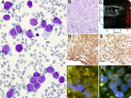
A 16-year-old girl was admitted for fever and severe malaise. Multiple ecchymoses and asymmetry of labia majora were observed. The hemogram showed a white blood cell count of 8.7 × 10⁹/L, hemoglobin 82 g/L, and platelets 33 × 109/L. A blood smear showed 3% blasts. Fibrinogen was 100 mg/dL. A bone marrow aspirate showed 83% immature noncohesive blast-like cells (panels A-B) negative for myeloperoxidase. Marked bleeding at the marrow and venipuncture sites occurred. Flow cytometry showed CD45– and CD56++ (panel C, red dots). No metaphases were obtained. A provisional diagnosis of acute undifferentiated leukemia was made, and she received a 3 + 7 idarubicin-cytarabine regimen. Twenty days later, a biopsy of the vulva was performed. Positive immunostains for actin (panel D) and desmin (panel E), as well as MyoD1, myogenin, vimentin, Alcian blue, and CD56 were observed. Fluorescent in situ hybridization analysis revealed t(2;13)(q35;q14) (panels Fi-ii). A final diagnosis of rhabdomyosarcoma (alveolar subtype) was established. The patient has received four cycles of a vincristine, actinomycin D, and cyclophosphamide schedule so far.
Leukemic phase and bone marrow metastases can be an uncommon presentation of rhabdomyosarcoma as well as other neural/neuroendocrine tumors, small cell carcinoma, and Ewing sarcoma. Those diagnoses should be pursued when atypical or inconsistent data are obtained after a bone marrow aspirate.
For additional images, visit the ASH IMAGE BANK, a reference and teaching tool that is continually updated with new atlas and case study images. For more information visit http://imagebank.hematology.org.


This feature is available to Subscribers Only
Sign In or Create an Account Close Modal