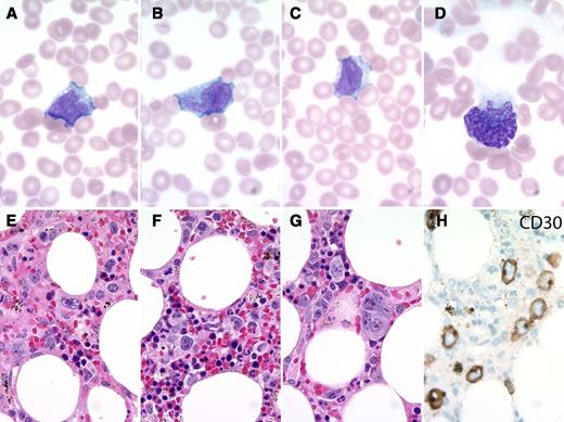
A 90-year-old woman presented with fevers and pancytopenia. The peripheral blood smear (see panels A-D) showed rare large atypical cells with convoluted nuclei and visible nucleoli. A few stripped multilobulated nuclei were present at the feathered edge. Bone marrow biopsy showed large atypical cells ranging from hallmark cells to larger pleomorphic cells (see panels E-H). These expressed CD3, CD4, CD7 (dim), CD30 (membranous and Golgi), TIA-1, granzyme B, and perforin. They were negative for CD8, CD20, PAX5, and ALK1. The patient entered hospice and died a week later.
Large pleomorphic lymphoma cells are rarely seen in the peripheral blood even with bone marrow involvement. The differential diagnosis in this case includes systemic involvement by a primary cutaneous CD30+ lymphoproliferative disorder, peripheral T-cell lymphoma NOS, ALK+ anaplastic large cell lymphoma (ALCL), and ALK− ALCL. The latter was favored given the clinical features and immunophenotype. A small-cell variant of ALK+ ALCL can sometimes present with leukemic involvement, but this morphology is not accepted for ALK− ALCL. Due to the lack of potential clinical impact in this patient, fluorescence in situ hybridization for DUSP22 and TP63 rearrangements was not pursued. However, the lymphoma cells do not have the typical morphology of DUSP22 rearranged cases, and immunohistochemistry for p63 was negative.
For additional images, visit the ASH IMAGE BANK, a reference and teaching tool that is continually updated with new atlas and case study images. For more information visit http://imagebank.hematology.org.


This feature is available to Subscribers Only
Sign In or Create an Account Close Modal