Key Points
Ibrutinib demonstrates durable responses and sustained single-agent activity in relapsed or refractory MCL at median 26.7-month follow-up.
Ibrutinib shows a favorable benefit-risk profile over time, with a manageable safety profile.
Abstract
Ibrutinib, an oral inhibitor of Bruton tyrosine kinase, is approved for patients with mantle cell lymphoma (MCL) who have received one prior therapy. We report the updated safety and efficacy results from the multicenter, open-label phase 2 registration trial of ibrutinib (median 26.7-month follow-up). Patients (N = 111) received oral ibrutinib 560 mg once daily, and those with stable disease or better could enter a long-term extension study. The primary end point was overall response rate (ORR). The median patient age was 68 years (range, 40-84), with a median of 3 prior therapies (range, 1-5). The median treatment duration was 8.3 months; 46% of patients were treated for >12 months, and 22% were treated for ≥2 years. The ORR was 67% (23% complete response), with a median duration of response of 17.5 months. The 24-month progression-free survival and overall survival rates were 31% (95% confidence interval [CI], 22.3-40.4) and 47% (95% CI, 37.1-56.9), respectively. The most common adverse events (AEs) in >30% of patients included diarrhea (54%), fatigue (50%), nausea (33%), and dyspnea (32%). The most frequent grade ≥3 infections included pneumonia (8%), urinary tract infection (4%), and cellulitis (3%). Grade ≥3 bleeding events in ≥2% of patients were hematuria (2%) and subdural hematoma (2%). Common all-grade hematologic AEs were thrombocytopenia (22%), neutropenia (19%), and anemia (18%). The prevalence of infection, diarrhea, and bleeding was highest for the first 6 months of therapy and less thereafter. With longer follow-up, ibrutinib continues to demonstrate durable responses and favorable safety in relapsed/refractory MCL. The trial is registered to www.ClinicalTrials.gov as #NCT01236391.
Introduction
Mantle cell lymphoma (MCL) is a distinct subtype of non-Hodgkin lymphoma (NHL), accounting for <10% of lymphoma cases.1,2 Patients are typically white (∼2:1), male (∼2.5:1), and elderly, with a median age of onset of 68 years, and usually present with extensive disease, including widespread lymphadenopathy and bone marrow involvement.3 MCL is not curable, and relapse is common,4 with the majority of patients dying from progressive disease (PD).5 In addition, the commonly used chemotherapeutic regimens can cause myelosuppression that can make treatment particularly difficult in elderly patients. Therefore, novel agents with improved efficacy and less toxicity are needed.
Bruton tyrosine kinase (BTK) is a critical signaling molecule in the B-cell receptor signaling pathway and functions via the activation of pathways mediating B-cell growth, adhesion, and survival.6 Ibrutinib is a first-in-class, once-daily, orally dosed, covalent inhibitor of BTK approved by the U.S. Food and Drug Administration and European Medicines Agency for the treatment of patients with MCL who have received at least one prior therapy. It is also approved for the treatment of patients with chronic lymphocytic leukemia (CLL) who have received at least one prior therapy and for CLL patients carrying the 17p deletion. Recently, ibrutinib has been approved by the Food and Drug Administration for the treatment of patients with Waldenström macroglobulinemia.7
Previous results of the international, multicenter, open-label, phase 2 trial that led to this accelerated approval of ibrutinib in relapsed/refractory (R/R) MCL demonstrated a high overall response rate (ORR) of 68% (21% complete response [CR]) and a median duration of response (DOR) of 17.5 months at a median follow-up of 15.3 months.8 In addition, ibrutinib showed a favorable safety profile relative to the current standard of care and a low rate of discontinuation resulting from adverse events (AEs). The present analysis is a longer-term follow-up (median, 26.7 months) of the safety and efficacy of ibrutinib in the registration trial and provides additional insights into the long-term use of ibrutinib in patients with R/R MCL.
Methods
Patient eligibility
Details of patient eligibility have been previously described.8 Key eligibility criteria included pathologically confirmed MCL (documented cyclin D1 overexpression or t[11;14]), with measurable disease (≥2 cm in the longest diameter and measurable in 2 perpendicular dimensions per computed tomography [CT]); Eastern Cooperative Oncology Group performance status (ECOG PS) ≤2; between 1 and 5 prior regimens for MCL; and documented failure to achieve at least partial response (PR) or disease progression after the most recent treatment.
Study design
Details of the study design have been previously published.8 In brief, this was an open-label, multicenter, phase 2 study (NCT01236391) conducted at 18 sites (9 US and 9 EU sites). Patients with R/R MCL were enrolled without randomization and classified as either bortezomib-treated (≥ 2 cycles) or bortezomib-naïve (<2 cycles or no prior bortezomib therapy). Patients were administered fixed continuous doses of oral ibrutinib 560 mg per day until disease progression or unacceptable toxicity. Ibrutinib dosing was withheld for any grade ≥3 nonhematologic toxicity. Treatment was restarted after complete resolution, or improvement to grade 1 or to baseline values within 14 days. If the AE was considered unrelated to the study drug, in the investigator’s opinion, ibrutinib could be restarted at the 560-mg dose (and then reduced to 420 mg if the AE recurred). If the AE was related to the drug, in the investigator’s opinion, ibrutinib treatment was restarted at a reduced dose of 420 mg per day. The study was conducted in accordance with Good Clinical Practice guidelines, as provided by the International Conference on Harmonization and principles of the Declaration of Helsinki. Institutional review boards approved the study protocol at the respective sites, and all patients provided written informed consent.
Efficacy and safety assessments
The primary end point was investigator-assessed ORR, defined as either a PR or a CR according to the Revised International Working Group Criteria for NHL.9 Tumor assessment was performed during screening using CT scans (repeated at cycles 3, 5, and 7, and then every 3 cycles until PD), positron emission tomography (PET) scans, and bone marrow biopsy. A PET scan was essential for confirmation of a CR. Subgroup analysis was performed based on baseline characteristics and risk factors.
The secondary end points included DOR, progression-free survival (PFS), overall survival (OS), and safety. Kaplan-Meier methods were used to estimate these time-to-event end points.
Safety assessments included the monitoring and recording of AEs and serious adverse events (SAEs), and measurements of protocol-specified laboratory parameters. Severity of AEs was graded according to the National Cancer Institute Common Terminology Criteria for Adverse Events, version 4.0. AEs of interest were evaluated over 6-month time intervals (1-6, 7-12, 13-18, 19-24, and >24 months). Prevalence of AEs was determined based on the number of patients with an AE occurring during a given time interval (either a new episode or an ongoing episode from the prior 6-month period continuing into the current interval). Bleeding events, including major bleeding, were also evaluated during these intervals. Major bleeding was defined as any hemorrhagic event of grade ≥3 or that resulted in intraocular bleeding causing loss of vision, the need for a transfusion of ≥2 units of red cells or an equivalent amount of whole blood, hospitalization, or prolongation of hospitalization.
Results
Patients
A total of 115 bortezomib-naïve and bortezomib-treated patients with R/R MCL were enrolled,8 111 of which received ≥1 dose of ibrutinib and were evaluable. Four patients did not receive ibrutinib treatment and were excluded from the analysis.
Patient baseline characteristics and disposition
Patient baseline characteristics are shown in supplemental Table 1. The median age was 68 years (range, 40-84), 77% were male, 49% had a high-risk Mantle Cell Lymphoma International Prognostic Index score, 89% had an ECOG PS of 0 to 1, and bone marrow and/or extranodal disease involvement was reported in 72% of the patients. More than half (55%) of the patients received ≥3 prior regimens, and 45% of the patients had refractory disease (defined as failure to achieve at least PR to the last therapy before study entry).
The median treatment duration was 8.3 months; 51 patients (46%) were treated for >1 year, and 22 (20%) were treated for ≥2 years. The primary reason for treatment discontinuation included PD (56%), AEs (11%), withdrawal of consent (5%), and physician’s decision (3%).
Efficacy
The investigator-assessed ORR for all patients was 67% (95% CI: 57.1%, 75.3%), with a CR rate of 23% (95% CI: 15.1%, 31.4%) (supplemental Figure 1). Forty-eight patients (43%) who had received prior bortezomib demonstrated an ORR of 65% (95% CI: 49.5%, 77.8%), and 27 (24%) patients who had received prior lenalidomide demonstrated an ORR of 59% (95% CI: 38.8%, 77.6%).
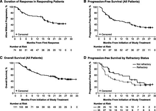
The median time to initial response for all patients was 1.9 months (range, 1.4-13.7), and the median time to CR was 5.5 months (range, 1.7-24.7). Consistent with previous analysis,8 response was durable with a median DOR of 17.5 months (95% CI: 14.9, not estimable [NE]; Figure 1A) and was independent of patient characteristics and risk factors. For patients with a best response of PR, the estimated median DOR was 14.9 months (95% CI: 6.6, 17.5), and for those with CR, the median DOR was not reached (95% CI: 20.3, NE). At a median follow-up of 26.7 months, the median PFS was 13 months (95% CI: 7.0, 17.5; Figure 1B), and the median OS was 22.5 months (95% CI: 13.7, NE; Figure 1C). The 24-month Kaplan-Meier estimates of PFS and OS were 31% (95% CI, 22.3, 40.4) and 47% (95% CI, 37.1, 56.9), respectively.
Kaplan-Meier analysis of time-to-event end points. (A) Duration of response in all responding patients. (B) Progression-free survival (all patients). (C) Overall survival (all patients). (D) Progression-free survival by refractory disease status.
Kaplan-Meier analysis of time-to-event end points. (A) Duration of response in all responding patients. (B) Progression-free survival (all patients). (C) Overall survival (all patients). (D) Progression-free survival by refractory disease status.
Subgroup analysis by baseline parameters and risk factors
There were no apparent differences in ORRs with respect to tumor volume; however, CR, as well as DOR, OS, and PFS, were generally higher for patients with less bulky tumors (Table 1). Median OS was not reached for nonrefractory patients (95% CI: 13.2, NE) compared with 13 months (95% CI: 6.3, NE) for refractory patients. In addition, median PFS was substantially prolonged for nonrefractory patients vs refractory patients (16.6 months [95% CI: 6.0, 24.4]) vs 6.6 months [95% CI: 3.5, 17.5]) (Table 1 and Figure 1D). Overall, the most prolonged DOR, PFS, and OS were observed in patients with lower disease burden and less refractory disease.
Efficacy outcomes by subgroup
| Efficacy outcome . | Tumors (<5 cm) (N = 68) . | Tumors (≥5 cm) (N = 43) . |
|---|---|---|
| ORR %, (95% CI) | 69% (56.7%, 79.8%) | 63% (46.7%, 77.0%) |
| CR %, (95% CI) | 27% (16.5%, 38.6%) | 16% (6.8%, 30.7%) |
| Median DOR (range), mo | NE (14.9, NE) | 16 (5.6, 22.6) |
| Median PFS (range), mo | 16.6 (8.3, 22.1) | 7.3 (5.2, 16.6) |
| Median OS (range), mo | NE (17.9, NE) | 15.6 (6.3, NE) |
| . | Bulky tumors (<10 cm) (N = 102) . | Bulky tumors (≥10 cm) (N = 9) . |
|---|---|---|
| ORR %, (95% CI) | 67% (56.6%, 75.7%) | 67% (29.9%, 92.5%) |
| CR %, (95% CI) | 24% (15.7%, 33.0%) | 11% (0.3%, 48.2%) |
| Median DOR (range), mo | 22.6 (14.9, NE) | 14.9 (3.0, 18.8) |
| Median PFS (range), mo | 13.9 (6.6, 17.5) | 7.4 (1.7, 19.2) |
| Median OS (range), mo | 22.8 (15.6, NE) | 13.7 (2.7, NE) |
| . | Prior treatments <2 (N = 22) . | Prior treatments ≥2 (N = 89) . |
|---|---|---|
| ORR %, (95% CI) | 82% (59.7%, 94.8%) | 63% (52.0%, 72.9%) |
| CR %, (95% CI) | 27% (10.7%, 50.2%) | 21% (13.4%, 31.3%) |
| Median DOR (range), mo | 16.5 (3.7, NE) | 17.5 (14.8, NE) |
| Median PFS (range), mo | 17.5 (4.9, NE) | 10.9 (6.0, 16.7) |
| Median OS (range), mo | 21.8 (10.0, NE) | 22.7 (13.0, NE) |
| . | Nonrefractory (N = 29) . | Refractory (N = 35) . |
|---|---|---|
| Median DOR (range), mo | 18.8 (9.2, NE) | 15.8 (3.4, NE) |
| Median PFS (range), mo | 16.6 (6.0, 24.4) | 6.6 (3.5, 17.5) |
| Median OS (range), mo | NE (13.2, NE) | 13.0 (6.3, NE) |
| . | Nonrefractory (N = 29) . | Refractory (N = 35) . |
|---|---|---|
| Median DOR (range), mo | 18.8 (9.2, NE) | 15.8 (3.4, NE) |
| Median PFS (range), mo | 16.6 (6.0, 24.4) | 6.6 (3.5, 17.5) |
| Median OS (range), mo | NE (13.2, NE) | 13.0 (6.3, NE) |
CI, confidence interval; CR, complete response; DOR, duration of response; NE, nonevaluable; ORR, overall response rate; OS, overall survival; PFS, progression-free survival.
Safety
Summary of treatment-emergent AEs.
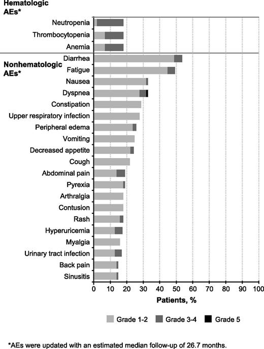
The most common AEs regardless of attribution in ≥30% of patients were diarrhea (54%), fatigue (50%), nausea (33%), and dyspnea (32%). Approximately two-thirds of the cases of dyspnea occurred in patients with a history of chronic obstructive pulmonary disease, heart failure, and chronic lung infection and/or anxiety, or they occurred concurrently with AEs that can affect respiratory function (eg, pneumonia or other respiratory infection, edema/fluid retention, malignant pleural effusion, grade 3 anemia). The most common grade ≥3 hematologic AEs were neutropenia (17%), thrombocytopenia (13%), and anemia (11%) (Figure 2). No hematologic event led to ibrutinib discontinuation; dose reduction occurred in 7 cases of neutropenia (6%). The most common hematologic and nonhematologic SAEs (>2%) are listed in Table 2.
Treatment-emergent adverse events (≥15% of patients) regardless of attribution.
Summary of serious adverse events (≥2% of patients) regardless of attribution
| . | Total (N = 111) . | ||
|---|---|---|---|
| SAE*, n (%) . | Any grade . | Grade 3-4 . | Grade 5 . |
| Disease progression† | 11 (10%) | 3 (3%) | 8 (7%) |
| Pneumonia | 8 (7%) | 7 (6%) | 1 (1%) |
| Atrial fibrillation | 7 (6%) | 6 (5%)‡ | 0 |
| Urinary tract infection | 4 (4%) | 3 (3%) | 0 |
| Febrile neutropenia | 3 (3%) | 3 (3%) | 0 |
| Abdominal pain | 3 (3%) | 3 (3%) | 0 |
| Acute renal failure | 3 (3%) | 2 (2%) | 1 (1%) |
| Subdural hematoma | 3 (3%) | 2 (2%) | 0 |
| Pyrexia | 3 (3%) | 1 (1%) | 0 |
| Confusional state | 3 (3%) | 1 (1%) | 0 |
SAEs were updated with an estimated median follow-up of 26.7 months.
Mantle cell lymphoma reported as a SAE by investigators.
One additional patient had a grade 3 atrial fibrillation that was not considered an SAE.
Eighteen patients (16%) had grade 5 AEs within 30 days of the last dose of ibrutinib. Eight of these fatal AEs were reported as MCL by investigators, and six were considered to be associated with disease progression (dyspnea, acute renal failure, malignant pleural effusion, ileus paralytic, Pneumocystis jirovecii pneumonia, and respiratory failure). The other 4 fatal AEs were pneumonia, sepsis, hypovolemic shock (3 weeks after discontinuation of ibrutinib resulting in PD and 12 days after starting R-hyper-CVAD), and cardiac arrest in a patient with a history of type 2 diabetes, hypertension, and heart failure. The autopsy of this patient revealed multiple pulmonary emboli and hypertensive heart disease.
Diarrhea.
Diarrhea was reported in 54% of patients during the study period, with the highest prevalence in the first 6 months. No SAE of diarrhea was reported after the first 6-month time interval (Table 3). Four cases of colitis were reported, all of which were associated with Clostridium difficile infection. Overall, the majority of diarrhea events were grade 1 (35%), and no patients discontinued therapy as a result of diarrhea. Among the 6 patients (5%) with grade 3 diarrhea (no grade 4 events), 4 patients had one dose interruption lasting 5 to 7 days, and the 3 patients with treatment-related diarrhea had their dose reduced to 420 mg once daily. Patients received antidiarrheal treatments (probiotics, Pepto-Bismol, loperamide, diphenoxylate/atropine), and no recurrence of grade 3 diarrhea was observed. The remaining 2 patients with grade 3 diarrhea stopped treatment because of unrelated concurrent PD and death.
Prevalence of select AEs by 6-month intervals
| Select AEs* n (%) . | 1-6 mo (n = 111) . | 7-12 mo (n = 72) . | 13-18 mo (n = 51) . | 19-24 mo (n = 41) . | >24 mo (n = 22) . |
|---|---|---|---|---|---|
| Any diarrhea | 49 (44%) | 21 (29%) | 15 (29%) | 8 (20%) | 6 (27%) |
| Grade 3† | 5 (5%) | 0 | 0 | 1 (2%) | 0 |
| SAE | 1 (1%) | 0 | 0 | 0 | 0 |
| Any infection | 76 (69%) | 43 (60%) | 30 (59%) | 22 (54%) | 9 (41%) |
| Grade ≥3 | 20 (18%) | 11 (15%) | 6 (12%) | 5 (12%) | 1 (5%) |
| SAE | 16 (14%) | 9 (13%) | 4 (8%) | 5 (12%) | 1 (5%) |
| Any bleeding | 46 (41%) | 17 (24%) | 17 (33%) | 14 (34%) | 5 (23%) |
| Major bleeding | 6 (5%) | 1 (1%) | 3 (6%) | 2 (5%) | 2 (9%) |
AE, adverse event
AEs were updated with an estimated median follow-up of 26.7 mo
No grade 4 or 5 diarrhea.
Infection.
All-grade infections occurred in 78% of patients during the study period, with grade ≥3 infections occurring in 28%. SAEs of infection occurred in 20% of patients. Both all-grade and grade ≥3 infections generally decreased over time (Table 3). Upper respiratory tract infection was the most commonly reported infectious AE (28%), followed by urinary tract infection (16%) and sinusitis (15%). Pneumonia was the most common grade ≥3 infection (8%). One case of reported pneumonia was associated with atypical infection (Pneumocystis jirovecii pneumonia). Other atypical infections included 1 cryptococcal infection and 1 case of histoplasmosis. No clinically significant decreases were observed in immunoglobulin levels over time (supplemental Figure 2).
Bleeding.
Overall, 50% of patients experienced bleeding events during the total study period, including bruising of any grade. Grade ≥3 bleeding events occurred in 6% of cases; no fatal bleeding events were reported. All-grade bleeding was highest in the first 6-month interval (41%; Table 3) and decreased thereafter; major bleeding occurred with similar prevalence rates across evaluated time intervals (Table 3). The most frequent bleeding event of any grade was contusion (18%), followed by epistaxis (11%) and petechiae (10%). No grade ≥3 bleeding events of contusion, bruising, epistaxis, or petechiae were reported. Grade ≥3 bleeding events of hematuria and subdural hematoma were each reported in 2% of patients (Table 4), indicating that the incidence of grade ≥3 bleeding events was low with continuous ibrutinib therapy. Subdural hematomas were reported in 4 patients (4%): grade 1 in one patient, grade 2 in one patient, and grade 3 in two patients. All cases were associated with falls, head trauma, or both, and all 4 patients had received either aspirin or warfarin within 2 days before or on the day of the event.8 Concomitant use of warfarin (or other vitamin K antagonist) was initially allowed in this study, but subsequently restricted as a precaution based on new study data. Overall, 61 patients in this study (55%) had received concomitant treatment with anticoagulants or antiplatelet agents during the study (supplemental Table 2). As expected, bleeding events were reported more frequently in patients receiving these agents (69% any grade, 8% grade 3-4) vs those not receiving these treatments (28% any grade, 4% grade 3-4). Among the 7 patients who experienced grade ≥3 bleeding events, 5 had been taking aspirin or nonsteroidal antiinflammatory drugs for 6 months to 2 years before the event. One patient experienced a grade 2 subdural hematoma after 576 days on ibrutinib and 89 days on warfarin. Both drugs were discontinued because of this event, and the patient died as a result of MCL progression 4 months after treatment discontinuation. No other patients on warfarin and no patients on heparin (including low-molecular-weight heparin) experienced grade ≥3 bleeding events. The only grade 4 bleeding event was splenic hematoma (occurring at the time of disease progression) in a patient with MCL-related splenomegaly and prestudy thrombocytopenia. Overall, treatment discontinuation for bleeding events occurred in 2 patients (both subdural hematomas), and no episodes of bleeding led to ibrutinib dose reduction.
Summary of bleeding events (≥2% of patients) regardless of attribution
| Bleeding event*, n (%) . | Any grade . | Grade ≥3 . |
|---|---|---|
| Any bleeding | 56 (50%) | 7 (6%)† |
| Contusion | 20 (18%) | 0 |
| Epistaxis | 12 (11%) | 0 |
| Petechiae | 11 (10%) | 0 |
| Hematuria | 7 (6%) | 2 (2%) |
| Ecchymosis | 6 (5%) | 0 |
| Increased tendency to bruise | 6 (5%) | 0 |
| Purpura | 4 (4%) | 0 |
| Subdural hematoma | 4 (4%) | 2 (2%) |
| Traumatic hematoma | 3 (3%) | 1 (1%) |
| Hematospermia | 2 (2%) | 0 |
AEs were updated with an estimated median follow-up of 26.7 months.
Other grade ≥3 bleeding events were grade 4 splenic and grade 3 lower gastrointestinal hemorrhage, both occurring in the context of disease progression.
Atrial fibrillation.
Atrial fibrillation (AF) was observed in 12 patients (11%), with grade 3 events in 7 (6%), and no grade 4 or 5 events. An SAE of AF was also reported in 7 patients (6%), and 1 case led to dose reduction. Among these 12 patients, 10 had cardiovascular disease that included AF (n = 1), and coronary artery disease (n = 4), hypertension (n = 8), hyperlipidemia (n = 6), and myocardial infarction (n = 1) were notable in the past medical history. The median time to first onset of AF was 140 days (range, 41-520) after starting ibrutinib, including one AF event that occurred 2 weeks after ibrutinib discontinuation. Seven patients had a single AF episode of short duration (2-12 days). AF was controlled with β-blockers and/or antiarrhythmics (amiodarone, dronedarone), and 1 patient received ablation therapy. Anticoagulants (warfarin and low-dose heparin) were administered in 6 of the 12 patients who experienced AF. No patient discontinued therapy as a result of AF, and 4 of the 7 patients with grade 3 AF (including the patient who received ablation therapy) continued into the extension study after 840 to 949 days of ibrutinib treatment.
Second primary malignancies.
Second primary malignancies (SPMs) were diagnosed in 5 patients (5%), which included metastatic adenocarcinoma of the bladder and metastatic neoplasm in the same patient (n = 1), cutaneous squamous cell carcinoma (n = 3), and basal cell carcinoma (n = 1).
Renal insufficiency/failure.
Based on laboratory measurements, increases in serum creatinine (above upper limit of normal) from baseline occurred in 39 patients (35%). No patient had a shift to postbaseline toxicity grade 3 or 4. Renal failure was reported as an AE in 8 patients (7%). Grade ≥3 renal failure was noted in 5 patients (5%), which included 4 patients with grade 3 renal failure and one patient with grade 5 renal failure. The fatal event occurred 13 days after ibrutinib discontinuation for PD and was caused by hydronephrosis as a result of obstructive lymphadenopathy. All 4 cases of grade 3 renal failure were confounded with preexisting hypertension and/or renal dysfunction and concurrent conditions such as dehydration and disease progression.
Tumor lysis syndrome.
One male patient with blastoid MCL and bulky disease >10 cm at baseline who achieved PR on ibrutinib treatment experienced an AE of tumor lysis syndrome (TLS) at the time of PD after approximately 17 months of therapy. TLS along with acute kidney failure occurred within 24 hours of the patient receiving high-dose methylprednisolone. Ibrutinib had been discontinued 4 days before the event, suggesting that the development of TLS was unlikely to be associated with ibrutinib therapy. Two additional patients were determined to have laboratory (chemical) TLS but did not fit the criteria for clinical TLS.10 Patient 1 had uric acid and phosphate levels of 11.2 mg/dL and 5.3 mg/dL, respectively, at the time of PD, and patient 2 had uric acid and phosphate levels of 8.4 mg/dL and 4.6 mg/dL, respectively, at cycle 2 of ibrutinib (per Cairo and Bishop classification [2004],10 criteria for laboratory TLS is uric acid >8 mg/dL and phosphate >4.5 mg/dL).
Discussion
Single-agent oral ibrutinib produced rapid and durable responses in our heavily treated patient population of R/R MCL at a median follow-up of 26.7 months. The ORR of 67% and the DOR of 17.5 months observed in our study are the highest reported for a single agent in R/R MCL. The median PFS of 13 months and OS of 22.5 months in this long-term follow-up analysis remain consistent with our prior report8 and further confirm the efficacy profile of ibrutinib in this setting. In addition, the ORR did not differ across patients with respect to the number of prior therapies or presence of refractory disease; however, less tumor bulk and nonrefractory disease were associated with longer PFS and OS.
Bortezomib and lenalidomide have been approved for the treatment of patients with R/R MCL and have shown response rates of 33% and 28%, respectively. The efficacy and safety of ibrutinib demonstrated in this longer-term follow-up compares favorably with that of the approved agents. In addition, the treatment discontinuation rate as a result of AEs in our study was low (11%) compared with the pivotal trials with bortezomib (26%) and lenalidomide (19%).11,12 The heavily pretreated R/R MCL population in our study comprised 43% of patients exposed to bortezomib and 24% of patients exposed to lenalidomide. The durable responses and clinical activity of oral ibrutinib observed suggest that ibrutinib is effective in patients previously treated with bortezomib and lenalidomide.
The safety profile of ibrutinib was manageable, with no unforeseen AEs observed with additional follow-up and few discontinuations and dose reductions as a result of AEs. Diarrhea remains the most common AE associated with ibrutinib therapy, but was primarily low grade and was treated, when necessary, with over-the-counter medications. No patient discontinued therapy because of unmanageable diarrhea.
High-grade bleeding events were infrequent and were similar throughout the treatment periods. Use of anticoagulant/antiplatelet agents in this study was high (55%), which is reflective of an elderly MCL population with underlying comorbid conditions. As expected, anticoagulant/antiplatelet use increased the risk of bleeding. However, most of the bleeding events were low grade, and only 2 patients discontinued therapy because of bleeding; both had received concomitant therapy with anticoagulants. It should be noted that anticoagulant and antiplatelet use is not contraindicated with ibrutinib therapy. However, an appropriate benefit-to-risk assessment should be undertaken when evaluating ibrutinib treatment in patients requiring these agents, which would be particularly important, for example, when evaluating a patient with AF. In our study, AF did not result in discontinuation of study therapy, and appropriate anti-arrhythmic management allowed for prolonged treatment with ibrutinib. Therefore, prudent assessment of the risk of bleeding and appropriate medical management can allow most patients with AF to continue ibrutinib therapy.
Infections are common in NHL because of the immunosuppression associated with chemotherapy along with the inherent properties of the disease, which lead to an immunocompromised state. In this study, the most common infections included those of the respiratory and urinary tracts, and the majority were addressed in an outpatient setting and were self-limiting; importantly, infections related to atypical organisms were infrequent, and SAEs of infection were relatively low. Notably, the incidence rates of infection, diarrhea, and bleeding in this trial are comparable with the rates observed in the RESONATE study that compared the safety and efficacy of single-agent ibrutinib with ofatumumab in R/R CLL/SLL.13 No substantial differences in rates of grade ≥3 infection, bleeding, or diarrhea were observed between the 2 arms of the RESONATE trial, suggesting that ibrutinib is not associated with increased frequency of these AEs vs ofatumumab.
Grade 3-4 hematologic events were experienced in <20% of patients, and hematopoietic growth factors and transfusions were used in a minority of patients. All cases of renal failure reported confounding risk factors and/or had alternative etiology. Less than 10% of an oral dose of ibrutinib is recovered in the urine as metabolites,7 indicating that renal excretion is not a major route of ibrutinib clearance. Furthermore, no significant differences in the incidence of renal insufficiency was observed between the 2 arms in the RESONATE study,13 indicating that ibrutinib was not associated with increased renal toxicity vs ofatumumab.
SPMs were reported in 5 patients (5%). Similar to previous reports in CLL, skin appears to be the most prevalent site.13 SPMs are common in MCL, and a study of 156 patients reported a 5-year cumulative incidence rate of 11%, which could be attributed to prior therapies, genetic predisposition, or increasing age.14 Only 2 patients experienced laboratory TLS, and no clear cases of clinical TLS were observed, suggesting that this syndrome is not a prominent feature of the ibrutinib toxicity profile.
In summary, we have presented extended (26.7-month follow-up) data, which continue to demonstrate a favorable benefit-risk profile of ibrutinib in R/R MCL patients. Approximately one-third of patients remain progression-free at 24 months.
Presented in part at the 56th American Society of Hematology Meeting and Exposition, San Francisco, CA, December 6-9, 2014.
The online version of this article contains a data supplement.
The publication costs of this article were defrayed in part by page charge payment. Therefore, and solely to indicate this fact, this article is hereby marked “advertisement” in accordance with 18 USC section 1734.
Acknowledgments
The authors thank the patients who participated in the study and their supportive families, the investigators and clinical research staff from the study centers, and Swati Ghatpande for medical writing assistance.
This study was sponsored by Pharmacyclics LLC.
Authorship
Contribution: M.L.W., S.R., and D.M.B. contributed to the conception and design of the manuscript; all authors and Pharmacyclics, Inc. provided study materials or patients; M.L.W., S.R., D.M.B., M.C., and D.L. contributed to collection and assembly of data, and data analysis and interpretation; D.M.B., M.L.W., S.R., M.C., and D.L. analyzed the data, and all authors had access to primary clinical trial data; and all authors contributed to review and revisions to manuscript content and final approval of manuscript.
Conflict-of-interest disclosure: D.M.B., D.L., L.B., and M.C. are employees of Pharmacyclics and have stock ownership in Pharmacyclics. M.L.W., S.R., and M.D. have served as consultants for, and received honoraria from, Pharmacyclics/Janssen. A.G. has served as a consultant for, and received research funding and honoraria from, Pharmacyclics/Janssen. J.C.B. and M.D. have served as consultants for, and received research funding from, Pharmacyclics. P.J. has served as a consultant for Pharmacyclics and received research funding and honoraria from Janssen-Cilag. W.W.J. has served as a consultant for, and received research funding from, Amgen and Novartis. P.M. has received honoraria from Janssen. R.A. has received honoraria from Gilead and Celgene. S.S. has received research funding and honoraria from Pharmacyclics/Janssen. B.S.K. and K.A.B. have received research funding from Pharmacyclics/Janssen. R.H.A. has received research funding from Pharmacyclics/Janssen, Seattle Genetics, Genentech, Celgene, and Takeda Pharmaceuticals.
Correspondence: Michael L. Wang, The University of Texas MD Anderson Cancer Center, Department of Lymphoma/Myeloma, 1515 Holcombe Blvd, Houston, TX 77030; e-mail: miwang@mdanderson.org.


