Key Points
When contact activation is inhibited, Factor XI:C levels only correlate with thrombin generation if platelets are present.
Thrombin generation measured in platelet-rich plasma with contact activation inhibition identifies bleeding phenotype in FXI deficiency.
Abstract
Individuals with Factor XI (FXI) deficiency have a variable bleeding tendency that does not correlate with FXI:C levels or genotype. Comparing a range of sample conditions, we tested whether the thrombin generation assay (TGA) could discriminate between control subjects (n = 50) and FXI-deficient individuals (n = 97), and between those with bleeding tendency (n = 50) and without (n = 24). The comparison used platelet-rich plasma (PRP) and platelet-poor plasma (PPP), either with or without corn trypsin inhibitor (CTI) to prevent contact activation, over a range of tissue factor (TF) concentrations. When contact activation was inhibited and platelets were absent, FXI:C levels did not correlate with thrombin generation parameters, and control and FXI-deficient individuals were not distinguished. In all other sample types, the best discrimination was obtained using TF 0.5 pM and assay measures: endogenous thrombin potential (ETP) and peak height. We showed that although a number of conditions could distinguish differences between the groups tested, TGA measured in PRP with CTI best differentiated between bleeders and nonbleeders. These measures provided high sensitivity and specificity (peak height receiver operating characteristic [ROC] area under the curve [AUC] = 0.9362; P < .0001) (ETP ROC AUC = 0.9362; P < .0001). We conclude that by using sample conditions directed to test specific pathways of FXI activation, the TGA can identify bleeding phenotype in FXI deficiency.
Introduction
Spontaneous bleeding is rare in individuals with Factor XI (FXI) deficiency, with most bleeding episodes occurring in the setting of surgery or trauma.1,2 However, the bleeding tendency is variable: some patients with major FXI deficiency (FXI:C ≤15 IU/dL) do not exhibit excessive bleeding, whereas others with partial deficiency (FXI:C 16-60 IU/dL) report significant hemorrhagic symptoms.3,4 Furthermore, no clear correlation exists between clinical bleeding risk and FXI:C level, FXI antigen level, activated partial thromboplastin time (APTT), or genotype.1,2,4,5 In the absence of an effective test to predict bleeding risk, FXI-deficient individuals are at risk of receiving unnecessary FXI replacement (carrying significant risks of thrombosis6,7 or transfusion-related complications) or of being undertreated (with risks of hemorrhage).
FXI was originally described in the waterfall model of coagulation as part of the contact activation pathway.8 The observation that individuals with deficiencies of other contact pathway proteins (FXII, prekallikrein, and high-molecular-weight kininogen) did not exhibit bleeding symptoms, whereas some patients with FXI deficiency did, led to 2 conclusions: (1) the contact pathway may not be essential for normal hemostasis, and (2) in vivo FXI may be activated via a route that did not require FXIIa.3,5 It was subsequently shown that thrombin generation could be amplified by a positive feedback loop that involved the activation of FXI by thrombin (independent of FXIIa), leading to a revised model of coagulation.9-12 The thrombin generation assay (TGA) has been used to research the role of FXI in hemostasis,12-14 and reduced thrombin generation has been demonstrated in some FXI-deficient individuals.15,16 However, conflicting conclusions have been reached about the ability of TGA to differentiate between FXI-deficient individuals with a history of bleeding (“bleeders”) and those without (“nonbleeders”).15-18 Given the different routes by which FXI can be activated, it is plausible that the sample conditions tested (ie, with or without platelets and with or without in vitro contact activation) will greatly affect which coagulation pathways are measured and therefore may significantly alter the ability of the test parameters to correlate with clinical bleeding.
We studied clinical samples from a large number of FXI-deficient patients (n = 97) with a wide range of FXI:C levels. Here we demonstrate that differing sample conditions affect the ability of FXI:C levels to influence thrombin generation and the ability of TGA to determine bleeding tendency. We also show that there is a clinically useful correlation between TGA parameters and bleeding phenotype when platelets are present and contact activation inhibited. We propose that by using these conditions TGA can identify FXI-deficient individuals with increased bleeding tendency.
Methods
Patients and controls
Ninety-seven adults were recruited from Manchester Royal Infirmary, 19 with major FXI deficiency (FXI:C ≤15 IU/dL) and 78 with partial FXI deficiency (FXI:C 16-60 IU/dL). The control group comprised healthy adults with no personal or family history of thrombosis or bleeding disorders and no relevant medications (n = 50). Regional research ethical committee approval was obtained (REC #11/NW/0612) and all subjects gave written informed consent.
Bleeding history
FXI-deficient individuals were divided into bleeders and nonbleeders based on their experience of tonsillectomy and/or dental extraction before diagnosis of FXI deficiency. High rates of bleeding have been reported in FXI deficiency in association with these procedures.2,4,19 Bleeders (n = 24) were defined as those requiring blood product transfusion or return to theater/dentist for resuturing or packing. Nonbleeders (n = 50) were those who had not experienced excessive bleeding. Individuals who had not undergone either of these procedures by the time of diagnosis were termed “indeterminate” (n = 23) and were not included in the main analysis.
Blood sample collection and plasma preparation
Blood samples were taken into S-Monovette tubes (Sarstedt, Leicester, United Kingdom) containing 0.106 M trisodium citrate (1:9, V:V) alone or in combination with corn trypsin inhibitor (CTI) (Haematologic Technologies Inc., Essex Junction, VT) (final concentration 20 µg/mL whole blood). Platelet-rich plasma (PRP) was prepared by centrifugation at 180g for 10 minutes at room temperature, adjusted to 150 × 109/L using autologous platelet-poor plasma (PPP). PPP was collected from the upper half of the plasma supernatant after double centrifugation at 3000g for 15 minutes at room temperature. PPP samples were frozen at −80°C and measured in batches.
Calibrated automated thrombin generation assays
Thrombin generation was measured using the calibrated automated thrombinography (CAT) method,20 with tissue factor (TF) of 5 pM, 1 pM, and 0.5 pM. TF concentration (Innovin, Dade Behring, Marburg, Germany) was determined using the Actichrome TF activity assay (American Diagnostica Inc., Greenwich, CT) and diluted in working buffer (20 mM HEPES, 140 mM NaCl, 5 mg/mL bovine serum albumin, pH 7.35 [Severn Biotech, Kidderminster, UK]). Synthetic phospholipids: phosphatidylcholine, phosphatidylethanolamine and phosphatidylserine (Avanti Polar Lipids, Alabaster, AL) were prepared using an extrusion method.21 Final phospholipid concentration was 4 µM (20 mol% phosphatidylserine, 20 mol% phosphatidylethanolamine, 60 mol% phosphatidylcholine).
TF/phospholipids or TF alone were mixed with PPP and PRP samples, respectively, in a 96-well plate (Greiner Bio-One, Stonehouse, UK) and heated to 37°C for 10 minutes. A starting reagent (2.5 mM fluorogenic substrate [Z-Gly-Gly-Arg-AMC; Bachem, Bubendorf, Switzerland], 0.1 M CaCl2 in 20 mM HEPES, bovine serum albumin 60 mg/mL, pH 7.35 [Severn Biotech]) was automatically dispensed into each well. Thrombin generation was measured using a Fluoroscan Ascent fluorometer (Thermolab Systems OY, Helsinki, Finland) and calculated using Thrombinoscope version 3.0.0.29 (Thrombinoscope, Maastricht, The Netherlands). Intra- and interassay coefficient of variation was <10%.
To confirm that contact activation was abolished using 20 µg/mL of CTI (whole-blood concentration), thrombin generation was compared between PPP samples with or without CTI from 5 controls and 5 FXI-deficient individuals in the absence of TF. Thrombin generation was detected in all controls and 3 FXI-deficient samples without CTI, but was not seen in samples containing CTI. In addition, 3 control individuals were tested using PRP samples. Thrombin generation was observed when CTI was absent, but not in the presence of CTI.
FXI dependence in the TGA measured at TF 0.5 pM was demonstrated using PPP and PRP samples with and without CTI from 3 normal individuals. Samples were incubated with or without a neutralizing anti-human FXI antibody (Haematologic Technologies Inc., Essex Junction, VT) at final plasma concentration of 100 µg/mL for 30 minutes at 37°C. This concentration had previously been demonstrated to inhibit FXI activity to FXI:C levels <1 IU/dL in all 3 individuals using identical conditions. The presence of anti-human FXI antibody reduced ETP and peak height measurements in all sample types, confirming FXI dependence (supplemental Figure 1 and supplemental Table 1, available on the Blood Web site).
Laboratory analysis and Factor XI assays
Prothrombin time (PT), activated partial thromboplastin time (APTT), and fibrinogen measurements were performed using the STA-R Evolution anaylzer using STA-Néoplastine R, STA-Cephascreen, STA-CaCl2, and STA-Fibrinogen 5 reagents (Diagnostica Stago,Theale, United Kingdom [UK]). Plasma FXI activity (FXI:C) was measured using a 1-stage clotting assay on a CS2000i instrument (Sysmex, Milton Keynes, UK) using Dade Actin FS APTT reagent and FXI-deficient plasma (Siemens, Marburg, Germany). Platelet counts were measured on a Sysmex XE-5000 analyzer using Sysmex reagents (Sysmex, Milton Keynes, UK).
Statistical analysis
GraphPad Prism v6 (GraphPad, San Diego, CA) was used to determine data distribution and perform significance testing. TGA comparisons between multiple groups used analysis of variance (ANOVA) testing with the Bonferroni multiple comparison test or the Kruskal-Wallis test with Dunn’s multiple comparison test. Correlation tests used Pearson’s or Spearman’s correlation coefficient. Direct comparison between PRP and PPP samples used the paired Student t test or the Wilcoxon matched-pairs signed-rank test. Receiver operator characteristic (ROC) curve analysis assessed the ability of assays to differentiate between bleeders and nonbleeders. A P value <.05 was considered significant.
Results
In the absence of platelets, FXI:C levels only correlate with thrombin generation when the contact activation pathway is not inhibited
The ability of TGA to differentiate between normal subjects (n = 50) and FXI-deficient individuals (n = 97) was investigated using PPP samples to exclude any contribution of platelets to thrombin generation. The influence of FXI:C levels on thrombin generation was assessed using conditions that either permitted in vitro contact activation (samples without CTI) or prevented contact activation (samples with CTI) using TF triggers (5 pM, 1 pM, and 0.5 pM). For analysis, FXI-deficient patients were divided into 3 groups: partial deficiency (FXI:C 16-60 IU/dL) (n = 78), major deficiency 1 (FXI:C 3-15 IU/dL) (n = 10), and major deficiency 2 (FXI:C ≤2 IU/dL) (n = 9).
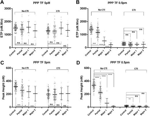
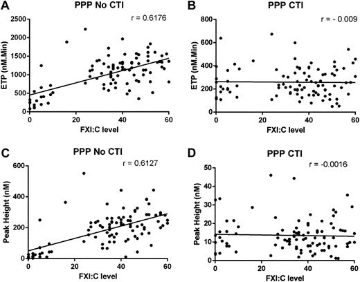
When in vitro contact activation was permitted and TF was high (5 pM), no difference in ETP was observed between controls and FXI-deficient groups. However, at low TF concentrations, a statistically significant difference in all TGA parameters was seen between controls and each of the 3 FXI-deficient groups. Best discrimination was seen for ETP and peak height values at TF 0.5 pM (Figure 1 and supplemental Figures 2 and 3). Using these conditions, all TGA parameters correlated significantly with FXI:C levels with the strongest correlation observed with ETP and peak height measurements (ETP: r = 0.6176, P < .0001; peak height: r = 0.6127, P < .0001; time to peak: r = −0.3812, P = .0001; and lag time: r = −0.2965, P = .0032) (Figure 2). In contrast, when in vitro contact activation was inhibited, TGA parameters did not discriminate effectively between controls and FXI-deficient groups (Figure 1 and supplemental Figures 2 and 3) and no significant correlation was demonstrated between TGA results and FXI:C levels (Figure 2). In the absence of platelets, FXI:C levels significantly influenced thrombin generation only when in vitro contact activation pathways were intact.
Comparison of ETP and peak height measured in PPP samples in control subjects and FXI-deficient individuals. Discrimination between 50 control subjects and FXI-deficient patients divided into 3 groups: partial deficiency (FXI:C 16-60 IU/dL, n = 78), major deficiency 1 (Major 1) (FXI:C 3-15 IU/dL, n = 10), and major deficiency 2 (Major 2) (FXI:C ≤2 IU/dL, n = 9) using the TG assay triggered at TF 5 pM (A,C) and TF 0.5 pM (B,D). ETP (A-B) and peak height measurements (C-D) are compared in PPP samples with or without CTI as indicated. *P < .5, **P < .01, ***P < .001, ****P < .0001; ns, not significant.
Comparison of ETP and peak height measured in PPP samples in control subjects and FXI-deficient individuals. Discrimination between 50 control subjects and FXI-deficient patients divided into 3 groups: partial deficiency (FXI:C 16-60 IU/dL, n = 78), major deficiency 1 (Major 1) (FXI:C 3-15 IU/dL, n = 10), and major deficiency 2 (Major 2) (FXI:C ≤2 IU/dL, n = 9) using the TG assay triggered at TF 5 pM (A,C) and TF 0.5 pM (B,D). ETP (A-B) and peak height measurements (C-D) are compared in PPP samples with or without CTI as indicated. *P < .5, **P < .01, ***P < .001, ****P < .0001; ns, not significant.
Correlation plots between FXI:C levels and ETP and peak height parameters measured in PPP samples. Correlation plots demonstrating the relationship between FXI:C levels and ETP measurements (A-B) and between FXI:C levels and peak height measurements (C-D) in PPP samples without CTI (A,C) and with CTI (B,D). Thrombin generation was measured with a TF trigger of 0.5 pM in PPP samples from patients with FXI deficiency (n = 97).
Correlation plots between FXI:C levels and ETP and peak height parameters measured in PPP samples. Correlation plots demonstrating the relationship between FXI:C levels and ETP measurements (A-B) and between FXI:C levels and peak height measurements (C-D) in PPP samples without CTI (A,C) and with CTI (B,D). Thrombin generation was measured with a TF trigger of 0.5 pM in PPP samples from patients with FXI deficiency (n = 97).
When contact activation is inhibited, correlation between FXI:C levels and thrombin generation is only demonstrated in the presence of platelets
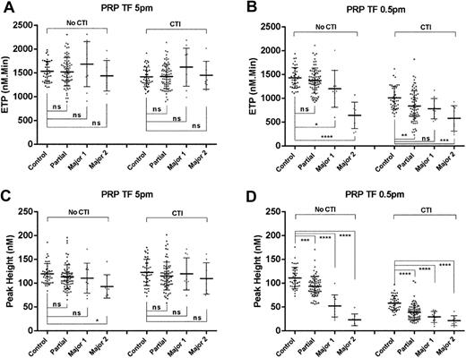
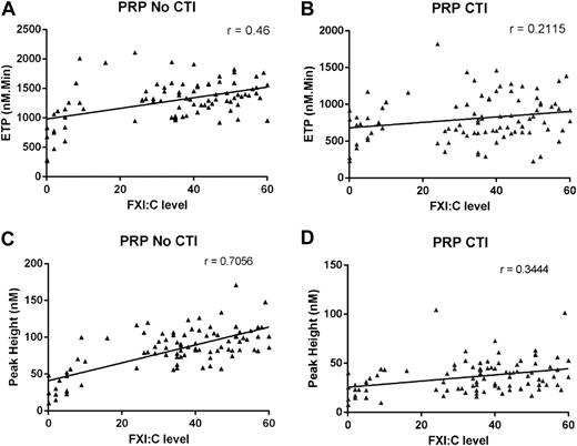
The experiments described in the previous section were performed using PRP samples. When contact activation was permitted (samples without CTI), the TGA results closely resembled those obtained using PPP: at high TF concentrations thrombin generation did not differentiate clearly between controls and FXI-deficient groups, whereas at low TF (0.5 pM) a significant difference in TGA parameters was again seen between control and FXI-deficient samples, with the best discrimination between groups determined using peak height measurements (Figure 3 and supplemental Figures 4 and 5). Using these conditions, all TGA parameters correlated with FXI:C levels (peak height r = 0.7056, ETP r = 0.46, time to peak r = −0.4972, and lag time r = −0.5644; all P < .0001) (Figure 4). In contrast, when in vitro contact activation was inhibited, the results differed markedly from those tested in PPP. The presence of platelets now allowed peak height and ETP measurements to differentiate between controls and each of the FXI-deficient groups. The addition of CTI in PRP samples resulted in a less strong correlation between FXI:C levels and TGA parameters (similar to the reduction seen with CTI presence in PPP samples). However, in PRP with CTI samples, a significant correlation was retained (peak height r = 0.3444, P = .0009; ETP r = 0.2115, P = .045) (Figure 4), although time to peak and lag time did not correlate with FXI:C levels. Using paired PPP and PRP samples in the presence of CTI, we showed that the presence of platelets significantly increased ETP and peak height results within the control group and each FXI-deficient group (all P < .008) (supplemental Figure 6). Thus in the absence of FXIIa, FXI:C levels can influence thrombin generation when platelets are present.
Comparison of ETP and peak height measured in PRP samples in control subjects and FXI-deficient individuals. Discrimination between 41 control subjects and FXI-deficient patients divided into 3 groups: partial deficiency (FXI:C 16-60 IU/dL, n = 72), major deficiency 1 (Major 1) (FXI:C 3-15 IU/dL, n = 10), and major deficiency 2 (Major 2) (FXI:C ≤2 IU/dL, n = 8) using the TG assay triggered at TF 5 pM (A,C) and TF 0.5 pM (B,D). ETP (A-B) and peak height measurements (C-D) are compared in PRP samples with or without CTI. *P < .5, **P < .01, ***P < .001, ****P < .0001; ns, not significant.
Comparison of ETP and peak height measured in PRP samples in control subjects and FXI-deficient individuals. Discrimination between 41 control subjects and FXI-deficient patients divided into 3 groups: partial deficiency (FXI:C 16-60 IU/dL, n = 72), major deficiency 1 (Major 1) (FXI:C 3-15 IU/dL, n = 10), and major deficiency 2 (Major 2) (FXI:C ≤2 IU/dL, n = 8) using the TG assay triggered at TF 5 pM (A,C) and TF 0.5 pM (B,D). ETP (A-B) and peak height measurements (C-D) are compared in PRP samples with or without CTI. *P < .5, **P < .01, ***P < .001, ****P < .0001; ns, not significant.
Correlation plots between FXI:C levels and ETP and peak height parameters measured in PRP samples. Correlation plots demonstrating the relationship between FXI:C levels and ETP measurements (A-B) and between FXI:C levels and peak height measurements (C-D) using samples without CTI (A,C) or with CTI (B,D). Thrombin generation was measured with a TF trigger of 0.5 pM in PRP samples from patients with FXI deficiency (n = 90).
Correlation plots between FXI:C levels and ETP and peak height parameters measured in PRP samples. Correlation plots demonstrating the relationship between FXI:C levels and ETP measurements (A-B) and between FXI:C levels and peak height measurements (C-D) using samples without CTI (A,C) or with CTI (B,D). Thrombin generation was measured with a TF trigger of 0.5 pM in PRP samples from patients with FXI deficiency (n = 90).
Standard measures of coagulation are unable to identify bleeding phenotype in FXI deficiency
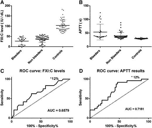
FXI-deficient subjects were divided into bleeder (n = 24) and nonbleeder (n = 50) groups (see Methods), and standard measures of coagulation were compared between both groups and with control results (n = 50) (Table 1).
Comparison of standard coagulation measurements between controls and FXI-deficient patients divided into nonbleeder and bleeder groups
| Coagulation measures (normal range) . | Controls (n = 50) . | Nonbleeders (n = 50) . | Bleeders (n = 24) . | P nonbleeder vs control . | P bleeder vs control . | P bleeder vs nonbleeder . |
|---|---|---|---|---|---|---|
| PT (12.5-15.3 s) | 13 ± 0.68 | 13.44 ± 0.76 | 13.55 ± 0.76 | ** | ** | ns |
| Fibrinogen (1.82-4.5s) | 2.94 ± 0.51 | 3.42 ± 0.69 | 3.35 ± 0.99 | ** | ns | ns |
| Platelet count (150-400 × 109/L) | 251 ± 47 | 262 ± 69 | 262 ± 62 | ns | ns | ns |
| APTT (24.6-34.9 s) | 29.79 ± 1.8 | 39.4 ± 12.6 | 53.65 ± 29 | **** | **** | ns |
| FXI:C level (60-140 IU/dL) | 104 ± 27.9 | 36.5 ± 16 | 27 ± 18.1 | **** | **** | ns |
Comparison of results (mean ± SD) between groups was performed using ANOVA test with the Bonferroni multiple comparison test or the Kruskal-Wallis test with Dunn’s multiple comparison test as appropriate to data distribution (**P < .01, ****P < .0001). Note that different mean levels are shown for APTT and FXI:C levels between bleeder and nonbleeder groups. However, as a result of substantial variability in these measurements (indicated by standard deviation), no significant difference was detected by ANOVA analysis, and significant overlap was observed using frequency plots of the measurements.
ns, not significant.
A statistically significant difference was observed in PT results between control subjects and both FXI-deficient groups, and for fibrinogen levels between controls and nonbleeders. However, the differences were small and PT and fibrinogen values for all groups lay within the reference range. No significant difference in PT, fibrinogen, and platelet count results was seen between the bleeder and nonbleeder groups.
For both APTT results and FXI:C levels, a highly significant difference was observed between control and the FXI-deficient groups; however, no significant difference was seen between bleeders and nonbleeders (Table 1 and Figure 5). ROC curve confirmed that APTT (AUC = 0.6579) and FXI:C level assays (AUC = 0.781) were unable to differentiate between bleeders and nonbleeders with adequate sensitivity and specificity for clinical use (Figure 5).
Comparison of FXI:C levels and APTT results among controls, bleeders, and nonbleeders in FXI deficiency. Comparison of FXI:C levels (A) and APTT results (B) in control individuals (n = 50), nonbleeder FXI-deficient (n = 50), and bleeder FXI-deficient (n = 24) groups. Line and error bars represent mean ± standard deviation. ROC curve analysis to test the ability of FXI:C (C) and APTT (D) assays to identify bleeders from nonbleeders in FXI deficiency. *Specificity of test at 100% sensitivity.
Comparison of FXI:C levels and APTT results among controls, bleeders, and nonbleeders in FXI deficiency. Comparison of FXI:C levels (A) and APTT results (B) in control individuals (n = 50), nonbleeder FXI-deficient (n = 50), and bleeder FXI-deficient (n = 24) groups. Line and error bars represent mean ± standard deviation. ROC curve analysis to test the ability of FXI:C (C) and APTT (D) assays to identify bleeders from nonbleeders in FXI deficiency. *Specificity of test at 100% sensitivity.
Thrombin generation in the presence of platelets identifies FXI-deficient patients with bleeding tendency when contact activation is prevented
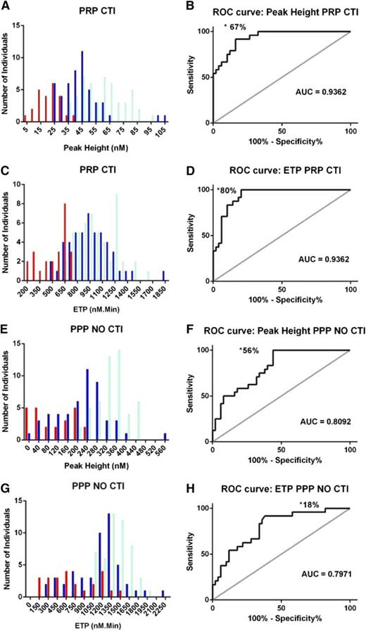
TGA parameters measured in different sample conditions (TF 0.5 pM) were compared for their ability to identify past bleeding tendency among FXI-deficient individuals. The parameters of lag time and time to peak did not effectively distinguish between the bleeder and nonbleeder groups under any conditions (supplemental Table 2). In contrast, peak height and ETP measurements demonstrated a significant ability to discriminate between the 2 groups. Highly significant differences were observed between the bleeder and nonbleeder groups under 2 distinct sample conditions: (1) peak height and ETP parameters in the presence of platelets when in vitro contact activation was inhibited (PRP with CTI samples); and (2) ETP parameters in the absence of platelets and presence of in vitro contact activation (PPP without CTI) (Table 2). Frequency distribution plots were used to determine whether these measurements could be used to discriminate between bleeders and nonbleeders. When thrombin generation was measured in PPP without CTI samples, considerable overlap of results was seen between bleeder and nonbleeder groups, suggesting poor performance of TGA as a predictive test using these conditions. For PRP samples with CTI, a much clearer distinction between bleeder and nonbleeder groups was seen (Figure 6A,C). ROC curve analysis confirmed that the TGA performed under these conditions had the best sensitivity and specificity for distinguishing between the 2 groups (peak height AUC = 0.9362, P < .0001; ETP AUC = 0.9362, P < .0001) (Figure 6B,D). In these conditions, a test sensitivity of 100% was achievable using an ETP cutoff value of 755 nM*min (test specificity of 80%) or a peak height cutoff value of 38.82 nM (test specificity of 67%). In contrast, corresponding test specificities of 18% and 56% for ETP and peak height measurements, respectively, were seen in PPP without CTI samples (Table 3). Based on these observations, we suggest that measurement of thrombin generation in PRP samples with CTI may be used to identify clinical bleeding phenotype in individuals with FXI deficiency.
Comparison of ETP and peak height measurements (TF0.5 pM) between controls and FXI-deficient patients divided into nonbleeder and bleeder groups
| TG parameter and sample conditions . | Controls (n = 50) . | Nonbleeders (n = 50) . | Bleeders (n = 24) . | P nonbleeder vs control . | P bleeder vs control . | P bleeder vs nonbleeder . |
|---|---|---|---|---|---|---|
| ETP (nM*mins) | ||||||
| PRP CTI | 1020 ± 234 | 957 ± 262 | 522 ± 166 | ns | **** | **** |
| PRP no CTI | 1433 ± 215 | 1395 ± 294 | 1143 ± 420 | ns | ** | ** |
| PPP CTI | 302 ± 103 | 275 ± 125 | 209 ± 138 | ns | *** | * |
| PPP no CTI | 1407 ± 223 | 1165 ± 414 | 706 ± 416 | ** | **** | **** |
| Peak height (nM) | ||||||
| PRP CTI | 58.6 ± 15.3 | 44.2 ± 16 | 21.8 ± 8.1 | *** | **** | **** |
| PRP No CTI | 111 ± 22.2 | 89.9 ± 27.5 | 67.7 ± 32.4 | *** | **** | ** |
| PPP CTI | 18.2 ± 7.9 | 15.2 ± 9.6 | 9.3 ± 7.1 | ns | **** | ** |
| PPP No CTI | 335 ± 60.7 | 217 ± 104 | 105 ± 84.1 | **** | **** | ** |
Comparison of results (mean ± standard deviation) between groups was performed using ANOVA test with the Bonferroni multiple comparison test or the Kruskal-Wallis test with Dunn’s multiple comparison test (*P < .05, **P < .01, ***P < .001, ****P < .0001).
ns, not significant.
Frequency distribution plots and ROC curve analysis to test the ability of the TGA to differentiate between bleeders and nonbleeders with FXI deficiency using different sample conditions. (A,C,E,G) Frequency distribution plots of ETP and peak height measurements in control (light blue), nonbleeder (dark blue), and bleeder (red) groups measured in PRP with CTI and PPP without CTI samples at TF 0.5 pM. (B,D,F,H) ROC curve analysis to test the ability of the TGA to distinguish bleeders from nonbleeders with FXI deficiency using ETP and peak height results measured in PRP with CTI and PPP without CTI samples at TF 0.5 pM. *Specificity of test at 100% sensitivity.
Frequency distribution plots and ROC curve analysis to test the ability of the TGA to differentiate between bleeders and nonbleeders with FXI deficiency using different sample conditions. (A,C,E,G) Frequency distribution plots of ETP and peak height measurements in control (light blue), nonbleeder (dark blue), and bleeder (red) groups measured in PRP with CTI and PPP without CTI samples at TF 0.5 pM. (B,D,F,H) ROC curve analysis to test the ability of the TGA to distinguish bleeders from nonbleeders with FXI deficiency using ETP and peak height results measured in PRP with CTI and PPP without CTI samples at TF 0.5 pM. *Specificity of test at 100% sensitivity.
Comparison of test specificity and sensitivity when TGA is used to differentiate between bleeder and nonbleeder groups in FXI deficiency under different sample conditions
| Sensitivity . | 100% . | 95% . | 90% . | 85% . |
|---|---|---|---|---|
| TG parameters and test conditions | Specificity | |||
| Peak height PRP CTI | 67 | 73 | 84 | 84 |
| ETP PRP CTI | 80 | 80 | 82 | 85 |
| Peak height PPP no CTI | 56 | 56 | 56 | 56 |
| ETP PPP no CTI | 18 | 42 | 63 | 64 |
| Sensitivity . | 100% . | 95% . | 90% . | 85% . |
|---|---|---|---|---|
| TG parameters and test conditions | Specificity | |||
| Peak height PRP CTI | 67 | 73 | 84 | 84 |
| ETP PRP CTI | 80 | 80 | 82 | 85 |
| Peak height PPP no CTI | 56 | 56 | 56 | 56 |
| ETP PPP no CTI | 18 | 42 | 63 | 64 |
When TGA data from FXI-deficient individuals who had not undergone dental extraction or tonsillectomy by the time of diagnosis (Indeterminate group, n = 23) were included in the frequency distribution plots, their results were found to be distributed within the bleeder and nonbleeder groups, consistent with a mixed population (supplemental Figure 7).
Discussion
In this study we have shown that by using specific sample conditions, TGA can identify bleeding phenotype in FXI deficiency. TGA may therefore form the basis for a future predictive test for bleeding risk in this disorder.
The concentration of TF used to initiate the TGA is recognized to be an important assay variable: using high TF (5 pM) we found that FXI:C levels did not consistently influence the thrombin generation response. This is in keeping with previous studies showing that when TF concentration is high, thrombin is generated principally through activation of the coagulation factors that comprise the extrinsic pathway and is not dependent on FXI.22-24 At low TF (0.5 pM), a significant role for FXI was confirmed; however, the extent to which FXI influenced thrombin generation in our system was shown to depend on whether in vitro contact activation was permitted and whether platelets were present in the reaction.
It is recognized that at low TF concentrations, in vitro contact activation has a substantial effect on thrombin generation: auto-activation of FXII occurring during sample preparation results in the presence of FXIIa and FXIa in the reaction mixture before initiation of the TGA. In our study, when in vitro contact activation was permitted, a strong correlation was observed between FXI:C levels and all TGA parameters. This is not surprising because both assays test the role of FXI within the contact activation pathway.8 In these conditions, preformed FXIIa and FXIa can contribute to the initiation phase of coagulation and therefore influence lag time and time to peak parameters. The relevance of the contact activation pathway to hemostasis in vivo is thought to be limited; it is therefore possible that assays that test the contribution of FXI via this pathway may not be good predictors of bleeding tendency in the clinical setting.
To inhibit in vitro contact activation, whole blood may be collected into tubes containing CTI. In addition to being a strong inhibitor of FXIIa, CTI may also be a competitive inhibitor of FXIa at higher concentrations of CTI. We used a low CTI concentration of 20 µg/mL (whole blood), which does not significantly inhibit FXIa.25 When platelets were absent (PPP with CTI), FXI:C levels did not correlate with thrombin generation, and FXI-deficient individuals could not be discriminated from control subjects. However, in the presence of platelets (PRP with CTI), ETP and peak height (but not lag time and time to peak) parameters showed a significant correlation with FXI:C levels and effectively distinguished between control and FXI-deficient individuals. These results suggest that when FXIIa is inhibited, FXI does not contribute significantly to the initiation phase of coagulation (lag time) but is important in influencing the total amount and maximal velocity of net thrombin generation (propagation phase) when platelets are present. This finding is consistent with the now recognized role of platelets and FXI in the propagation of thrombin via a positive feedback loop in which FXI is activated by thrombin in the presence of platelets.
Naito and Fujikawa9 and Gailani and Broze10 were the first to report that thrombin could upregulate its own generation through activation of FXI. That finding was initially made within a purified system9,10 and later confirmed in plasma supplemented with dextran sulfate or nonphysiologically high concentrations of cofactors or thrombin.26,27 Several studies have since demonstrated, in both the presence and absence of platelets, that FXI can be activated independently of FXII.12-14,22,23,28 These studies differ in their methodology, with some using artificially made FXI-deficient plasma/coagulation systems or samples from FXI-deficient individuals. CTI was only used in some studies and the source of in vitro FXI replacement also varied. Our study design differed in that we used clinical samples from FXI-deficient individuals with no addition of FXI in vitro. Although our anti-human FXI antibody studies suggested a small effect of FXI on thrombin generation in PPP with CTI sample conditions, we were unable to demonstrate a significant correlation between FXI:C levels and TGA parameters in these conditions, but we did observe a significant correlation in PRP with CTI samples.
Two studies using CTI concluded that FXIIa-independent activation of FXI by thrombin could occur without a requirement for nonphysiologic cofactors if activated platelets were present,13,14 and that in contrast to other reports, exogenous thrombin was unable to propagate further thrombin generation unless both FXI and platelets were present.13 A recent study has demonstrated that platelet releasates enhance the activation of FXI by thrombin, and that polyphosphate polymers equivalent in size to those secreted by activated human platelets can potently increase thrombin-mediated activation of FXI and also auto-activation of FXI.29 This confirms that the presence of platelets in TGA samples introduces additional variables that may play a significant role in determining thrombin generation (eg, amount of polyphosphate polymers, Von Willebrand factor, factor V, or coagulation factor inhibitors secreted from activated platelets).
We went on to explore whether the TGA parameters measured in different sample conditions had different correlation with bleeding phenotype in FXI-deficient patients. Consistent with published work, standard coagulation assays including FXI:C levels did not allow discrimination between bleeder and nonbleeder groups.4,5,17,30 In contrast, TGA parameters (ETP and peak height) identified statistically significant differences between bleeder and nonbleeder groups in all sample conditions. However, only in PRP with CTI samples did the 2 groups become well separated on frequency plots. The clinical value of this separation was confirmed by ROC analysis. Using an ETP cutoff providing an assay sensitivity of 100% (all patients at risk of hemorrhage would be identified) resulted in a specificity of 80%. If used clinically to direct treatment plans, 20% of nonbleeders would be incorrectly identified as bleeders and would potentially receive unnecessary treatment. However, 80% of nonbleeders would be prevented from receiving unnecessary treatment.
Previous studies of the ability of the TGA to identify bleeding phenotype in FXI deficiency have been inconsistent.15-18 Two studies concluded that the TGA was unable to differentiate between bleeder and nonbleeder groups. However, those studies either did not inhibit contact activation (PPP without CTI at TF 1 pM, PRP without CTI at TF 0.5 pM)17 or were performed in the absence of platelets (PPP with CTI at TF 1 pM),18 and in both studies the cohorts were smaller than our own and different criteria were used to determine bleeding tendency. In contrast, a third study concluded that thrombin generation measured with TF 1 pM in PPP without CTI samples was able to distinguish between bleeders and nonbleeders.16 In our own work, using the same sample conditions as the aforementioned 3 studies but using a lower TF (0.5 pM), we detected a statistically significant difference in TGA parameters between the bleeder and nonbleeder groups. However, ROC curve analysis of our data showed that despite the differences identified, the TGA did not have clinically useful sensitivity or specificity in separating the 2 groups. We therefore conclude that the TGA measured in samples without platelets and contact activation inhibition could not be used as an effective clinical test to determine bleeding phenotype in FXI deficiency.
Rugeri et al measured thrombin generation in PRP with CTI samples at low TF (0.5 pM) and reported a significant difference between TGA parameters in bleeders and nonbleeders.15 Our study confirms this finding in a larger cohort and, in addition, demonstrates through ROC curve analysis that the assay has sufficient sensitivity and specificity to be applied at a clinical level to identify bleeding tendency in FXI deficiency.
Zucker et al measured thrombin generation at TF 1 pM in PPP with CTI samples from 16 FXI-deficient individuals (FXI <9 IU/dL) and did not find a significant difference in thrombin generation between bleeder and nonbleeder groups. They did, however, report that bleeders had significantly reduced fibrin network density and decreased clot stability compared with the nonbleeders but no difference in clot formation under these sample conditions. The authors suggest that in individuals with FXI deficiency, other factors may be important in modifying the bleeding risk through the alteration of fibrin network structure.18
Our research provides an explanation for the apparent inconsistency in previous reports by presenting evidence that the measured influence of FXI on thrombin generation and the ability of TGA to identify bleeders are heavily dependent on the sample conditions used and therefore the corresponding FXI activation pathways being tested. We have clearly shown that thrombin generation measured in the presence of platelets and absence of contact activation correlates well with known bleeding history in FXI deficiency. The reason platelets are required to distinguish between the bleeder and nonbleeder phenotypes in clinical samples is not directly addressed in this study. We speculate, however, that our findings reflect the likelihood that the major site for FXI activation in vivo is the platelet surface and that other platelet-associated factors may be important in determining thrombin generation. The conditions may therefore more closely reflect the in vivo environment and the contribution of other elements of the clotting pathway. This offers an explanation as to why FXI:C levels derived from a contact activation–driven assay in the absence of platelets do not correlate with the bleeding phenotype. Our study supports the potential use of the TGA as a clinical tool for the prediction of bleeding risk in FXI-deficient individuals undergoing surgery. Ongoing work aims to achieve standardization of the TGA technique,31,32 which would be required before a prospective multicenter study could be performed to validate this.
The online version of this article contains a data supplement.
There is an Inside Blood Commentary on this article in this issue.
The publication costs of this article were defrayed in part by page charge payment. Therefore, and solely to indicate this fact, this article is hereby marked “advertisement” in accordance with 18 USC section 1734.
Acknowledgments
The authors thank all patients and controls who participated in the study, and Roger Luddington for his training in the TGA technique.
This work constitutes part of a PhD program undertaken by G.N.P. at the University of Manchester.
This study was supported by a Fellowship Project Award from the Bayer Hemophilia Awards Program (G.N.P.), a grant from LFB Biotechnologies, and a Wycherley Fellowship grant (Charity grant #9107, Manchester Royal Infirmary).
Authorship
Contribution: G.N.P., P.H.B.B.-M., J.B., and A.M.C. designed the study; C.R.M.H. contributed to aspects of experimental design; G.N.P. performed the experimental work; G.N.P., P.H.B.B.-M., and J.B. analyzed the data; G.N.P. and J.B. interpreted the data and wrote the manuscript; and all authors read and approved the manuscript.
Conflict-of-interest disclosure: P.H.B.B.-M. has participated in an advisory board for BPL. G.N.P. has received honoraria from LFB and Bayer, outside of the submitted work. The remaining authors declare no competing financial interests.
Correspondence: Gillian N. Pike, Department of Haematology, Manchester Royal Infirmary, Central Manchester University Hospitals NHS Trust, Manchester, M13 9WL, UK; e-mail: gill.pike@cmft.nhs.uk.

