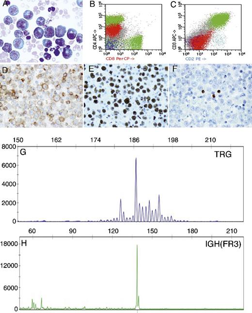
An 80-year-old HIV-negative man was found to have a large right pleural effusion. Cytology smears (panel A, Diff-Quik) showed numerous large atypical lymphoid cells with plasmablastic features. Flow cytometry showed these atypical cells were dimly positive for CD4 (panel B) but negative for the rest of the T-cell (panels B, C) and B-cell antigens (data not shown). Immunohistochemistry using the cell block showed the atypical cells were positive for CD4 (panel D), Kaposi sarcoma herpesvirus (KSHV) (panel E), and Epstein-Barr virus (in situ hybridization) but negative for the rest of the T-cell antigens and all B-cell antigens including octamer binding protein 2 (panel F and data not shown). Polymerase chain reaction analyses showed monoclonal TRG (panel G) and IGH (framework 3) (panel H) rearrangements. Of note, there was a single peak with little or no polyclonal background observed in the IGH assay. Based on the aforementioned findings, a primary effusion lymphoma (PEL) of T cells was diagnosed.
PEL is a body cavity–based KSHV-driven aggressive lymphoma of B-cell origin in HIV-positive patients in the vast majority of cases. PELs of T cells have rarely been reported. The diagnostic pitfalls in this unique case include (1) lack of T-cell–specific and T-cell–associated antigens (except CD4) and (2) the presence of IGH pseudo-monoclonality due to extremely low numbers of B cells.
For additional images, visit the ASH IMAGE BANK, a reference and teaching tool that is continually updated with new atlas and case study images. For more information visit http://imagebank.hematology.org.


This feature is available to Subscribers Only
Sign In or Create an Account Close Modal