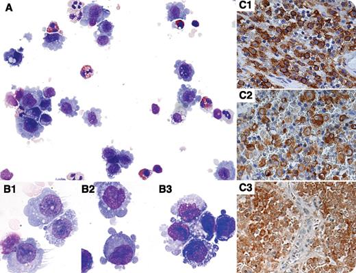
A 4-year-old boy presented with 3 weeks of right-sided knee pain and difficulty walking. On examination, a decrease in right hip abduction was noted without erythema, swelling, fluctuance, or warmth of the right hip or knee. Radiographic studies demonstrated a 4-cm poorly marginated lesion in the right supraacetabular region that was biopsied. Cytospin preparations of the biopsy showed numerous abnormal histiocytic cells admixed with eosinophils, small lymphocytes, and macrophages (panel A). The histiocytic cells showed prominent dendritic projections (panel B1), cytoplasmic blebbing (panel B2), and folded nuclei (panel B3). Flow cytometry demonstrated an atypical population expressing bright CD1a, CD4, CD13, CD33, CD14, CD64, and HLA-DR. The diagnosis of Langerhans cell histiocytosis (LCH) was confirmed by histologic sections, which showed numerous histiocytic cells with grooved nuclei in an eosinophil-rich background. Immunohistostains were positive for CD1a (panel C1), Langerin (panel C2), and BRAF V600E (panel C3).
LCH is the most commonly diagnosed histiocytic neoplasm. Recent evidence suggests that LCH may arise from abnormal bone marrow-derived dendritic cell precursors that are driven by oncogenic mutations. The BRAF V600E mutation is present in ∼50% of cases. The diagnosis of LCH is based on detecting morphologically abnormal CD1a+ histiocytes in an eosinophil-rich background. Rarely, these diagnostic features are present in cytologic preparations combined with flow cytometry.
For additional images, visit the ASH IMAGE BANK, a reference and teaching tool that is continually updated with new atlas and case study images. For more information visit http://imagebank.hematology.org.


This feature is available to Subscribers Only
Sign In or Create an Account Close Modal