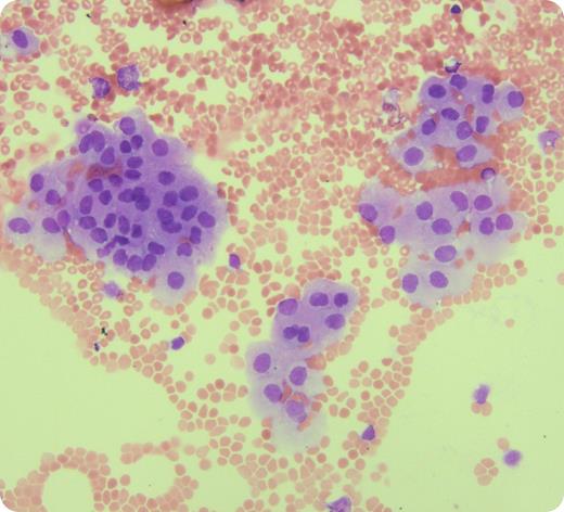
A 74-year-old man with stage IV diffuse large B-cell lymphoma underwent a lumbar puncture for staging and prophylactic intrathecal methotrexate. No neurological symptoms were reported. The cerebrospinal fluid (CSF) sample was macroscopically clear and colorless. Cytology showed numerous erythrocytes, small lymphocytes, and neutrophils consistent with blood contamination. No abnormal lymphocytes were seen and flow cytometry showed no lymphoma. Additionally, many clusters of large cells were seen with plentiful cytoplasm, round to oval nucleus, and open chromatin. They were identified as normal ependymal cells. Although morphology may suggest a malignant origin, we wish to highlight the importance of recognizing these as normal CSF cells by hematopathologists/cytologists.
Ependymal cells are CSF-producing cells from the cerebrospinal surface epithelium and have a distinct morphologic appearance which differs from lymphoma or carcinoma cells. They are seen in normal CSF samples as part of traumatic artifact from the lumbar puncture needle as well as the CSF of patients with central nervous system infections and/or subarachnoid hemorrhage and in hydrocephalic children. Correctly identifying ependymal cells in this case correlated with other features of a traumatic tap in an otherwise normal CSF. This case highlights the need for understanding all normal cellular components in a CSF cytological examination to recognize artifact and distinguish from malignancy.
For additional images, visit the ASH IMAGE BANK, a reference and teaching tool that is continually updated with new atlas and case study images. For more information visit http://imagebank.hematology.org.


This feature is available to Subscribers Only
Sign In or Create an Account Close Modal