On page 830 in the 26 July 2012 issue, there is an error in the Figure 7C panel labeled “E6AP” for the J-1 cell line. The E6AP lanes have been replaced with the correct lanes, which were adjacent on the same gel. The conclusion from this result remains the same. The corrected Figure 7 is shown.
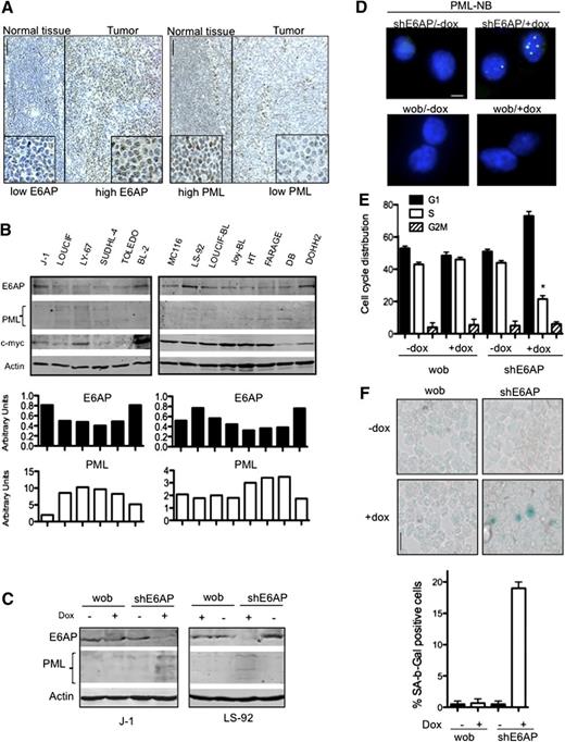
E6AP levels are elevated in human Burkitt lymphomas and downregulation of E6AP restores PML-induced cellular senescence. (A) Representative images of E6AP (left panel) and PML (right panel) immunostaining in human Burkitt lymphomas. Expression of E6AP is relatively low in normal lymphoid tissue, but the infiltrating Burkitt lymphoma cells show elevated levels of E6AP accompanied by reduced levels of PML; N = 20; Magnification ×200. (B) Immunoblot analysis to determine the levels of E6AP, PML, and c-Myc in a panel of cell lines derived from Burkitt lymphoma derived (J-1, LOUCIF, LY-67, BL-2, MC116, LS-92, LOUCIF-BL, JOY-BL), DLBCL (SUDHL-4, FARAGE, TOLEDO, DB, HT), or follicular lymphoma (DOHH2). Probing for actin was used as a loading control. The expression levels of E6AP and PML normalized against the levels of actin were quantified and are presented on the graphs (bottom panel). (C) Immunoblot analysis of the indicated proteins in J-1 and LS-92 Burkitt lymphoma derived cells transduced with inducible lentiviral constructs containing wobble E6AP or shRNA for E6AP that were treated with (+) or without (−) doxycyclin. Cells were analyzed 7 days after doxycyclin (dox) induction. Probing for actin was used as a loading control. (D) Immunofluorescence staining of PML-NBs in J-1 Burkitt lymphoma cells transduced with the aforementioned shRNA expression constructs on day 7 after dox treatment. Magnification ×1000. (E) Cell-cycle analyses of J-1 Burkitt lymphoma cells transduced with the aforementioned shRNA expression constructs performed on day 7 after dox induction; *P < .001. Values represent means ± SD. (F) SA-β-gal levels were determined in cytospins of J-1 Burkitt lymphoma cells transduced with the aforementioned shRNA expression constructs on day 7 after dox treatment. Magnification ×400. Quantification of SA-β-gal positive cells: > 400 cells/cytospin, n = 3, cells were counted in 4 randomly selected fields. Values represent means ± SD, and were derived from 3 independent experiments performed in triplicate.
E6AP levels are elevated in human Burkitt lymphomas and downregulation of E6AP restores PML-induced cellular senescence. (A) Representative images of E6AP (left panel) and PML (right panel) immunostaining in human Burkitt lymphomas. Expression of E6AP is relatively low in normal lymphoid tissue, but the infiltrating Burkitt lymphoma cells show elevated levels of E6AP accompanied by reduced levels of PML; N = 20; Magnification ×200. (B) Immunoblot analysis to determine the levels of E6AP, PML, and c-Myc in a panel of cell lines derived from Burkitt lymphoma derived (J-1, LOUCIF, LY-67, BL-2, MC116, LS-92, LOUCIF-BL, JOY-BL), DLBCL (SUDHL-4, FARAGE, TOLEDO, DB, HT), or follicular lymphoma (DOHH2). Probing for actin was used as a loading control. The expression levels of E6AP and PML normalized against the levels of actin were quantified and are presented on the graphs (bottom panel). (C) Immunoblot analysis of the indicated proteins in J-1 and LS-92 Burkitt lymphoma derived cells transduced with inducible lentiviral constructs containing wobble E6AP or shRNA for E6AP that were treated with (+) or without (−) doxycyclin. Cells were analyzed 7 days after doxycyclin (dox) induction. Probing for actin was used as a loading control. (D) Immunofluorescence staining of PML-NBs in J-1 Burkitt lymphoma cells transduced with the aforementioned shRNA expression constructs on day 7 after dox treatment. Magnification ×1000. (E) Cell-cycle analyses of J-1 Burkitt lymphoma cells transduced with the aforementioned shRNA expression constructs performed on day 7 after dox induction; *P < .001. Values represent means ± SD. (F) SA-β-gal levels were determined in cytospins of J-1 Burkitt lymphoma cells transduced with the aforementioned shRNA expression constructs on day 7 after dox treatment. Magnification ×400. Quantification of SA-β-gal positive cells: > 400 cells/cytospin, n = 3, cells were counted in 4 randomly selected fields. Values represent means ± SD, and were derived from 3 independent experiments performed in triplicate.


This feature is available to Subscribers Only
Sign In or Create an Account Close Modal