To the editor:
The costs of conducting a clinical trial from phase 1 through phase 3 completion are staggering at greater than $100 million,1 and the cost per patient in a trial has ballooned from ∼$25 000 in 2000 to ∼$100 000 in 2012.2 These high costs underscore the necessity for the information obtained from these trials to be evaluated with the utmost care. During clinical trials, blood from patients is often collected and analyzed for pharmacodynamic and biomarker changes to measure response or toxicity. Traditionally, blood collected into heparinized green-top tubes (GTTs) followed by Ficoll-Hypaque (Ficoll) density gradient separation methods has been used to isolate peripheral blood mononuclear cells (PBMCs).3,4 Recently, one-step closed systems, such as Vacutainer cell processing tubes (CPTs), have been used for PBMC isolation and shipment. We evaluated whether use of these tubes could have an impact on cellular proteins.
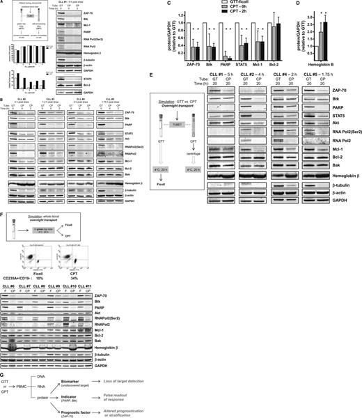
A 10-mL glass GTT contains 158 USP units of freeze-dried sodium heparin. The glass sodium heparin CPT used in this study has a 8-mL draw capacity and contains a minimum of 132 USP units of sodium heparin in 1 mL of phosphate-buffered saline solution, 3 g of polyester gel, and 2 mL of Ficoll. Blood from patients with chronic lymphocytic leukemia (CLL) was collected into a Vacutainer GTT or CPT, both containing sodium heparin, and processed immediately (Figure 1A). CPT- and Ficoll-isolated cells had similar levels of apoptosis as measured by flow cytometry using annexin V/propidium iodide staining; however, erythrocyte contamination was greater in CPT-isolated cells (28% vs 11%; n = 5; P = .0036). More strikingly, there was substantial decline in protein levels in CPT-isolated cells, and immunoblot analysis revealed that ZAP-70, Btk, PARP, STAT5, Akt, phospho-RNA Pol2(Ser2), RNA Pol2, Mcl-1, and β-tubulin protein levels were substantially lower in CPT-isolated CLL cells (Figure 1A-B). However, Bcl-2 family member proteins (Bcl-2 and Bak) remained stable as did loading controls glyceraldehyde-3-phosphate dehydrogenase (GAPDH) and β-actin, and there were increased levels of hemoglobin β in CPT-isolated cells, consistent with erythrocyte contamination. When normalized to GAPDH, the decline in ZAP-70, Btk, PARP, STAT5, and Mcl-1 protein levels ranged from 0.1- to 0.5-fold of that of GTTs and was statistically significant, whereas Bcl-2 protein levels were not significantly affected (Figure 1C). In contrast, normalized hemoglobin β was increased to twofold to threefold and was statistically significant (Figure 1D).
Blood leukemia cell isolation methods have an impact on intracellular protein integrity. All studies were performed using fresh primary lymphocytes isolated from blood obtained from patients with CLL. Patients gave informed consent to participate in this laboratory protocol, which was approved by the Institutional Review Board of The University of Texas MD Anderson Cancer Center. Informed consent was provided according to the Declaration of Helsinki. (A) Immediate processing of whole blood on-site at the clinical trial location. Blood from the same patient (n = 5) was collected directly into a Vacutainer GTT (#366480; Becton Dickinson, Franklin Lakes, NJ) and a CPT (#362753; Becton Dickinson) simultaneously. Each tube contained sodium heparin and was kept at ambient temperature until processing. Briefly, for the GTT processing using Ficoll, the blood was mixed with 2 volumes of phosphate-buffered saline (PBS) and then overlaid gently onto 10 mL of Ficoll-Hypaque and centrifuged for 20 minutes at 20°C. The PBMCs were isolated, washed twice with PBS, and counted. The cell number was determined by using a Coulter Channelyzer (Coulter Electronics, Hialeah, FL). The CPT was centrifuged at 1800g for 20 minutes at ambient temperature, and then the tube was inverted several times to recover the isolated cells above the polyester gel barrier per the manufacturer’s recommendation. Isolated cells were washed twice with cold PBS and counted. For measurement of apoptosis levels, CLL cells (n = 5) isolated by Ficoll (white bars) or CPT (black bars) were incubated with annexin V, fluorescein isothiocyanate (FITC), and propidium iodide and were analyzed by flow cytometry. Similarly, to measure percentages of erythrocytes and B cells, CLL cells were stained with CD19-phycoerythrin (PE) and CD235A-FITC and analyzed by flow cytometry. Immunoblot analysis was performed as previously described5 by using the following antibodies: Akt (BD Pharmingen); Bak (Millipore); Bcl-2 (Dako, Carpinteria, CA); Btk (Abcam, Cambridge, MA); hemoglobin β, β-actin, and β-tubulin (Sigma); glyceraldehyde-3-phosphate dehydrogenase (GAPDH; Novus Biologicals, Littleton, CO); Mcl-1 and STAT5 (Santa Cruz Biotechnology, Santa Cruz, CA); PARP (Enzo Life Sciences International, Plymouth Meeting, PA); total RNA Pol2 (8WG16) and phospho-RNA Pol2 (Ser2) (Covance, Emeryville, CA); and ZAP-70 (Cell Signaling Technologies, Beverly, MA). Immunoblot analysis of cellular proteins of PBMCs isolated from blood collected in GTT (lane GT) or CPT (lane CP) from patient #1 is shown. Protein levels were quantified relative to GAPDH, then normalized to protein levels in Ficoll-isolated PBMCs from GTT-collected blood (lane GT). (B) Immunoblot analysis of cellular proteins of PBMCs isolated from blood collected in GTTs (lane GT) or CPTs (lane CP) from patients #2 to #5. (C-D) Quantified cellular protein levels in GTT- and CPT-isolated cells. Immunoblots shown in Figure 1A-B were analyzed by using Li-Cor Odyssey software (Li-Cor Inc., Lincoln, NE), and protein levels were normalized to GAPDH loading control. Protein levels in CPT-isolated cells (lanes CP) were expressed relative to levels in GTT-isolated cells (lane GT), and asterisks denote statistical significance (*P < .05) as determined by using GraphPad Prism software (GraphPad Software, Inc. San Diego, CA). (E) Simulation of blood drawn into a GTT or CPT and then shipped overnight to an off-site reference laboratory. Blood was collected as described in (A), the CPT was processed by centrifugation, and the PBMCs were kept in the CPT at 4°C for 20 hours to simulate overnight transport on icepacks. The GTT was kept at 4°C for 20 hours, and then the PBMCs were isolated using the Ficoll method. Immunoblot analysis of cellular proteins of PBMCs isolated from blood collected in GTTs (lane GT) or CPTs (lane CP) from patients #1, #2, #4, and #5 are shown. (F) Isolation of primary leukemia cells drawn into GTTs and then shipped overnight to an off-site reference laboratory. Blood from patients with CLL (n = 6) was collected into GTTs and kept at 4°C for 20 hours to simulate shipment on wet ice to a reference laboratory. Half the sample was then processed using Ficoll and half was transferred into a CPT containing sodium heparin and centrifuged. Shown is the flow cytometry analysis of CLL cells from patient #11 isolated by Ficoll (left) or CPT (right) and stained with CD235A-FITC/CD19-PE. Immunoblot analysis of CLL primary cells from patients #6 to #11 isolated by Ficoll (lane F) or CPT (lane CP) is shown. (G) Implications and consequences of cellular protein loss on CLL biology, diagnostics, and therapeutic research.
Blood leukemia cell isolation methods have an impact on intracellular protein integrity. All studies were performed using fresh primary lymphocytes isolated from blood obtained from patients with CLL. Patients gave informed consent to participate in this laboratory protocol, which was approved by the Institutional Review Board of The University of Texas MD Anderson Cancer Center. Informed consent was provided according to the Declaration of Helsinki. (A) Immediate processing of whole blood on-site at the clinical trial location. Blood from the same patient (n = 5) was collected directly into a Vacutainer GTT (#366480; Becton Dickinson, Franklin Lakes, NJ) and a CPT (#362753; Becton Dickinson) simultaneously. Each tube contained sodium heparin and was kept at ambient temperature until processing. Briefly, for the GTT processing using Ficoll, the blood was mixed with 2 volumes of phosphate-buffered saline (PBS) and then overlaid gently onto 10 mL of Ficoll-Hypaque and centrifuged for 20 minutes at 20°C. The PBMCs were isolated, washed twice with PBS, and counted. The cell number was determined by using a Coulter Channelyzer (Coulter Electronics, Hialeah, FL). The CPT was centrifuged at 1800g for 20 minutes at ambient temperature, and then the tube was inverted several times to recover the isolated cells above the polyester gel barrier per the manufacturer’s recommendation. Isolated cells were washed twice with cold PBS and counted. For measurement of apoptosis levels, CLL cells (n = 5) isolated by Ficoll (white bars) or CPT (black bars) were incubated with annexin V, fluorescein isothiocyanate (FITC), and propidium iodide and were analyzed by flow cytometry. Similarly, to measure percentages of erythrocytes and B cells, CLL cells were stained with CD19-phycoerythrin (PE) and CD235A-FITC and analyzed by flow cytometry. Immunoblot analysis was performed as previously described5 by using the following antibodies: Akt (BD Pharmingen); Bak (Millipore); Bcl-2 (Dako, Carpinteria, CA); Btk (Abcam, Cambridge, MA); hemoglobin β, β-actin, and β-tubulin (Sigma); glyceraldehyde-3-phosphate dehydrogenase (GAPDH; Novus Biologicals, Littleton, CO); Mcl-1 and STAT5 (Santa Cruz Biotechnology, Santa Cruz, CA); PARP (Enzo Life Sciences International, Plymouth Meeting, PA); total RNA Pol2 (8WG16) and phospho-RNA Pol2 (Ser2) (Covance, Emeryville, CA); and ZAP-70 (Cell Signaling Technologies, Beverly, MA). Immunoblot analysis of cellular proteins of PBMCs isolated from blood collected in GTT (lane GT) or CPT (lane CP) from patient #1 is shown. Protein levels were quantified relative to GAPDH, then normalized to protein levels in Ficoll-isolated PBMCs from GTT-collected blood (lane GT). (B) Immunoblot analysis of cellular proteins of PBMCs isolated from blood collected in GTTs (lane GT) or CPTs (lane CP) from patients #2 to #5. (C-D) Quantified cellular protein levels in GTT- and CPT-isolated cells. Immunoblots shown in Figure 1A-B were analyzed by using Li-Cor Odyssey software (Li-Cor Inc., Lincoln, NE), and protein levels were normalized to GAPDH loading control. Protein levels in CPT-isolated cells (lanes CP) were expressed relative to levels in GTT-isolated cells (lane GT), and asterisks denote statistical significance (*P < .05) as determined by using GraphPad Prism software (GraphPad Software, Inc. San Diego, CA). (E) Simulation of blood drawn into a GTT or CPT and then shipped overnight to an off-site reference laboratory. Blood was collected as described in (A), the CPT was processed by centrifugation, and the PBMCs were kept in the CPT at 4°C for 20 hours to simulate overnight transport on icepacks. The GTT was kept at 4°C for 20 hours, and then the PBMCs were isolated using the Ficoll method. Immunoblot analysis of cellular proteins of PBMCs isolated from blood collected in GTTs (lane GT) or CPTs (lane CP) from patients #1, #2, #4, and #5 are shown. (F) Isolation of primary leukemia cells drawn into GTTs and then shipped overnight to an off-site reference laboratory. Blood from patients with CLL (n = 6) was collected into GTTs and kept at 4°C for 20 hours to simulate shipment on wet ice to a reference laboratory. Half the sample was then processed using Ficoll and half was transferred into a CPT containing sodium heparin and centrifuged. Shown is the flow cytometry analysis of CLL cells from patient #11 isolated by Ficoll (left) or CPT (right) and stained with CD235A-FITC/CD19-PE. Immunoblot analysis of CLL primary cells from patients #6 to #11 isolated by Ficoll (lane F) or CPT (lane CP) is shown. (G) Implications and consequences of cellular protein loss on CLL biology, diagnostics, and therapeutic research.
To simulate conditions under which blood is drawn into CPTs and then shipped in CPTs overnight, we analyzed PBMCs from CPTs kept at 4°C overnight after processing (Figure 1E). The results were consistent with a substantial decline in proteins in CPT-isolated CLL cells as described for Figure 1A-B. Our data suggest that irrespective of processing time, collection of blood into CPTs may not be optimal for detection of proteins by immunoblot.
If CPTs are not used for blood collection, blood samples collected in GTTs are often shipped overnight to reference laboratories where CPTs may be used for processing, and the question remains whether this affects cellular protein integrity. To simulate this scenario, blood from 6 patients was collected into GTTs and stored at 4°C overnight; then, half the blood in the tube was processed by Ficoll and the remaining half was transferred into a CPT and processed (Figure 1F). Even using the CPT only for processing and not for overnight shipment resulted in greater erythrocyte contamination in the CPT-isolated cells as well as substantial decline in several CLL-relevant proteins, including ZAP-70, Btk, PARP, RNA Pol2, Mcl-1, and β-tubulin.
These results are alarming, and in the context of CLL, the decline in ZAP-70 is of particular relevance, given its application as a prognostic factor.6,7 Moreover, new kinase (Btk,8 PI3K,9 cyclin-dependent10 ) inhibitors and Bcl-2 antagonists are increasingly used in the clinic for patients with CLL, and loss of cellular proteins may have significant clinical implications (Figure 1G). Although the current work focused on CLL, we feel that these observations would apply to other hematologic and perhaps solid malignancies, and we propose immediate processing of blood collected in GTTs using the Ficoll method to better preserve cellular protein integrity. We hope that the data presented here will be taken into consideration during clinical trial design and decision making.
Authorship
Acknowledgments: The authors thank Benjamin Hayes for obtaining blood samples and Susan C. Smith for providing information on patient characteristics. This work was supported in part by a grant from the Translational Research Program of the Leukemia and Lymphoma Society of America (LLS R6011-14), a Chronic Lymphocytic Leukemia (CLL) Research Consortium grant from the National Institutes of Health (PO1-CA081534), and a CLL-Global Research Foundation Alliance Grant.
Contribution: L.S.C. designed research, performed experiments, analyzed results, and wrote the manuscript; M.J.K. identified patients for this study, obtained consent for these studies, and reviewed the manuscript; and V.G. conceptualized and supervised research, analyzed data, and reviewed the manuscript.
Conflict-of-interest disclosure: The authors declare no competing financial interests.
Correspondence: Varsha Gandhi, Department of Experimental Therapeutics, The University of Texas MD Anderson Cancer Center, Houston, TX 77030; e-mail: vgandhi@mdanderson.org.


This feature is available to Subscribers Only
Sign In or Create an Account Close Modal