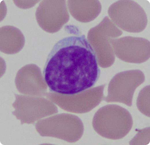
The patient is a 92-year-old woman who presented to the hematology clinic 3 years earlier for evaluation of persistent lymphocytosis. At that time, her white blood cell count was 15 200/µL, hemoglobin was 12.8 g/dL, and platelets were 245 000/µL. Her absolute lymphocyte count was 7870/µL. She did not have lymphadenopathy or hepatosplenomegaly on physical examination. Flow cytometry was performed and revealed a monoclonal B-cell proliferation that was CD5 and CD10 negative. Fluorescence in situ hybridization testing for t(11;14) and rearrangements/additions of chromosomes 11q, 12, 13q, and 17p13.1 were negative. The monoclonal lymphocytosis was monitored with serial surveillance. At the most recent clinic visit, her absolute lymphocyte count was 21 310/µL, and morphologic examination of the peripheral smear revealed mononuclear cells with Auer rod–like cytoplasmic inclusions. Flow cytometry redemonstrated the monoclonal B-cell proliferation, without any evidence of myeloid differentiation. A cytochemical stain for myeloperoxidase was negative.
This case highlights a rare example where the presence of Auer rod–like inclusions does not automatically denote myeloid differentiation. To the best of our knowledge, the presence of Auer rod–like lymphocyte inclusions has been previously reported only in a single case of a peripheralizing marginal zone lymphoma from Japan.
For additional images, visit the ASH IMAGE BANK, a reference and teaching tool that is continually updated with new atlas and case study images. For more information visit http://imagebank.hematology.org.


This feature is available to Subscribers Only
Sign In or Create an Account Close Modal