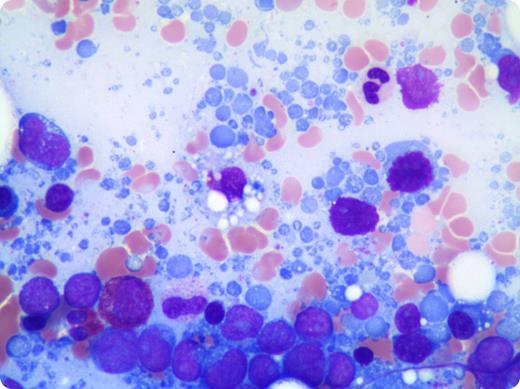
A 79-year-old woman presented with hypercalcemia and a mediastinal mass identified by computed tomography scan. Further imaging revealed several intracranial lesions that were not intraparenchymal, as well as a large periaortic mass with diffuse abdominal lymphadenopathy. A biopsy of the mediastinal mass yielded a diagnosis of B-cell lymphoma, unclassifiable, with features intermediate between diffuse large B-cell lymphoma and Burkitt lymphoma. A staging bone marrow examination was performed and revealed extensive involvement by an aggressive appearing large B-cell lymphoma. A striking number of lymphoglandular bodies were present in the bone marrow aspirate smears.
Lymphoglandular bodies are cytoplasmic fragments, commonly observed and reported in fine-needle aspiration or cytology preparations of lymphoid tissue, often in association with lymphoid malignancies. They have been less frequently reported in bone marrow aspirate smears. Although not a specific finding, lymphoglandular bodies have been shown to be present in higher numbers in bone marrows involved by B-cell lymphoid malignancies compared with T-cell lymphoid malignancies and myeloid leukemias. Their pale, lightly basophilic color with smooth borders, occasional blebs, and lack of granulation allow them to be distinguished from platelets.
For additional images, visit the ASH IMAGE BANK, a reference and teaching tool that is continually updated with new atlas and case study images. For more information visit http://imagebank.hematology.org.


This feature is available to Subscribers Only
Sign In or Create an Account Close Modal