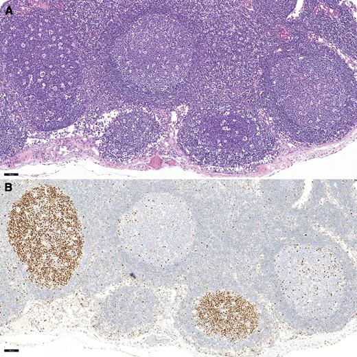
A 12-year-old boy presented with right lower quadrant pain and a 5-lb weight loss. A 4.5-cm ileocecal mass was excised and diagnosed as Epstein-Barr virus–negative Burkitt lymphoma (BL). Numerous pericecal lymph nodes revealed preserved nodal architecture with follicles with intact mantle zones and numerous tingible body macrophages imparting a “starry sky” appearance (panel A: original magnification, ×10; hematoxylin and eosin staining). Closer inspection revealed patchy follicular colonization by BL, which also characteristically imparts a starry sky appearance due to high cellular turnover. This was confirmed by an immunostain for MYC (panel B: original magnification, ×10).
BL is a mature, follicular-derived B-cell neoplasm that harbors a MYC translocation in 100% of cases. Characteristic homing of neoplastic cells to follicular centers may appear deceptively reactive because reactive germinal centers also harbor a starry sky. BL localized to follicles should not be considered an early lesion because it is invariably associated with the presence of aggressive lymphoma elsewhere.
For additional images, visit the ASH IMAGE BANK, a reference and teaching tool that is continually updated with new atlas and case study images. For more information visit http://imagebank.hematology.org.


This feature is available to Subscribers Only
Sign In or Create an Account Close Modal