Key Points
JAK2 (V617F)-mutated essential thrombocythemia and polycythemia vera are different phenotypes in the evolution of a single neoplasm.
CALR-mutated essential thrombocythemia is a distinct disease entity not only at the molecular level, but also with respect to clinical outcomes.
Abstract
Patients with essential thrombocythemia may carry JAK2 (V617F), an MPL substitution, or a calreticulin gene (CALR) mutation. We studied biologic and clinical features of essential thrombocythemia according to JAK2 or CALR mutation status and in relation to those of polycythemia vera. The mutant allele burden was lower in JAK2-mutated than in CALR-mutated essential thrombocythemia. Patients with JAK2 (V617F) were older, had a higher hemoglobin level and white blood cell count, and lower platelet count and serum erythropoietin than those with CALR mutation. Hematologic parameters of patients with JAK2-mutated essential thrombocythemia or polycythemia vera were related to the mutant allele burden. While no polycythemic transformation was observed in CALR-mutated patients, the cumulative risk was 29% at 15 years in those with JAK2-mutated essential thrombocythemia. There was no significant difference in myelofibrotic transformation between the 2 subtypes of essential thrombocythemia. Patients with JAK2-mutated essential thrombocythemia and those with polycythemia vera had a similar risk of thrombosis, which was twice that of patients with the CALR mutation. These observations are consistent with the notion that JAK2-mutated essential thrombocythemia and polycythemia vera represent different phenotypes of a single myeloproliferative neoplasm, whereas CALR-mutated essential thrombocythemia is a distinct disease entity.
Introduction
In the World Health Organization (WHO) classification of tumors of hematopoietic and lymphoid tissues, Philadelphia-negative myeloproliferative neoplasms (MPNs) include polycythemia vera (PV), essential thrombocythemia (ET), and primary myelofibrosis (PMF).1 These disorders have overlapping clinical features and a common molecular basis. In fact, three-quarters of these patients carry the unique JAK2 (V617F) mutation,2,3 which is present in about 95% of subjects with PV and in about 60% of those with ET or PMF.4 Somatic mutations of JAK2 exon 12 are found in the remaining 5% of patients with PV,5 whereas mutations of MPL exon 10 are present in about 5% of those with ET or PMF.6 We and others recently found that most patients with ET or PMF with nonmutated JAK2 and MPL carry a somatic mutation of CALR, the gene encoding calreticulin.7,8
How a single mutation, JAK2 (V617F), can be associated with different phenotypes has been partly clarified in the past few years. Campbell et al9 found that patients with JAK2 (V617F)-mutated ET had multiple features resembling PV and concluded that JAK2 (V617F)-mutated ET and PV form a biological continuum in which the degree of erythrocytosis is determined by physiological and genetic modifiers. The JAK2 (V617F) mutant allele burden is significantly higher in patients with PV than in those with ET,10 and evidence of JAK2 (V617F) homozygosity, as a result of mitotic recombination of chromosome 9p, is mainly found in the former.11,12 Tiedt et al13 generated JAK2 (V617F) transgenic mice and studied the relationship between ratio of mutant to wild-type JAK2 and phenotypic manifestation: they clearly showed that lower ratios were associated with an ET phenotype and higher ratios with a PV phenotype.
The recent identification of somatic mutations in CALR that are mutually exclusive with JAK2 and MPL mutations has provided a new powerful tool for studying MPNs. In the present work, we compared hematologic and clinical features of patients with JAK2-mutated ET with those of patients with CALR-mutated ET and related the findings to features observed in patients with PV.
Patients and methods
Study population and definitions
This study was approved by the institutional Ethics Committee (Comitato di Bioetica, Fondazione Istituto di Ricovero e Cura a Carattere Scientifico [IRCCS] Policlinico San Matteo, Pavia, Italy). The procedures followed were in accordance with the Helsinki Declaration of 1975, as revised in 2000, and samples were obtained after patients had provided written informed consent.
We identified 1235 consecutive patients diagnosed with ET or PV at the Department of Hematology Oncology, Fondazione IRCCS Policlinico San Matteo, and University of Pavia, Italy, between 1980 and 2012, for which at least 1 DNA sample was available (Table 1). Diagnosis of PV or ET was performed in accordance with the criteria in use at the time of first observation, as previously described.14 In 2002, we adopted the WHO criteria for diagnosis of MPN15 ; in 2009, we implemented the 2008 revision.1 Post-PV or post-ET myelofibrosis was diagnosed according to the criteria of the International Working Group of Myelofibrosis Research and Treatment,16 whereas evolution into acute myeloid leukemia was defined according to the WHO criteria.1 Thrombotic events were defined as described in detail by the Cytoreductive Therapy in Polycythemia Vera Collaborative Group.17
Initial study population including 1235 consecutive patients diagnosed with ET or PV for which at least 1 DNA sample was available*
| MPN . | JAK2 (V617F) mutated (%) . | JAK2 exon 12 mutated (%) . | MPL exon 10 mutated (%) . | CALR exon 9 mutated (%) . | Nonmutated JAK2/MPL/CALR (%) . | All genotypes . |
|---|---|---|---|---|---|---|
| ET | 466 (62) | — | 28 (4) | 176 (24) | 75 (10) | 745 |
| PV | 468 (96) | 22 (4) | — | — | — | 490 |
| All patients | 1235 |
Population included patients followed at the Department of Hematology Oncology, Fondazione IRCCS Policlinico San Matteo and University of Pavia, Italy, between 1980 and 2012.
For the assessment of bone marrow fibrosis, paraffin sections were stained with Gomori’s silver impregnation technique, and fibrosis was assessed semiquantitatively following the European consensus guidelines.18
In the evaluation of transformation of ET to PV, or polycythemic transformation of ET, we could not routinely use the WHO criteria for diagnosis of PV1 because bone marrow biopsy was not regularly available during follow-up. We therefore adopted the criteria of the British Committee for Standards in Hematology for diagnosis of PV, described in the supplemental Methods on the Blood Web site.19,20 Basically, according to these latter criteria, the combination of erythrocytosis and JAK2 mutation allows diagnosis of PV.20
Because we excluded from outcome analysis patients with PV carrying a JAK2 exon 12 mutation (22 subjects) and patients with ET carrying an MPL substitution (28 subjects), 1185 of the initial 1235 subjects were eventually studied as detailed in supplemental Table 1. Of these 1185 patients, 674 had been studied in targeted resequencing investigations of our original article on somatic mutations of calreticulin.7 These 674 cases have been included in the present work with the purpose of analyzing features that were not examined in the original article, including clinical relevance of mutant allele burden, incidence of polycythemic or myelofibrotic transformation, and risk of leukemic evolution. To evaluate the clinical relevance of the mutant allele burden, we also examined an additional cohort of 83 patients with secondary myelofibrosis that was extracted from our database and is reported in supplemental Table 1.
JAK2, MPL, and CALR mutation analysis
Granulocyte JAK2 (V617F) mutation status and mutant allele burden were assessed using a quantitative polymerase chain reaction–based allelic discrimination assay on a Rotor-Gene 6000 real-time analyzer (Qiagen), as previously described.10,21
Patients with PV without JAK2 (V617F) were studied for JAK2 exon 12 mutations as previously described.22 Patients with ET and nonmutated JAK2 were further evaluated for MPL exon 10 mutations using a high-resolution melt assay.23 Patients with nonmutated JAK2 and MPL were studied for CALR exon 9 mutations as previously reported7 or by Sanger sequencing, as described in the supplemental Methods.
Statistical analysis
Numerical variables have been summarized by their median and range, and categorical variables by count and relative frequency (%) of each category. Comparisons of quantitative variables between groups of patients were carried out by the nonparametric Wilcoxon rank-sum test. The Wilcoxon signed-rank test was applied to compare measures of quantitative variables repeated in different phases of the disease. Association between categorical variables (2-way tables) was tested by the Fisher’s exact test. Correlation between numerical variables was tested by the nonparametric Spearman’s ρ (ρ) coefficient.
The incidence of polycythemic transformation of ET was estimated with a Poisson model, and comparison of incidence rates was carried out via the likelihood ratio test. The cumulative incidence of polycythemic, myelofibrotic, and leukemic transformation, and that of thrombotic events was estimated with a competing risk approach, considering death for any cause as a competing event.24 The comparison of cumulative incidence curves in different groups of patients was carried out using the Pepe-Mori test,25 whereas the effect of quantitative covariates (eg, JAK2 allele burden at diagnosis) was estimated by applying the Fine-Gray regression model.26 Overall survival (OS) was estimated using the Kaplan-Meier product limit method, and survival curves were compared by the log-rank test. Multivariate analysis of OS was carried out by Cox regression.
All P values were considered statistically significant when <.05 (2-tailed). Statistical analyses were performed using Stata 12.1 (StataCorp LP, College Station, TX) software.
Results
Presenting hematologic and clinical features of JAK2 (V617F)-mutated ET, JAK2 (V617F)-mutated PV, and CALR-mutated ET
We studied 468 patients with PV and 717 with ET. As reported in Table 1, this latter group included 466 patients with JAK2 mutation, 176 with CALR mutation, and 75 with nonmutated JAK2, MPL, and CALR. Overall, 23 types of CALR mutation were detected (supplemental Table 2): the 52-bp deletion (mutation type 1) and the 5-bp insertion (mutation type 2) were the most frequent alterations, representing 46% and 38% of all cases, respectively. We found 4 novel deletions and named them type 37, 38, 39, and 40 (supplemental Table 2) in accordance with our previously reported nomenclature.7 Consistent with the thus far described CALR mutations,7 all the novel deletions caused a change from a negatively to a positively charged C-terminal peptide and loss of the endoplasmic reticulum retention motif KDEL at the C-terminal end.
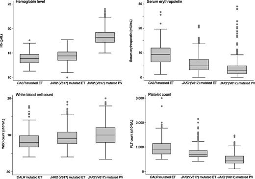
Table 2 reports the demographic and clinical characteristics at diagnosis of the patients studied according to their genotype, whereas the main hematologic parameters are summarized in Figure 1. Patients with CALR-mutated ET were significantly younger than those with JAK2-mutated ET (P =. 001) or PV (P < .001). Compared with patients with CALR-mutated ET, those with JAK2-mutated ET had higher hemoglobin (Hb) level and white blood cell (WBC) count, and lower platelet (PLT) count and serum erythropoietin (Epo) level (P < .001 in all comparisons). Patients with PV had higher values for Hb level and WBC count, lower values for PLT count and serum Epo, and higher frequency of splenomegaly compared with both CALR- and JAK2-mutated ET patients (P values shown in Table 2). The incidence of thrombosis at diagnosis was significantly higher in patients with PV than in those with CALR-mutated ET (P = .001), but not different between patients with PV and those with JAK2-mutated ET (P = .08).
Demographic, hematologic, and clinical features at diagnosis of patients with ET, subdivided according to JAK2 or CALR mutation status, and of patients with PV
| . | ET . | PV (C) . | P . | |||
|---|---|---|---|---|---|---|
| . | CALR mutated (A) . | JAK2 mutated (B) . | ||||
| No. . | 176 . | 466 . | 468 . | (A) vs (B) . | (B) vs (C) . | (A) vs (C) . |
| Sex (male/female) | 90/86 (51%/49%) | 167/299 (36%/64%) | 233/235 (50%/50%) | .001 | <.001 | .791 |
| Age at onset, years, median (range) | 45 (15-83) | 50 (15-92) | 57 (13-86) | .001 | <.001 | <.001 |
| Hemoglobin, g/dL, median (range) | 13.8 (11.3-17.6) | 14.4 (10-17.7) | 18.2 (15.0-24.0) | <.001 | <.001 | <.001 |
| WBC count, ×109/L, median (range) | 8.0 (4.0-17.9) | 9.0 (4.0-28.0) | 10.0 (3.4-55.3) | <.001 | <.001 | <.001 |
| PLT count, ×109/L, median (range) | 883 (500-3000) | 700 (456-2148) | 464 (109-1472) | <.001 | <.001 | <.001 |
| Serum erythropoietin, mU/mL, median (range) | 9.4 (1.2-27) | 4.7 (0-47) | 2.7 (0-66) | <.001 | <.001 | <.001 |
| Splenomegaly, no. (%) | 4 (2.3%) | 30 (6.4%) | 105 (22.4%) | .046 | <.001 | <.001 |
| Lactate dehydrogenase, mU/mL, median (range) | 199 (78-472) | 200 (77-540) | 217 (104-758) | .83 | <.001 | .003 |
| Circulating CD34+ cells, ×106/L, median (range) | 4.1 (0.6-18) | 4 (0-15.3) | 3.4 (0-261.3) | .50 | .037 | .039 |
| Thrombosis at diagnosis, no. (%) | 5 (2.8%) | 33 (7.1%) | 49 (10.5%) | .059 | .082 | .001 |
Hematologic parameters in patients with CALR-mutated ET, JAK2 (V617F)-mutated ET, and JAK2 (V617F)-mutated PV. Data are shown in a box plot depicting the upper and lower adjacent values (highest and lowest horizontal line, respectively), upper and lower quartile with median value (box), and outside values (dots). Of note, patients with JAK2 (V617F)-mutated ET had markedly lower serum Epo values than those of patients with CALR exon 9–mutated ET, despite the fact that the difference in median Hb levels was <1 g/dL.
Hematologic parameters in patients with CALR-mutated ET, JAK2 (V617F)-mutated ET, and JAK2 (V617F)-mutated PV. Data are shown in a box plot depicting the upper and lower adjacent values (highest and lowest horizontal line, respectively), upper and lower quartile with median value (box), and outside values (dots). Of note, patients with JAK2 (V617F)-mutated ET had markedly lower serum Epo values than those of patients with CALR exon 9–mutated ET, despite the fact that the difference in median Hb levels was <1 g/dL.
Granulocyte mutant allele burden in patients with JAK2 (V617F)-mutated MPNs
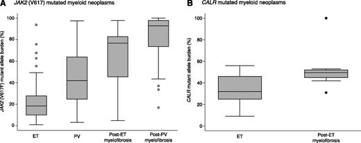
The median JAK2 (V617F) mutant allele burden at presentation was significantly lower in JAK2-mutated ET than in PV (18% vs 42%, P < .001), as shown in Figure 2A. Patients with myelofibrosis secondary to ET or PV had significantly higher values for JAK2 (V617F) mutant allele burden than those with the primary MPN (18% vs 77% in ET and post-ET myelofibrosis, respectively, P < .001; 42% vs 93% in PV and post-PV myelofibrosis, respectively, P < .001) (Figure 2A). A mutant allele burden greater than 50% was observed in 2% of patients with JAK2-mutated ET, 41% of those with PV, 72% of those with post-ET myelofibrosis, and 93% of those with post-PV myelofibrosis (Fisher exact test, P < .001 for ET vs PV, ET vs post-ET myelofibrosis, and PV vs post-PV myelofibrosis).
Granulocyte mutant allele burden in JAK2 (V617F)-mutated and in CALR-mutated myeloid neoplasms. Data are shown in a box plot depicting the upper and lower adjacent values (highest and lowest horizontal line, respectively), upper and lower quartile with median value (box), and outside values (dots). (A) This analysis includes 250 patients with ET and 212 patients with PV at presentation, and 18 patients with post-ET myelofibrosis and 55 with post -PV myelofibrosis. In these JAK2 (V617F)-mutated myeloid neoplasms, progression from the primary disease to secondary myelofibrosis appears to be related to the mutant allele burden. In particular, the proportion of patients with values >50% increases progressively, indicating an increasingly higher proportion of cells that are homozygous for the mutation as a result of copy neutral loss of heterozygosity of chromosome 9p. Most patients with post-ET or post-PV myelofibrosis have values for granulocyte JAK2 (V617F)-mutant allele burden greater than 75%, consistent with a dominant population of homozygous cells. (B) This analysis includes 38 patients with ET at presentation and 10 patients with post-ET myelofibrosis. Also within these CALR-mutated myeloid neoplasms, progression to secondary myelofibrosis appears to be associated with a significant increase in the mutant allele burden. However, only 1 patient with post-ET myelofibrosis had a value consistent with a dominant population of homozygous cells. This might suggest that the higher mutant allele burden in patients with post-ET myelofibrosis most often reflects the progressive expansion of a heterozygous clone that eventually achieves full dominance in the bone marrow.
Granulocyte mutant allele burden in JAK2 (V617F)-mutated and in CALR-mutated myeloid neoplasms. Data are shown in a box plot depicting the upper and lower adjacent values (highest and lowest horizontal line, respectively), upper and lower quartile with median value (box), and outside values (dots). (A) This analysis includes 250 patients with ET and 212 patients with PV at presentation, and 18 patients with post-ET myelofibrosis and 55 with post -PV myelofibrosis. In these JAK2 (V617F)-mutated myeloid neoplasms, progression from the primary disease to secondary myelofibrosis appears to be related to the mutant allele burden. In particular, the proportion of patients with values >50% increases progressively, indicating an increasingly higher proportion of cells that are homozygous for the mutation as a result of copy neutral loss of heterozygosity of chromosome 9p. Most patients with post-ET or post-PV myelofibrosis have values for granulocyte JAK2 (V617F)-mutant allele burden greater than 75%, consistent with a dominant population of homozygous cells. (B) This analysis includes 38 patients with ET at presentation and 10 patients with post-ET myelofibrosis. Also within these CALR-mutated myeloid neoplasms, progression to secondary myelofibrosis appears to be associated with a significant increase in the mutant allele burden. However, only 1 patient with post-ET myelofibrosis had a value consistent with a dominant population of homozygous cells. This might suggest that the higher mutant allele burden in patients with post-ET myelofibrosis most often reflects the progressive expansion of a heterozygous clone that eventually achieves full dominance in the bone marrow.
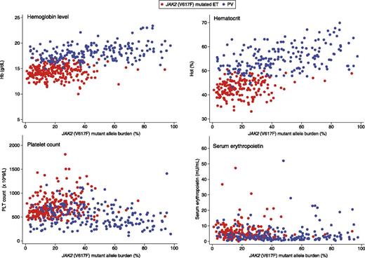
Grouping together patients with ET carrying JAK2 (V617F) and those with PV, significant associations were found between mutant allele burden and hematologic parameters (Hb level, hematocrit, PLT count, and serum Epo) as illustrated in Figure 3. These observations are consistent with the notion that JAK2-mutated ET and PV form a biological continuum in which the mutant allele burden acts as a determinant of phenotypic expression.
Relationship between granulocyte JAK2 (V617F)-mutant allele burden and hematologic parameters in patients with ET or PV. The mutant allele burden was directly correlated with Hb level (ρ = 0.53, P < .001) and hematocrit (ρ = 0.61, P < .001), and inversely correlated with PLT count (ρ = −0.18, P < .001) and serum Epo level (ρ = −0.23, P < .001). These correlations suggest that the mutant allele burden is a determinant of the phenotypic features of JAK2 (V617F)-mutated MPN.
Relationship between granulocyte JAK2 (V617F)-mutant allele burden and hematologic parameters in patients with ET or PV. The mutant allele burden was directly correlated with Hb level (ρ = 0.53, P < .001) and hematocrit (ρ = 0.61, P < .001), and inversely correlated with PLT count (ρ = −0.18, P < .001) and serum Epo level (ρ = −0.23, P < .001). These correlations suggest that the mutant allele burden is a determinant of the phenotypic features of JAK2 (V617F)-mutated MPN.
Granulocyte mutant allele burden in patients with CALR-mutated MPNs
We assessed the mutant allele burden in 110 of 148 patients with ET who carried a type 1 or type 2 CALR mutation (supplemental Table 2) and in 10 patients with post-ET myelofibrosis carrying the same alterations. The median mutant allele burden was 42% in patients with ET vs 50% in those with post-ET myelofibrosis (P = .018). We then restricted the analysis to patients with CALR-mutated ET evaluated at diagnosis. As shown in Figure 2B, the median mutant allele burden was 32% in patients with ET at clinical onset vs 50% in those with post-ET myelofibrosis (P = .002).
To have a relative estimation of the mutant clones, we compared the mutant allele burden at diagnosis in the 2 subtypes of ET. The median mutant allele burden of CALR-mutated patients was significantly higher than that of JAK2-mutated patients (32% vs 18%, P < .001).
Polycythemic transformation of ET and evidence of a prepolycythemic phase mimicking ET in JAK2 (V617F)-mutated PV
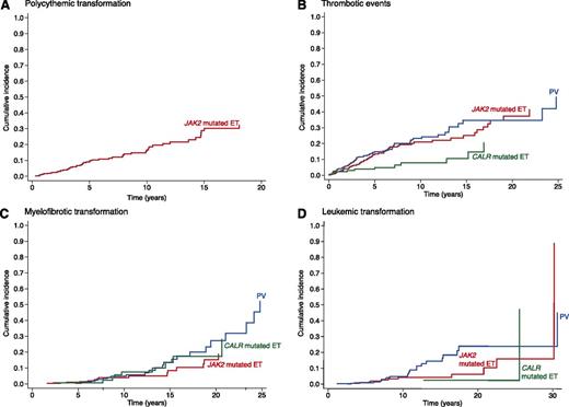
During follow-up, based on the diagnostic criteria of the British Committee for Standards in Hematology,19,20 polycythemic transformation occurred in 53 patients with JAK2-mutated ET (incidence 19 cases per 1000 person-years, 95% confidence interval [CI] 14-25) vs none of the 176 patients with CALR-mutated ET (incidence 0 per 1000 person-years, 95% CI 0-3). The median time to progression was 54 (range 4-220) months, and the cumulative incidence is reported in Figure 4A. Progression to PV was significantly associated with higher JAK2 mutant allele burden at diagnosis (Cox regression, hazard ratio [HR] = 1.04, 95% CI 1.02-1.07, P = .001): the median mutant allele burden was 25.5% (range 8.2-75.7) in patients who developed PV vs 17.9% (range 1-93.8) in those who did not (P = .019).
Cumulative incidence of polycythemic transformation, thrombotic events, myelofibrotic transformation, and leukemic transformation in patients with CALR- or JAK2-mutated ET and in those with PV. The cumulative incidences were estimated with a competing risk approach, considering death for all causes as a competing event.24 (A) In patients with JAK2-mutated ET, the cumulative incidence of polycythemic transformation was 28.6% (95% CI 20.7-37.0) at 15 years. No progression to PV was observed in patients with CALR-mutated ET. (B) Patients with CALR-mutated ET showed a lower incidence of thrombosis than those with JAK2-mutated ET (10.5% vs 25.1% at 15 years, P = .001), or those with PV (10.5% vs 34.7% at 15 years, P < .001). By contrast, patients with JAK2-mutated ET and those with PV did not differ in terms of cumulative incidence of thrombosis (P = .314). These differences in risk of thrombosis remained statistically significant even after adjusting for age, as detailed in the text. (C) The 15-year cumulative incidence of myelofibrotic transformation was 13.4% (CI 95% 5.4-25.2) in CALR-mutated ET, 8.4% (CI 95% 3.9-15.3) in JAK2-mutated ET, and 13.6% (CI 95% 7.3-21.9) in PV, without any significant difference among these 3 subgroups even after adjusting for age. (D) The 15-year cumulative incidence of leukemic transformation was 2.5% (CI 95% 0.2-11.3) in CALR-mutated ET, 4.3% (CI 95% 1.9-8.2) in JAK2-mutated ET, and 14.6% (CI 95% 8.4-22.3) in PV. Although CALR-mutated patients showed a lower risk of leukemic transformation in comparison with both those with JAK2-mutated ET (P = .026) and those with PV (P < .001), no significant difference was observed after adjusting for age.
Cumulative incidence of polycythemic transformation, thrombotic events, myelofibrotic transformation, and leukemic transformation in patients with CALR- or JAK2-mutated ET and in those with PV. The cumulative incidences were estimated with a competing risk approach, considering death for all causes as a competing event.24 (A) In patients with JAK2-mutated ET, the cumulative incidence of polycythemic transformation was 28.6% (95% CI 20.7-37.0) at 15 years. No progression to PV was observed in patients with CALR-mutated ET. (B) Patients with CALR-mutated ET showed a lower incidence of thrombosis than those with JAK2-mutated ET (10.5% vs 25.1% at 15 years, P = .001), or those with PV (10.5% vs 34.7% at 15 years, P < .001). By contrast, patients with JAK2-mutated ET and those with PV did not differ in terms of cumulative incidence of thrombosis (P = .314). These differences in risk of thrombosis remained statistically significant even after adjusting for age, as detailed in the text. (C) The 15-year cumulative incidence of myelofibrotic transformation was 13.4% (CI 95% 5.4-25.2) in CALR-mutated ET, 8.4% (CI 95% 3.9-15.3) in JAK2-mutated ET, and 13.6% (CI 95% 7.3-21.9) in PV, without any significant difference among these 3 subgroups even after adjusting for age. (D) The 15-year cumulative incidence of leukemic transformation was 2.5% (CI 95% 0.2-11.3) in CALR-mutated ET, 4.3% (CI 95% 1.9-8.2) in JAK2-mutated ET, and 14.6% (CI 95% 8.4-22.3) in PV. Although CALR-mutated patients showed a lower risk of leukemic transformation in comparison with both those with JAK2-mutated ET (P = .026) and those with PV (P < .001), no significant difference was observed after adjusting for age.
To test the hypothesis that PV patients might have a silent prepolycythemic phase mimicking ET, we did an ad hoc search for any complete blood cell (CBC) count that had been performed before diagnosis of PV. Overall, 169 patients had a previous CBC performed at a median time of 22 months (range 1-305) before diagnosis of PV. Although previous values were normal in 27 of these 169 patients (16%), in the remaining subjects there was evidence of erythrocytosis and/or leukocytosis and/or thrombocytosis. In particular, 38 patients had evidence of isolated erythrocytosis and 35 had evidence of isolated thrombocytosis. The median time to PV onset was significantly shorter in patients showing at least 1 abnormality in CBC count than in those with normal CBC count (19 vs 48 months, P = .004).
Clinical course of JAK2 (V617F)-mutated ET, JAK2 (V617F)-mutated PV, and CALR-mutated ET
The median follow-up of the study population was 5.2 years (range 0-32 years).
No difference in OS was observed between patients with JAK2 (V617F)-mutated ET and those with PV (P = .359). Patients with ET carrying a CALR mutation had a better OS than PV patients (95.3% vs 82.5% at 15 years, P = .010), and showed a trend toward a better OS than ET patients carrying JAK2 (V617F) (95.3% vs 90.4% at 15 years, P = .085). However, after adjusting for age, no significant difference between these subgroups was found (P > .05 in both cases).
The incidence of thrombotic events was 30 (95% CI 23-37) cases per 1000 person-years in patients with PV, 25 (95% CI 19-32) in those with JAK2-mutated ET, and 10 (95% CI 5-17) in those with CALR-mutated ET. The cumulative incidence in these subgroups is reported in Figure 4B. Patients with a CALR mutation had a lower risk of thrombosis than either patients with JAK2-mutated ET (P = .001) or those with PV (P < .001), whereas no significant difference was found between JAK2-mutated ET and PV (P = .314). These differences in thrombotic risk remained statistically significant after adjusting for age. In fact, when comparing patients with JAK2-mutated ET and those with CALR-mutated ET, the subdistribution HR was 2.23 (95% CI 1.20-4.12, P = .011). Similarly, when comparing patients with PV and those with CALR-mutated ET, the subdistribution HR was 2.34 (95% CI 1.25-4.39, P = .008). Of note, there was no significant difference in the use of cytoreductive therapy between the 2 subtypes of ET. We also evaluated the cumulative incidence of major bleeding using a competitive risk approach and did not find any difference between the subgroups.
The incidence of myelofibrotic transformation was 8 (95% CI 5-12) cases per 1000 person-years in patients with PV, 5 (95% CI 3-8) in those with JAK2-mutated ET, and 7 (95% CI 3-13) in those with CALR-mutated ET. The cumulative incidence in these subgroups is reported in Figure 4C; no significant difference in the risk of myelofibrotic transformation was observed.
Leukemic transformation was observed in 23 patients with PV, 12 with JAK2-mutated ET, and 2 with CALR-mutated ET. All but 1 patient received cytoreductive therapy. The estimated incidence was 7 (95% CI 5-11) cases per 1000 person-years in patients with PV, 4 (95% CI 2-7) in those with JAK2-mutated ET, and 2 (95% CI 0-6) in those with CALR-mutated ET. The cumulative incidence in these subgroups is reported in Figure 4D; no significant difference was observed after adjusting for age.
Risk classification, IPSET, and JAK2 or CALR mutation status
Age older than 60 years and previous thrombosis are major predictors of vascular complications in ET and PV.27 We evaluated the distribution of risk groups (high risk: age ≥60 years and/or previous thrombosis; low risk: age <60 years and no previous thrombosis) in patients with ET or PV. The distribution of low and high risk was statistically different (P < .001) among the 4 groups analyzed: the percentage of high-risk patients was 51.3% in PV, 41.6% in JAK2-mutated ET, 23.3% in CALR-mutated ET, and 24% in ET with nonmutated JAK2, CALR, and MPL. When performing a comparison between 2 groups, patients with CALR-mutated ET belonged more frequently to the low-risk group than those with JAK2-mutated ET (P < .001), and patients with JAK2-mutated ET belonged more frequently to the low-risk group than those with PV (P = .003).
The International Prognostic Score for Essential Thrombocythemia (IPSET) model accounts for age (<60 vs ≥60 years), WBC count (<11 vs ≥11 × 109/L), and prior thrombosis (no vs yes), and stratifies ET patients into 3 risk categories (low, intermediate, and high) with significantly different survival.28 The distribution of IPSET categories was statistically different (P < .001) among the 3 groups analyzed: the percentage of high-risk patients was 13.5% in JAK2-mutated ET, 3.4% in CALR-mutated ET, and 4.1% in ET with nonmutated JAK2, CALR, and MPL. When performing a comparison between 2 groups, both patients with CALR mutations and those with nonmutated JAK2, CALR, and MPL belonged more frequently to a lower risk group than those with JAK2 mutation (P < .001 and P = .003, respectively).
In a multivariate analysis corrected for IPSET score, we observed no effect of JAK2 vs CALR mutation status on OS of patients with ET (P = .524). By contrast, using a similar analysis, we found that the JAK2 mutation maintained its impact on the risk of thrombosis: patients with JAK2-mutated ET had higher risk of thrombosis than those with CALR-mutated ET (HR 2.12, CI 95% 1.14-3.92, P = .017). This observation confirms the validity of the IPSET-thrombosis model, which includes JAK2 (V617F) (present vs absent) as a major predictor of the risk of thrombosis in ET.29
Discussion
ET is an MPN that involves primarily the megakaryocytic lineage and is mainly characterized by excessive PLT production.1 The WHO classification has defined as a diagnostic criterion a sustained PLT count ≥450 × 109/L, which may be reasonable from a clinical point of view but is likely arbitrary with respect to pathophysiology. In fact, in the initial development of ET, which may be long-lasting, PLT count may be higher with respect to baseline but still in the normal range. ET now has at least 3 biomarkers, ie, JAK2 (V617F), MPL (W515) substitutions, and CALR exon 9 mutations. Facing a patient with an elevated PLT count, the assessment of these biomarkers allows a correct diagnosis in about 90% of cases of ET. Only ∼10% of patients with a suspected ET do not carry any of the 3 biomarkers: these individuals may have as-yet-unknown mutant genes or even a nonclonal thrombocytosis.
The findings of this study indicate that the 3 biomarkers have considerable clinical relevance. In particular, while JAK2 (V617F)-mutated ET and PV are different expressions or phases of a single MPN, CALR-mutated ET is a distinct disease entity not only at the molecular level but also at the clinical level.
The concept that JAK2 (V617F)-mutated ET and PV are different expressions of a genotypic/phenotypic continuum, suggested by Campbell et al9 soon after the identification of the JAK2 mutation, is now supported by a large body of molecular evidence, recently analyzed in a comprehensive review.30 As illustrated in Figure 3, the hematologic parameters of patients with JAK2 (V617F)-mutated ET or PV form a continuum in which the mutant allele burden behaves as a determinant of phenotypic expression. More importantly, in this study, we found that a significant proportion of patients with JAK2-mutated ET, but none of those with a CALR mutation, progressed to PV during their clinical course. In addition, we were able to show that patients with PV may have an antecedent phase characterized by isolated thrombocytosis, ie, the “thrombocythemic” phase in the natural history of JAK2 (V617F)-mutated MPNs. The final phase of these disorders is clearly secondary myelofibrosis, which occurs in a nonnegligible portion of patients.31 Thus, ET, PV, and myelofibrosis most likely represent different phenotypes in the evolution of JAK2 (V617F)-mutated MPNs.
JAK2 (V617F)-mutated ET and PV have similar clinical outcomes and comparable excess mortality. We previously reported that life expectancy of patients with PV was reduced compared with the general population, whereas that of patients with ET appeared not to be significantly affected by the disease.14 In a study with a longer follow-up, Wolanskyi et al32 found that life expectancy of patients with ET was worse than that of the control population, and that ET becomes a more aggressive disease beyond the first decade of diagnosis.32 In a population-based study, Hultcrantz et al33 recently confirmed that patients with any MPN subtype have a significantly reduced life expectancy compared with the general population.
Part of the variability in clinical outcomes of patients with ET is likely related to the founding or initiating mutation: JAK2 (V617F), an MPL (W515) substitution, or a CALR exon 9 mutation. In a previous study,6 we found only minor phenotypic differences between ET patients carrying an MPL (W515) substitution and those carrying JAK2 (V617F). We rather found that acquired copy-neutral loss of heterozygosity of chromosome 1p, involving transition from heterozygosity to homozygosity for the MPL mutation, is a molecular mechanism associated with myelofibrotic transformation. This mechanism is similar to that associated with disease progression in JAK2 (V617F)-mutated MPNs. In fact, acquired copy-neutral loss of heterozygosity of chromosome 9p is responsible for the transition from heterozygosity to homozygosity for the JAK2 (V617F) mutation and in turn for higher JAK2 (V617F) mutant allele burden.3 The findings of the present study are consistent with previous observations indicating that this transition may be responsible for progression from JAK2 (V617F)-mutated ET to PV and from PV to secondary myelofibrosis. Physiological factors,9 type of hematopoietic cell involved,34 and cooperative genetic events35 may clearly play a role in these processes, but the mutant allele burden represents a major factor, as illustrated by findings reported in Figure 2A.
CALR-mutated ET appears to be substantially different from JAK2 (V617F) ET in terms of biologic, hematologic, and clinical features. Calreticulin has multiple functions including protein quality control and calcium signaling.36 The protein resides in the lumen of the endoplasmic reticulum, and its C-terminal domain is responsible for calcium buffering activity, which controls calcium homeostasis. The mechanism by which a mutant calreticulin, characterized by lower calcium-binding activity and devoid of the endoplasmic reticulum retention motif KDEL, determines abnormal proliferation of megakaryocytes and overproduction of PLTs is largely unclear at present. Nonetheless, we found that Janus kinase–signal transducer and activator of transcription signaling is involved in the cytokine-independent growth of mutant CALR-expressing Ba/F3 cells.7
Within the patients with CALR-mutated ET studied so far, only a few individuals had mutant allele burdens >75% (ie, a value that unequivocally indicates the existence of at least a subpopulation of cells homozygous for the mutation).7 In particular, only 1 patient with post-ET myelofibrosis had a value consistent with a fully dominant population of cells homozygous for the mutation (Figure 2B). By contrast, most patients with JAK2-mutated secondary myelofibrosis had very high mutant allele burdens (Figure 2A). Based on values reported in Figure 2A-B, patients with CALR-mutated ET apparently had bigger mutant clones that those with JAK2-mutated ET. In the former patients, the natural history of disease appears to be mainly characterized by the progressive expansion of a heterozygous clone that becomes dominant in the bone marrow and specifically activates megakaryocytes.
At the phenotypic level, CALR-mutated ET was associated with PLT counts greater than 1000 × 109/L in a substantial portion of patients. Despite this, the risk of thrombosis was significantly lower in patients with CALR-mutated ET than in those with JAK2-mutated ET (Figure 4B), even after adjustment for age. These observations are in close agreement with the findings of a recent study in 891 patients with WHO-defined ET,37 which clearly showed that JAK2 (V617F) represents a strong independent risk factor for thrombosis, whereas a PLT count greater than 1000 × 109/L is associated with a lower thrombotic risk. Absence of JAK2 (V617F) and markedly elevated PLT counts are the main features of CALR-mutated ET. These observations raise questions about the role of PLT count per se in the pathogenesis of vascular complications in ET patients. They reinforce the opinion that granulocyte/PLT activation38 and increased WBC count28 may be more relevant to the pathogenesis of thrombotic complications than the PLT count per se.
In conclusion, CALR-mutated ET is an MPN that affects relatively young individuals and is characterized by markedly elevated PLT count but relatively low thrombotic risk.
The online version of this article contains a data supplement.
There is an Inside Blood commentary on this article in this issue.
The publication costs of this article were defrayed in part by page charge payment. Therefore, and solely to indicate this fact, this article is hereby marked “advertisement” in accordance with 18 USC section 1734.
Acknowledgments
Studies performed at the Department of Hematology Oncology, Fondazione IRCCS Policlinico San Matteo, and Department of Molecular Medicine, University of Pavia, were supported by grants from the Associazione Italiana per la Ricerca sul Cancro (AIRC), Fondo per gli investimenti della ricerca di base (project no. RBAP11CZLK), and Ministero dell’Istruzione, dell’Università e della Ricerca (PRIN 2010-2011) (M.C.), and from Italian Ministry of Health (GR‐2010‐2312855) (E.R.). Funding was specifically received from the AIRC Special Program “Molecular Clinical Oncology 5 per mille” (AIRC Gruppo Italiano Malattie Mieloproliferative project #1005) (M.C.). The studies performed in Vienna were supported by funding from the MPN Research Foundation and the Austrian Science Fund (FWF23257-B12, FWF4702-B20) (R.K.).
Authorship
Contribution: M.C., E.R., and D.P. conceived this study, collected and analyzed data, and wrote the manuscript; C.E., I.C.C., E.S., C.A., and M.B. collected clinical data; T.K., A.S.H., J.D.M., N.C.C.T., C.M., M.C.R., T.B., and R.K. did molecular investigations; V.F., E.F., and C.P. did statistical analyses; and E.B. studied bone marrow biopsies.
Conflict-of-interest disclosure: T.K. and R.K. have pending patent applications on the use of calreticulin gene mutations for the diagnosis and therapeutic targeting of MPNs. The remaining authors declare no competing financial interests.
Correspondence: Mario Cazzola, Department of Hematology Oncology, Fondazione IRCCS Policlinico San Matteo, Viale Golgi 19, 27100 Pavia, Italy; e-mail: mario.cazzola@unipv.it.
References
Author notes
E.R. and D.P. contributed equally to this article.


This feature is available to Subscribers Only
Sign In or Create an Account Close Modal