Abstract
Acute myeloid leukemia (AML) with t(8;21) chromosomal translocation, leading to the RUNX1-RUNX1T1 fusion, belong to the favorable risk AML subset. However, relapse incidence may reach 30-40% in these patients. Minimal residual disease monitoring (MRD) based on the quantification of RUNX1-RUNX1T1 fusion transcript by real-time quantitative PCR (RQ-PCR) has been reported to be an independent prognostic factor for the risk of relapse. The specificity of the RUNX1-RUNX1T1 fusion and the high sensitivity of RQ-PCR techniques have made RUNX1-RUNX1T1 an ideal marker to assess treatment response in t(8;21) AML. Undetectable MRD could mean either that tumor cells persist in a latent state without RNA expression or that MRD level is below the sensitivity threshold. Studies in chronic myeloid leukemia showed that BCR-ABL DNA was still detectable in patients in long-term complete molecular response with undetectable BCR-ABL fusion transcript. Using a similar approach, we investigated the use of RUNX1-RUNX1T1 DNA as a MRD marker in t(8;21) AML, instead of RUNX1-RUNX1T1 mRNA. This approach allows linking results directly to the amount of leukemic cells, since each leukemic cell contains one copy of the RUNX1-RUNX1T1 sequence, while the level of RUNX1-RUNX1T1 mRNA may vary from a patient to another.
This study focuses on 17 patients with t(8;21) AML included in the CBF-2006 trial and for whom frozen material was available for further molecular analysis. Bone marrow and blood samples were collected at AML diagnosis and during follow-up, as defined in the CBF-2006 trial. Eight patients relapsed during follow-up and 9 were still in complete remission at the end of the study. Interestingly, 3 patients relapsed with a previously undetectable MRD (in blood and bone marrow samples). First, we identified the breakpoints in the RUNX1 and RUNX1T1 genes for each patient using long-range PCR approaches, coupled with next-generation sequencing (NGS) on Personal Genome Machine™ (PGM). The stability of the RUNX1-RUNX1T1 rearrangement at relapse was checked by Sanger sequencing. Then, we performed quantification of RUNX1-RUNX1T1 DNA by RQ-PCR using Taqman technology. For each patient, a primer pair and a probe were designed using the patient's unique RUNX1-RUNX1T1 breakpoint sequence. The forward and reverse primers were located in RUNX1 and RUNX1T1 genes, respectively, and the probe was located at the RUNX1-RUNX1T1 junction. Calibration curves were established using 10-fold dilutions of the diagnostic DNA of each patient in normal control DNA. Results were given as a ratio of chimeric DNA amount in the follow-up sample to chimeric DNA amount at diagnosis.
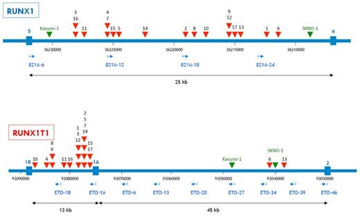
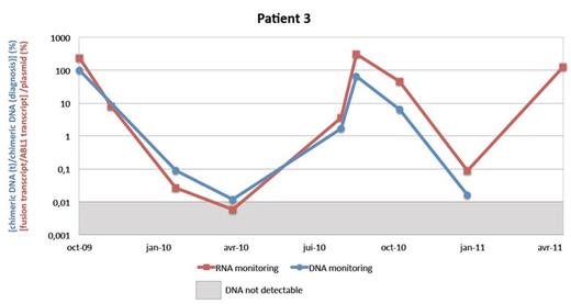
Chromosomal breakpoints were located in RUNX1 intron 5 for all patients. RUNX1T1 breakpoints were located in intron 1b for 15 patients, and in intron 1a for 2 patients (Fig. 1). Quantification failed for 1 patient which was further leave up. Between 2 and 7 follow-up samples were studied for the other patients (median 4.5). DNA monitoring was strongly correlated with RNA monitoring (Fig. 2). Sensitivity threshold, determined by the lowest diagnostic sample dilution which gives a signal, was 10-5 for 7 patients, 10-4 for 6 patients, and only 5.10-4 for 3 patients. MRD was detectable in 31 samples and undetectable in 30 samples by both methods, whereas MRD was detectable only on RNA in 7 samples, probably because of a lack of sensitivity of the RQ-PCR assay. Interestingly, RUNX1T1-RUNX1 DNA was detected in 3 samples from 2 patients who relapsed and for whom RUNX1T1-RUNX1 transcript was undetectable, despite a good RNA quality.
Overall, RUNX1-RUNX1T1 MRD levels on DNA and RNA were quite similar. The level of mRNA expression did not seem to change during follow-up when compared with the amount of DNA. MRD monitoring on genomic DNA is a useful method, but with sensitivity variations depending on the patient's breakpoint sequence and the efficiency of the RQ-PCR assay. DNA has potential advantages: it is more stable than RNA and a best substrate for collection, processing, transport and storage. Additionally, interpretation of the results is easier because it is closely related to the number of leukemic cells. However, this method greatly increases complexity, time of implementation, and cost of monitoring MRD, which limits its interest in routine practice.
No relevant conflicts of interest to declare.
Author notes
Asterisk with author names denotes non-ASH members.


This feature is available to Subscribers Only
Sign In or Create an Account Close Modal