Abstract
Clinical and epidemiological studies support a connection between obesity and thrombosis, involving elevated expression of the prothrombotic molecules plasminogen activator inhibitor-1 and tissue factor (TF) and increased platelet activation. Cardiovascular diseases and metabolic syndrome–associated disorders, including obesity, insulin resistance, type 2 diabetes, and hepatic steatosis, involve inflammation elicited by infiltration and activation of immune cells, particularly macrophages, into adipose tissue. Although TF has been clearly linked to a procoagulant state in obesity, emerging genetic and pharmacologic evidence indicate that TF signaling via G protein-coupled protease-activated receptors (PAR2, PAR1) additionally drives multiple aspects of the metabolic syndrome. TF–PAR2 signaling in adipocytes contributes to diet-induced obesity by decreasing metabolism and energy expenditure, whereas TF–PAR2 signaling in hematopoietic and myeloid cells drives adipose tissue inflammation, hepatic steatosis, and insulin resistance. TF-initiated coagulation leading to thrombin–PAR1 signaling also contributes to diet-induced hepatic steatosis and inflammation in certain models. Thus, in obese patients, clinical markers of a prothrombotic state may indicate a risk for the development of complications of the metabolic syndrome. Furthermore, TF-induced signaling could provide new therapeutic targets for drug development at the intersection between obesity, inflammation, and thrombosis.
Introduction
Evidence from clinical and epidemiological studies has clearly established that obesity predisposes to cardiovascular diseases, insulin resistance, and type 2 diabetes (T2D).1,2 Central obesity is specifically associated with cardiovascular mortality,3,4 and obesity is a significant risk factor for the development of arterial thrombosis and venous thromboembolism.5,6 Moreover, the incidence of venous thromboembolism correlates with epicardial fat thickness7 and abdominal obesity.8 Additional important risk factors for venous thromboembolism in obese patients include inflammation, decreased fibrinolysis, increased thrombin generation, and platelet hyperactivity.9 Interventions such as diet, exercise, and gastric bypass surgery not only result in weight loss and reduced visceral adipose tissue mass, but also improve insulin sensitivity, delay the onset of T2D, and prevent cardiovascular disease. Importantly, weight loss–induced metabolic improvements also reduce levels of prothrombotic mediators, such as plasminogen activator inhibitor-1 (PAI-1)10 and tissue factor (TF),11,12 and ameliorate chronic inflammation and platelet dysfunction.13 Here, we review the mechanisms causing the hypercoagulable state in obesity and examine the emerging evidence that the TF pathway plays complex roles in multiple aspects of obesity and the metabolic syndrome.
TF is a 47 kDa transmembrane glycoprotein that initiates the coagulation cascade by serving as the cell surface receptor for coagulation factor VIIa (FVIIa). TF belongs to the cytokine receptor family, but unlike cytokine receptors, the 23 amino acid cytoplasmic tail of human TF lacks tyrosine kinase recruitment motifs and is, instead, involved in the regulation of integrin function and cell migration.14-16 A soluble, alternatively spliced isoform of TF lacks the transmembrane-anchoring region of full-length TF and has a truncated extracellular domain and a unique carboxyl terminus. This alternatively spliced TF also ligates endothelial integrins and regulates angiogenesis and monocyte recruitment.17,18 The extrinsic coagulation pathway is triggered by the formation of cell surface- or microparticle-associated TF–FVIIa complexes, which leads to factor Xa (FXa)-mediated generation of the downstream coagulation protease thrombin, followed by fibrin deposition and platelet activation. Of relevance for hemostasis, TF forms a hemostatic envelope around vital organs, and is constitutively expressed in skin and gastrointestinal tract epithelia, uterus, placenta, brain, lungs, smooth muscle, and adventitial cells surrounding blood vessels. In contrast, myelomonocytic cells and, perhaps, platelets express low levels of inactive TF. Inflammatory mediators are able to activate these cryptic cell surface pools of TF through protein disulfide isomerase–dependent thiol-disulfide exchange,19 and they can induce transcriptional upregulation20 and messenger RNA (mRNA) splicing21 to increase the levels of blood-borne TF in the vascular compartment.
Evidence for a hypercoagulable state in obesity and T2D
Strong clinical evidence exists that the TF pathway is upregulated in obesity and the metabolic syndrome. Thus, obese patients have higher plasma concentrations of FVII,22 increased levels of thrombin and thrombin–antithrombin (TAT) complexes,23 and increased circulating monocyte TF procoagulant activity.24 Weight loss in morbidly obese patients significantly reduces thrombin generation potential12 and decreases levels of circulating TF; FVII; and prothrombin fragment F1.2, a marker of in vivo thrombin formation.11 Compared with nondiabetic control patients, patients with T2D display signs of hypercoagulability and increased plasma TF procoagulant activity,25,26 increased abundance of TF-positive microparticles,25,27 and higher TF activity of circulating monocytes.28,29
Elevated levels of blood-borne or circulating TF are a biomarker for the severity of microvascular disease in patients with T2D.30,31 Clinical and epidemiological studies of obese patients and patients with T2D have identified changes in markers of the prothrombotic state that are recapitulated in animal models, making such models highly valuable in deciphering the mechanisms by which the hemostatic system contributes to obesity and the metabolic syndrome. In mice, obesity promotes accelerated arterial thrombosis and is associated with elevated levels of circulating TAT complexes.32 In genetically or high-fat diet (HFD)-induced obese mice, TF activity is increased in the blood and adipose tissues, and TF mRNA levels are increased in adipocytes and adipose-infiltrating macrophages.33-35 Similar to humans, calorie restriction and weight loss result in reduced levels of plasma FVII coagulant activity and adipose tissue inflammation.36
Obesity is also associated with increased platelet activation,37 an event that has implications for both thrombosis and inflammation.38 Several platelet activation markers are elevated in obese patients and patients with T2D, including the mean platelet volume; circulating levels of platelet microparticles; thromboxane B2 metabolites; soluble P-selectin; and platelet-derived CD40L, a proinflammatory molecule that induces TF expression in monocytes39 and endothelial cells.40 Platelets respond to chronic inflammation by altering mRNA splicing and protein expression.38 In obesity, transcriptional changes in activated platelets amplify inflammatory processes through pleiotropic interactions with vascular, immune, and stromal cells. Furthermore, platelet function is modulated by several key regulators of body weight and metabolism. For example, platelet-dependent thrombosis is increased by leptin, the satiety hormone produced primarily by the adipose tissue, and is decreased by adiponectin, an insulin-sensitizing adipokine produced exclusively by adipocytes.37 Obesity is also thought to cause resistance to antiplatelet therapy.37 Insulin receptor–mediated signaling sensitizes platelets to the antiaggregating actions of prostaglandin I2 and nitric oxide; thus, insulin resistance in platelets contributes to platelet hyperactivity in obesity and T2D. In addition, adhesion-induced and thromboxane A2–dependent TF expression in platelets is inhibited by insulin in healthy patients but is increased in obese, insulin-resistant patients.41 Importantly, weight loss in obese patients decreases markers of platelet activation and increase their sensitivity to antiaggregating agents.37
TF regulation in obesity
Several hormonal and metabolic changes affect signaling and contribute to elevated TF expression in obesity. Induction of TF in monocytes has been studied in detail and shown to be dependent on the coordinated activation of Toll-like receptor (TLR)-dependent nuclear factor-кB (NF-кB) and Jun N-terminal kinase (JNK) signaling pathways.20 In the local environment of obese adipose tissues, increased lipolysis leads to the release of free fatty acids that activate NF-кB and JNK signaling cascades in adipocytes and adipose tissue macrophages (ATMs).42 A role for lipolysis in TF induction was confirmed by the finding that TF expression in adipocytes is increased by palmitate (F.S., unpublished observations), the most abundant plasma free fatty acid in obesity. Leptin, which is markedly increased in leptin-resistant obese individuals, is able to increase functional TF in human peripheral blood mononuclear cells and neutrophils via Janus kinase 2-dependent mechanisms.43 Treatment of 3T3-L1 adipocytes with leptin or injection of leptin into lean mice also induces adipose tissue TF expression (F.S., unpublished observations). TF expression is negatively regulated by phosphoinositide 3-kinase signaling,44 suggesting that downregulation of this pathway as a consequence of insulin resistance may favor TF upregulation in T2D.28
Increased expression of tumor necrosis factor α (TNF-α) and transforming growth factor β in obese adipose tissue induces TF mRNA in adipocytes and adipose tissue stromal vascular cells, which include macrophages33,34 and fibroblasts. Transforming growth factor β is known to upregulate TF in these cell types in other tissues.45 TF gene induction is also regulated epigenetically via histone acetylation. Inhibition of the protein deacetylase sirtuin-1 promotes TF expression and activity in endothelial cells via the NF-кB/p65 pathway and also induces arterial thrombi.46 Of note, sirtuin-1 expression is reduced in ATMs from HFD-induced obese mice and correlates with adipose macrophage inflammation.47 Interestingly, a number of mediators that protect from obesity and insulin resistance are known to decrease TF expression. For example, peroxisome proliferator-activated receptor-α (PPARα) agonists, which increase energy expenditure and fatty acid oxidation, inhibit TF expression in human monocytes and macrophages.48 Metformin, an antidiabetic agent that decreases weight and reduces atherothrombotic disease, suppresses TF in monocytes via an early growth response 1-dependent pathway.49 Obesity and T2D also correlate with low levels of adiponectin.50 In obese mice, TF–FVIIa signaling blunts the production of adiponectin by adipocytes and may contribute to decreased levels of this adipokine.35 Conversely, adiponectin inhibits TNF-α–induced TF expression in human endothelial cells through inhibition of NF-κB signaling.51
TF-initiated cell signaling events in inflammation
The TF pathway contributes to innate immunity at several levels. The procoagulant and hemostatic functions of TF play a role in defense against infectious microorganisms,52,53 and highly selective interactions of platelets or fibrin with leukocytes and macrophages are crucial for amplifying acute and chronic inflammatory responses.54,55 Indeed, thrombin generated by TF-initiated coagulation is a key signaling protease with pleiotropic proinflammatory effects. Thrombin–platelet signaling in hemostasis and thrombosis has served as a paradigm to understand the molecular mechanisms of protease activated receptor (PAR)-dependent signaling.56 Extracellular proteolysis of a PAR generates an endogenous tethered ligand that initiates G protein–coupled signaling. Thrombin cleaves PAR1, PAR3, and PAR4, whereas a variety of other proteases cleave and activate PAR2.57 PAR signaling is generally conserved between mouse and man, with the notable exception that PAR1 and PAR4 are the primary thrombin receptors in human platelets, whereas PAR3 and PAR4 serve this function in mouse platelets.
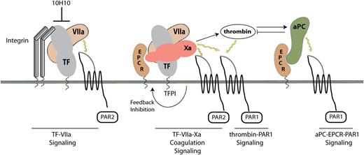
Several additional coagulation proteases can activate PARs in association with other cell surface receptors. This allows limited amounts of coagulation proteases to elicit profound cellular effects in the absence of significant levels of thrombin, such as those typically generated in the context of a hemostatic response. Specifically, TF interacts with upstream coagulation proteases to form 2 distinct cell signaling complexes that cleave PARs and directly influence inflammatory reactions58 (Figure 1). In the ternary TF–FVIIa–FXa coagulation initiation complex, FXa is the primary activator of PAR1 or PAR2. Formation of this ternary complex is inhibited by lipid-raft localized TF pathway inhibitor, which also regulates signaling by the complex.59 Importantly, the endothelial protein C receptor (EPCR) is required for FXa-dependent PAR cleavage by the ternary complex in both human and murine cells.60 EPCR has well-established roles as a coreceptor for anti-inflammatory and cytoprotective activated protein C (aPC) signaling through PAR1.61 EPCR binds with similar affinity to FVIIa and aPC.62 Although ligation of EPCR by aPC or FVIIa has important roles in regulating endothelial cell functions, emerging evidence indicates that EPCR plays a pivotal role in promoting inflammation as a cofactor for FXa in signaling by the TF–FVIIa–FXa complex on hematopoietic cells.63
Schematic overview of TF signaling complexes. The TF–FVIIa binary signaling complex signals via PAR2, whereas the ternary TF–FVIIa–FXa coagulation signaling complex activates both PAR2 and PAR1. TF pathway inhibitor inhibits the coagulation and signaling function of the ternary complex. Thrombin signals through PAR1 to induce inflammatory responses. Binding of aPC to EPCR switches the specificity of PAR1–thrombin signaling to mediate protective anti-inflammatory responses. Antibody 10H10 targets the TF–FVIIa binary signaling complex and disrupts the TF–integrin interaction.
Schematic overview of TF signaling complexes. The TF–FVIIa binary signaling complex signals via PAR2, whereas the ternary TF–FVIIa–FXa coagulation signaling complex activates both PAR2 and PAR1. TF pathway inhibitor inhibits the coagulation and signaling function of the ternary complex. Thrombin signals through PAR1 to induce inflammatory responses. Binding of aPC to EPCR switches the specificity of PAR1–thrombin signaling to mediate protective anti-inflammatory responses. Antibody 10H10 targets the TF–FVIIa binary signaling complex and disrupts the TF–integrin interaction.
In contrast to the ternary TF–FVIIa–FXa complex interaction with PAR1 or PAR2, the binary TF–FVIIa complex initiates an alternative direct TF signaling pathway by cleaving only PAR2 (Figure 1). In myelomonocytic cells and macrophages, TF is typically coagulation inactive (encrypted).64 This coagulation-inactive TF initiates binary TF–FVIIa signaling in association with integrin β1 cell adhesion receptors65 and regulates cell migration through a mechanism involving phosphorylation of the TF cytoplasmic domain.14 Interestingly, evidence that direct TF signaling contributes to both tumor progression and inflammation has come from studies with an antibody (10H10) that specifically inhibits this TF–FVIIa signaling complex without interfering with coagulation.65,66 Studies in mice carrying a cytoplasmic domain–deleted form of TF (TFΔCT), which display normal or even increased TF coagulant function, have provided evidence for noncoagulant roles of TF in angiogenesis15,67 and inflammation.66,68,69 Within this context, PAR2-deficient mice largely phenocopy many features of the TFΔCT mice,35,66,67 indicating that the TF cytoplasmic domain is required for pathological PAR2 signaling. Because tissue macrophages synthesize FVIIa70 and PAR2 can signal as a heterodimer with TLR4,71 it is tempting to speculate that TF–FVIIa may at least partly contribute to PAR2–TLR4 signaling for the regulation of innate immune responses to TLR ligands.
TF cytoplasmic tail-dependent signaling promotes obesity
Although the association of TF with thrombotic complications in cardiovascular diseases is clear, TF also makes coagulation-independent contributions to the pathophysiology of obesity. TFΔCT mice with normal procoagulant activity display attenuated weight gain, increased energy expenditure, and improved glucose homeostasis when fed an HFD. Furthermore, similar protection is afforded by knockout of PAR2 but not PAR1, and double-deficient TFΔCT/PAR2−/− mice show similar, but not more profound, reductions in HFD-induced obesity and insulin resistance.35 In obese adipose tissue, TF is expressed in multiple cell types including adipocytes and ATMs.33-35 Bone marrow chimeric mice showed that the obesity-resistant phenotype is dependent on TF cytoplasmic domain–PAR2 signaling in nonhematopoietic cells. In addition, treating obese mice with an antibody that blocks TF–FVIIa binding rapidly improves metabolism and fatty acid oxidation independently of weight reduction and changes in food intake or activity.
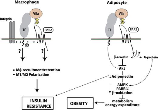
In cultured primary adipocytes, TF cytoplasmic tail–dependent signaling of the TF–FVIIa complex blunts both basal and insulin-mediated Akt (protein kinase B) activation and alters the expression of several Akt target genes causally linked to weight gain, including adiponectin, PPARα, uncoupling protein-2, PAI-1, and TNF-α.35 TF-dependent regulation of these genes in adipocytes in vivo has been confirmed by studies in which bone marrow chimeric mice expressing mouse TF only in nonhematopoietic cells were treated with a mouse TF-specific antibody. TF-regulated expression of adiponectin by adipocytes has central effects on body weight reduction and also regulates glucose uptake, adenosine 5-monophosphate (AMP)–activated protein kinase (AMPK) activation, and fatty acid oxidation.72 Thus, TF–VIIa-mediated suppression of adiponectin reduces energy expenditure, thereby contributing to an obese phenotype (Figure 2). In addition to signaling through G proteins, PAR2 also signals in a G protein–independent manner through cytosolic recruitment of β-arrestin 2. In adipocytes, PAR2 signaling through β-arrestin suppresses AMPK activation,73 but the significance of this pathway remains to be fully elucidated.
Contributions of macrophage and adipocyte TF signaling to obesity and insulin resistance. In adipocytes, FVIIa inhibits both basal and insulin-mediated activation of Akt through a mechanism that requires the TF cytoplasmic domain. Suppression of Akt activity increases insulin resistance and decreases adiponectin synthesis by adipocytes. Reduced systemic levels of adiponectin further blunt insulin signaling and additionally inhibits AMPK and PPARα pathways of energy expenditure and β-oxidation, causing obesity. PAR2 is known to suppress AMPK in a manner dependent on β-arrestin recruitment, and this receptor may therefore contribute to the TF–FVIIa signaling pathway through this specific link. PAR2 may also have an opposing effect through G protein–mediated activation of AMPK in the absence of β-arrestin.73 In adipose tissue, TF–FVIIa–PAR2 signaling may regulate macrophage recruitment and/or retention via phosphorylation-dependent crosstalk between integrins and the cytoplasmic domain of TF. TF–FVIIa–PAR2 signaling and/or TF–integrin interactions activate and sustain M1 polarization of ATMs, contributing to insulin resistance.
Contributions of macrophage and adipocyte TF signaling to obesity and insulin resistance. In adipocytes, FVIIa inhibits both basal and insulin-mediated activation of Akt through a mechanism that requires the TF cytoplasmic domain. Suppression of Akt activity increases insulin resistance and decreases adiponectin synthesis by adipocytes. Reduced systemic levels of adiponectin further blunt insulin signaling and additionally inhibits AMPK and PPARα pathways of energy expenditure and β-oxidation, causing obesity. PAR2 is known to suppress AMPK in a manner dependent on β-arrestin recruitment, and this receptor may therefore contribute to the TF–FVIIa signaling pathway through this specific link. PAR2 may also have an opposing effect through G protein–mediated activation of AMPK in the absence of β-arrestin.73 In adipose tissue, TF–FVIIa–PAR2 signaling may regulate macrophage recruitment and/or retention via phosphorylation-dependent crosstalk between integrins and the cytoplasmic domain of TF. TF–FVIIa–PAR2 signaling and/or TF–integrin interactions activate and sustain M1 polarization of ATMs, contributing to insulin resistance.
TF–PAR2 signaling in hematopoietic cells promotes adipose tissue inflammation and insulin resistance
Obesity-associated chronic inflammation plays an important role in the development of insulin resistance, and visceral adipose tissue is a crucial player in this process. Infiltration of macrophages and other immune cells into adipose tissue is more extensive in obese humans and mice than in lean individuals (Figure 3), and ATMs play a significant role in systemic insulin resistance, T2D, and the metabolic syndrome.74,75 Typically, macrophages participate in tissue homeostasis and express inflammation or angiogenesis regulatory genes when activated.76 Tissue macrophages display remarkable plasticity and can be activated by a variety of environmental cues to display an inflammatory phenotype (M1 macrophages) or a spectrum of alternatively activated phenotypes (M2 macrophages).
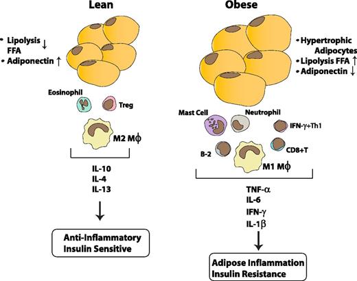
Obesity-associated adipose inflammation. In the lean state, immune cells in adipose tissues (primarily resident M2-like macrophages together with T regulatory (Treg) cells and eosinophils) synthesize IL-10, IL-4, and IL-13 and help to maintain an anti-inflammatory environment that contributes to the insulin-sensitive state. Obesity drives a shift in the number and phenotype of immune cells.74,75 Monocytes are recruited from the blood into the obese adipose tissue, where they become M1 polarized and produce proinflammatory cytokines, including TNF-α, IL-1β, and IL-6, which contribute to insulin resistance. Other changes contributing to the proinflammatory state include decreased numbers of eosinophils and Tregs, and increased numbers of neutrophils, B cells, mast cells, and interferon γ–producing Th1 and CD8+ T cells. The proinflammatory cytokines and chemokines act in autocrine, paracrine, and endocrine manners to promote inflammation and insulin resistance in adipose and other target tissues.
Obesity-associated adipose inflammation. In the lean state, immune cells in adipose tissues (primarily resident M2-like macrophages together with T regulatory (Treg) cells and eosinophils) synthesize IL-10, IL-4, and IL-13 and help to maintain an anti-inflammatory environment that contributes to the insulin-sensitive state. Obesity drives a shift in the number and phenotype of immune cells.74,75 Monocytes are recruited from the blood into the obese adipose tissue, where they become M1 polarized and produce proinflammatory cytokines, including TNF-α, IL-1β, and IL-6, which contribute to insulin resistance. Other changes contributing to the proinflammatory state include decreased numbers of eosinophils and Tregs, and increased numbers of neutrophils, B cells, mast cells, and interferon γ–producing Th1 and CD8+ T cells. The proinflammatory cytokines and chemokines act in autocrine, paracrine, and endocrine manners to promote inflammation and insulin resistance in adipose and other target tissues.
In obesity, ATMs adopt a proinflammatory phenotype (Figure 3) and secrete TNF-α, interleukin 1β (IL-1β), and IL-6, which reduce insulin sensitivity in target cells, specifically adipocytes, hepatocytes, and muscle cells. The initial cues for the recruitment of macrophages into the adipose tissue are chemokines produced by hypertrophic adipocytes, including monocyte chemoattractant protein-1, chemokine (C-X-C motif) ligand 1, and leukotriene B4. Once recruited, ATMs are retained and subsequently activated by binding of free fatty acids and other endogenous danger signals to TLRs, which propagates and sustains the inflammatory response.74,75 Thus, the switch in ATM phenotype from an alternatively activated M2-polarized anti-inflammatory state (surface phenotype F4/80+CD11b+CD11c–) in lean animals to an M1 proinflammatory state (F4/80+CD11b+CD11c+) in obese animals constitutes a key event in the development of insulin resistance (Figure 3). Moreover, M2 macrophage-derived anti-inflammatory genes such as IL-10 are expressed at higher levels in lean mice than in obese mice, where they counteract the effects of proinflammatory cytokines to improve the insulin sensitivity of target tissues.74,75
Although the number of M1 macrophages in adipose tissue is known to correlate with insulin resistance, it is becoming increasingly clear that ATMs do not display fixed phenotypes, but are able to express alternative cytokine profiles in response to changing environmental cues. Crucial regulators for the recruitment and phenotypic maturation of ATMs have been identified, but signals that sustain their proinflammatory phenotype in obesity remain an area of intense investigation. The NF-кB and JNK signaling pathways that promote the development of M1 ATMs also induce TF in monocytes.20 In the inflammation-prone epididymal visceral adipose tissue, TF expression is higher in proinflammatory CD11b+CD11c+ macrophages than in CD11b+CD11c-macrophages. Interestingly, HFD-fed chimeric mice lacking the TF cytoplasmic tail or PAR2 in hematopoietic cells have markedly fewer M1 ATMs compared with HFD-fed wild-type mice.35 Thus, TF signaling in myeloid cells specifically contributes to the recruitment and/or retention of proinflammatory ATMs (Figure 2). These data are consistent with studies showing that TF plays roles in the regulation of reverse endothelial transmigration by monocytes and the maturation of dendritic cells, as well as in cell migration via phosphorylation-dependent crosstalk between integrins and the TF cytoplasmic domain.14,77 In this context, it is of interest that mutation of the α4 chain of the α4β1integrin ligand for TF14 in hematopoietic cells provides protection from adipose tissue inflammation, as seen with TFΔCT mice.78
In addition to their roles in macrophage recruitment, TF and PAR2 also regulate the activation of myeloid cells.66,69 In microglia, lipopolysaccharide signaling in PAR2-expressing cells induces TNF-α and IL-6 production, whereas IL-10 is increased in PAR2-deficient cells.79 ATMs from mice lacking the TF cytoplasmic domain or PAR2 in hematopoietic cells show reduced production of the proinflammatory cytokine IL-6 and increased production of the anti-inflammatory cytokine IL-10 in response to HFD feeding (Figure 2). Because free fatty acids, and potentially other endogenous danger signals produced by stressed adipocytes, activate ATMs through TLR4,80 the attenuated inflammatory phenotype in PAR2−/− mice is consistent with studies demonstrating that PAR2 associates with and contributes to activation of TLR4.71
The inflammatory changes in mice deficient in hematopoietic TF signaling have minimal influence on adiposity and weight gain, but markedly improve insulin sensitivity, overall glucose homeostasis, and hepatic steatosis.35 Increasing evidence suggests that adipose dysfunction and not adiposity per se drives insulin resistance. For example, overexpression of adiponectin in obese mice leads to adipose tissue expansion without inflammation, and maintains insulin sensitivity despite profound obesity.81 Similarly, obese macrophage galactose-type C–type lectin 1-deficient mice are protected from glucose intolerance, insulin resistance, and steatosis despite having more visceral fat.82 Adipocyte-specific deletion of the nuclear receptor corepressor NCoR increases adipogenesis and improves systemic insulin sensitivity.83 Thus, adipose tissue expansion alone is not sufficient to cause insulin resistance in all contexts, and the loss of hematopoietic TF–PAR2 signaling may promote a state of “healthy” obesity. Indeed, a switch in inflammatory cytokine profile and improvement in insulin sensitivity can be achieved by targeting the hematopoietic compartment with the anti-TF antibody 10H10, which disrupts the TF–FVIIa–β1 integrin complex without inhibiting coagulation or downstream coagulation signaling.58,65 However, the precise contribution of TF–FVIIa vs TF–β1 integrin signaling pathways to the macrophage inflammatory phenotype in obesity remains to be determined (Figure 2). The improved insulin sensitivity observed in 10H10-treated mice was associated with a rapid downregulation of TNF-α and IL-6 and upregulation of IL-10 (Figure 2). This rapid phenotypic switch in ATMs in response to TF inhibition is reminiscent of the improved insulin sensitivity and repolarization of macrophages observed in mice after moving from an HFD to a diet low in fat,84 or after treating obese mice with ω-3 fatty acids85 or thiazolidinediones.86 These observations emphasize the plasticity of ATM phenotypes in vivo, and further show that conversion from an M1 proinflammatory to an M2 anti-inflammatory phenotype coincides with increased insulin sensitivity.
Although the role of proinflammatory ATMs in promoting insulin resistance is well documented, it is becoming increasingly clear that ATMs are embedded in regulatory networks involving other immune cells, including T cells, B cells, eosinophils, mast cells, and neutrophils, which participate in the chronic inflammatory response observed in obesity (Figure 3).74,87 Therefore, mast cell tryptase and neutrophil-secreted proteases represent additional potential mechanisms for activation of PAR2 in the adipose tissue.88 In support of this, neutrophils infiltrate obese adipose tissue early after the initiation of an HFD, and increased levels of neutrophil elastase activity are observed in plasma of obese humans and adipose tissues of obese mice.89 Genetic and pharmacologic studies indicate that neutrophil elastase promotes obesity via regulation of AMPK activity and fatty acid oxidation, and additionally decreases expression of high-molecular-weight adiponectin, enhances proinflammatory responses, and increases insulin resistance.89 Although elastase produces a disarming cleavage of PAR2, recent data indicate that the alternative tethered ligand of PAR2 retains biased agonist activity,88 suggesting that elastase may exert its obesity-promoting effects, at least in part, by modulating PAR2 signaling.
Adipocyte–macrophage interactions and prothrombotic responses
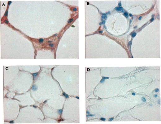
TF expressed by myelomonocytic cells typically remains in a noncoagulant or cryptic state. However, complement activation and danger signals associated with cell damage can rapidly induce TF procoagulant activity through a mechanism that requires thiol-disulfide exchange and protein disulfide isomerase.19,64 Extracellular adenosine triphosphate (ATP) triggers the macrophage P2X7 receptor to activate TF and generate prothrombotic microparticles,64 and this signaling pathway also triggers the release of the proinflammatory cytokines IL-1β and IL-18.90 In vivo, extracellular ATP can be released from apoptotic and necrotic cells, providing an interesting mechanism not only for TF activation but also for P2X7-dependent inflammation.91 The adipose tissue in obesity is characterized by increased adipocyte hypertrophy and cell death. Macrophages are closely associated with apoptotic adipocytes in “crownlike structures,” thus facilitating paracrine crosstalk specific for the obese adipose tissue. Indeed, apoptotic adipocytes contribute to the progression of adipose tissue inflammation.92 Release of free fatty acids because of increased lipolysis, typically observed in obese insulin-resistant adipose tissues, could increase the expression of TF as well as the P2X7 receptor.93 In this scenario, ATP released from adipocytes might trigger local coagulation and inflammation through activation of the P2X7 receptor. In support of possible crosstalk between adipocytes and macrophages, fibrin deposition is increased in obese adipose tissue in regions of crownlike structures (Figure 4). In addition, complement activation occurs in obese adipose tissue,94 which may provide an alternative mechanism for TF prothrombotic activation.19
Fibrin deposition in adipose tissue in obesity. Immunohistochemical staining for fibrin in paraffin sections of adipose tissue, showing increased fibrin deposition (reddish-brown color) in obese mice (A,B) compared with lean mice (C). (D) Negative control staining without the primary anti-fibrin antibody. Slides were counterstained with hematoxylin. Original magnification in all panels, ×400.
Fibrin deposition in adipose tissue in obesity. Immunohistochemical staining for fibrin in paraffin sections of adipose tissue, showing increased fibrin deposition (reddish-brown color) in obese mice (A,B) compared with lean mice (C). (D) Negative control staining without the primary anti-fibrin antibody. Slides were counterstained with hematoxylin. Original magnification in all panels, ×400.
Coagulation activation through TF expressed on nonhematopoietic cells induces PAI-1 expression by adipocytes and indirectly supports TNF-α and IL-6 production by macrophages.35 These results suggest that the adipose tissue microenvironment is prone to increased local fibrin deposition. Moreover, the elevated levels of glucose seen in diabetes cause posttranslational modifications of plasminogen that impair fibrinolysis,95 which may exacerbate the inefficient removal of fibrin and thereby enhance the retention and activation of proinflammatory ATMs. Indeed, integrin αMβ2–fibrin interactions are crucial for macrophage activation and inflammatory cytokine production, providing another unexplored pathway by which TF prothrombotic activity may sustain inflammation and the metabolic syndrome.54,96 Furthermore, thrombin increases the release of inflammatory cytokines and growth factors from adipocytes and inhibits insulin-stimulated Akt activation.97,98 In genetically obese (db/db) mice, treatment with a selective thrombin inhibitor ameliorates insulin resistance and ATM infiltration.98 In mice fed a diet deficient in methionine and choline, a model of hepatic steatosis, TF-dependent thrombin generation and PAR1 signaling drive macrophage and neutrophil accumulation as well as hepatic inflammation.99 Similarly, PAR1 deficiency decreases inflammation and steatosis in mice fed a Western diet.100 Although these data underscore the role of TF-mediated coagulation responses in various aspects of the metabolic syndrome, much remains to be elucidated, including the precise cellular targets of TF and the mechanisms eliciting these responses.
Conclusion
Obesity has reached epidemic proportions in Western societies and is a strong risk factor for the development of insulin resistance, T2D, and cardiovascular disease. Nevertheless, the molecular events that promote these conditions remain incompletely defined. Treatment options remain limited, in part because of the challenge of identifying suitable drug targets among the complex series of signaling pathways and cellular mediators that control the metabolic syndrome. Accumulating evidence supports a role for TF signaling in the development of multiple aspects of the metabolic syndrome, including obesity, insulin resistance, and hepatic steatosis. Although adipose tissue inflammation promotes systemic hypercoagulability and insulin resistance, TF signaling via G protein–coupled receptors adds a new dimension to the connections between obesity and the hemostatic system. Additional studies will be required to identify the specific proteases that activate adipocyte PAR2 and to decipher how these signaling events are coupled to β-arrestin pathways and suppression of downstream AMPK signaling in promoting the obese phenotype. The multicellular interactions that favor adipocyte–macrophage crosstalk and induction of adipose tissue inflammation also need to be unraveled. Further elucidation of the complex interactions between coagulation signaling cascades and the metabolic syndrome are likely to uncover additional targets that could be selectively modulated to correct the metabolic dysfunction and to reduce the thrombotic risk and cardiovascular complications of obesity.
Acknowledgments
We thank Azaam Samad for the preparation of figures.
This study was supported by National Institutes of Health, Heart, Lung and Blood Institute grants HL71146 and HL104232 (F.S.), HL77753 and HL31950 (W.R.), and a grant from the Diabetes National Research Group (F.S.).
Authorship
Contribution: F.S. and W.R. wrote the manuscript.
Conflict-of-interest disclosure: The authors declare no competing financial interests.
Correspondence: Fahumiya Samad, Torrey Pines Institute for Molecular Studies, 3550 General Atomics Court, San Diego, CA 92121; e-mail: fsamad@tpims.org.


This feature is available to Subscribers Only
Sign In or Create an Account Close Modal