Abstract
The vascular endothelial cells line the inner surface of blood vessels and function to maintain blood fluidity by producing the protease plasmin that removes blood clots from the vasculature, a process called fibrinolysis. Plasminogen receptors play a central role in the regulation of plasmin activity. The protein complex annexin A2 heterotetramer (AIIt) is an important plasminogen receptor at the surface of the endothelial cell. AIIt is composed of 2 molecules of annexin A2 (ANXA2) bound together by a dimer of the protein S100A10. Recent work performed by our laboratory allowed us to clarify the specific roles played by ANXA2 and S100A10 subunits within the AIIt complex, which has been the subject of debate for many years. The ANXA2 subunit of AIIt functions to stabilize and anchor S100A10 to the plasma membrane, whereas the S100A10 subunit initiates the fibrinolytic cascade by colocalizing with the urokinase type plasminogen activator and receptor complex and also providing a common binding site for both tissue-type plasminogen activator and plasminogen via its C-terminal lysine residue. The AIIt mediated colocalization of the plasminogen activators with plasminogen results in the rapid and localized generation of plasmin to the endothelial cell surface, thereby regulating fibrinolysis.
Introduction
The vascular endothelium consists of a single cell layer lining all vessels that separates the blood from the tissues. It is estimated to be composed of ∼ 1013 cells, representing a weight of 1.5 kg and an area of 4000 to 7000 m2.1 Endothelial cells play a role in primary hemostasis, coagulation, fibrinolysis, and regulation of vasomotor tone. In addition to regulating the flow of nutrients, the vascular endothelium regulates many diverse biologically active molecules. These functions of the endothelium are achieved through the presence of membrane-bound receptors for various proteins, lipid-transporting complexes, hormones, and metabolites, as well as through specific extracellular proteins and receptors that regulate cell-cell and cell-matrix interactions.2 Whereas exposure to inflammatory and/or septic stimuli rapidly leads to procoagulant behavior, unperturbed endothelial cells provide an anticoagulant environment. After vascular insult, endothelial cells express tissue factor and initiate the coagulation cascade that results in thrombin activation and fibrin clot deposition. At the same time, anticoagulant pathways and fibrinolysis are activated to avoid disseminated coagulation and to also limit fibrin accumulation.3-5 Fibrinogen is a large glycoprotein that constitutes the main component of a fibrin clot. Each fibrinogen molecule is composed of 2 sets of Aα-, Bβ-, and γ-polypeptide chains that form a protein containing 2 distal D regions connected to a central E region by a coiled-coil segment.6 Fibrin is produced on cleavage of the fibrinopeptides by thrombin, which results in the formation of double-stranded half-staggered oligomers that lengthen into protofibrils. The protofibrils then aggregate and branch, yielding a 3-dimensional clot network. Factor XIII, a transglutaminase, cross-links the fibrin, stabilizing the clot and protecting it from mechanical stress and proteolytic attack.7,8 The viscoelastic property of the fibrin clot is essential for its function as it prevents bleeding but still allows the penetration of cells.9 Endothelial cells constitute the inner lining of blood vessels and are responsible for the maintenance of blood fluidity through the inhibition of blood coagulation and promotion of blood clot dissolution, a process known as fibrinolysis.10-12 The fibrinolytic system is composed of the inactive zymogen plasminogen that is proteolytically processed to its active form, plasmin by the action of the plasminogen activators, tissue-type plasminogen activator (tPA), and urokinase-type plasminogen activator (uPA). The tPA and uPA cleave the Arg561-Val562 peptide bond of plasminogen, generating the disulfide bond-linked 2-chain protease, plasmin. tPA is a secretion product of endothelial cells, whereas uPA is produced by renal epithelial cells, monocytes, and endothelial cells stimulated with proinflammatory cytokines.13-18
Increasing evidence has shown that the cellular receptors for plasminogen play a major role in the regulation of fibrinolysis.19-21 Binding of plasminogen to the cells significantly increases the rate of plasmin activation because of colocalization of plasminogen with its activators, tPA and uPA. Indeed, certain plasminogen receptors can also bind tPA, directly stimulating plasmin activation. Cellular receptor-mediated binding of plasminogen also protects the newly generated plasmin from inactivation by α2-antiplasmin, promoting its proteolytic activity.22-25 The plasminogen receptors are typically highly and ubiquitously expressed in cells and show low affinity toward plasminogen.26 The low binding affinity between plasminogen and its receptors is thought to be essential to localize plasmin activity and limit excessive proteolysis. A main characteristic of the plasminogen receptors is the existence of a C-terminal lysine residue that binds to the kringle domains of plasminogen.19,27-29 A few plasminogen receptors have been reported that do not contain a C-terminal lysine residue; nevertheless, it is unclear as to the extent these proteins contribute to cellular plasmin generation.
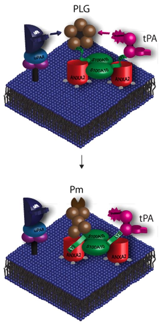
In this review article, we analyze in detail the role of AIIt and its ANXA2 and S100A10 subunits in plasmin generation and fibrinolysis. We develop the theme, based on an exhaustive analysis of the literature, that S100A10 functions as a key plasminogen receptor and that ANXA2 does not function as a plasminogen receptor but rather serves to anchor S100A10 to the cell surface (Figure 1).
AIIt-dependent plasminogen activation. AIIt is expressed on the cell surface of a variety of cells and functions as a plasminogen receptor. AIIt consists of 2 molecules of ANXA2 bound together by a dimer of the protein S100A10. ANXA2 contains phospholipid-binding sites that anchor S100A10 to the cell surface membrane, whereas the C-terminal lysine residue of S100A10 binds tPA and plasminogen. S100A10 has also been shown to colocalize plasminogen with the uPA/uPAR complex. S100A10-induced colocalization of plasminogen with its activators accelerates the proteolytic cleavage of plasminogen, resulting in plasmin generation and therefore enhanced fibrinolytic activity. Both ANXA2 and S100A10 bind to plasmin, protecting it from inactivation by α2-antiplasmin.
AIIt-dependent plasminogen activation. AIIt is expressed on the cell surface of a variety of cells and functions as a plasminogen receptor. AIIt consists of 2 molecules of ANXA2 bound together by a dimer of the protein S100A10. ANXA2 contains phospholipid-binding sites that anchor S100A10 to the cell surface membrane, whereas the C-terminal lysine residue of S100A10 binds tPA and plasminogen. S100A10 has also been shown to colocalize plasminogen with the uPA/uPAR complex. S100A10-induced colocalization of plasminogen with its activators accelerates the proteolytic cleavage of plasminogen, resulting in plasmin generation and therefore enhanced fibrinolytic activity. Both ANXA2 and S100A10 bind to plasmin, protecting it from inactivation by α2-antiplasmin.
Historical perspective
The specific roles of ANXA2 and S100A10 within the ANXA2 heterotetrameric complex in plasminogen binding and activation have evolved significantly since the initial proposal that the ANXA2 monomer functioned as a plasminogen receptor. Studies of the mechanism of interaction between tPA and plasminogen with their cell surface receptors initially established that both tPA and plasminogen interacted with the C-terminal residue of cell surface receptors in a reversible manner.30,31 Therefore, it was surprising that the ANXA2 monomer, a protein that lacked a C-terminal lysine residue, was initially identified as a plasminogen receptor on the surface of endothelial cells.32,33 However, ANXA2 was proposed to interact with plasminogen after a proteolytic cleavage event that liberated a new C-terminal lysine residue (Lys-307). The interaction of ANXA2 with tPA was reported to involve an interaction between Cys-8 of ANXA2 with tPA, suggesting the formation of an irreversible disulfide bond.34 Shortly after these reports, another group demonstrated the presence of S100A10 on the surface of endothelial cells and the reversible interaction of the C-terminal lysine of S100A10 with both tPA and plasminogen.35,36 This group established that S100A10 was present in a heterotetrameric complex with ANXA2 on the surface of many cells35,37-41 and demonstrated that purified AIIt bound tPA and plasminogen via the C-terminal lysine of the S100A10 subunit without the participation of the ANXA2 subunit.36,42,43 With the development of methods to deplete cells of ANXA2 and the report of defective fibrinolysis in the ANXA2 knockout mouse,44 it appeared that ANXA2 might play a role in cellular fibrinolysis. However, subsequent studies demonstrated that depletion of ANXA2 resulted in the concomitant depletion of S100A10,45 and multiple other reports demonstrated that the proposed cleaved form of ANXA2 was undetectable in several cellular model systems of fibrinolysis.37-41 Thus, it became apparent that the extracellular function of ANXA2 was most likely to transport and localize the plasminogen regulatory protein S100A10 to the cell surface. To this end, it was demonstrated that the phosphorylation of ANXA2 regulated its translocation to the extracellular surface.46-49 With the most recent development of an S100A10 knockout mouse and the demonstration of fibrinolytic defects in this mouse,41 the role of S100A10 as an important plasminogen receptor and fibrinolytic regulatory protein was firmly established.
The AIIt structure
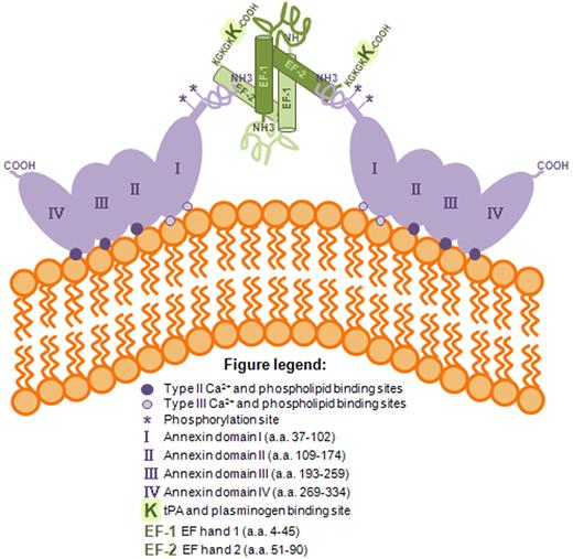
AIIt is composed of 2 molecules of ANXA2 linked together by a dimer of the protein S100A1050-52 (Figure 2). A cryo-electron microscopy low-resolution image of the structure of the AIIt complex bridging 2 phospholipid membranes has shown that the S100A10 subunits of AIIt are positioned in the center, with an ANXA2 molecule on each side, in contact with the membrane surfaces.53 A high-resolution crystal structure of the S100A10 dimer in complex with a peptide that corresponds to the N-terminus of ANXA2 showed that the first 10 amino acids of ANXA2 form an α-helix, which lies in a hydrophobic cleft formed by loop L2 and helix HIV of one monomer of S100A10 and helix HI of the other monomer of S100A10.54 The ANXA2 binding site on the S100A10 dimer primarily encompasses residues in the C-terminal region extending beyond the second EF hand.55,56
Schematic representation of the ANXA2 heterotetramer on the cell surface. ANXA2 belongs to the annexin family of phospholipid and calcium-binding proteins. Annexins bind to anionic phospholipids in a calcium (Ca2+)–dependent manner. All annexins share a conserved domain of 4 repeat sequences ∼ 70 residues long composed of 5 α-helices containing several Ca2+ binding sites. Within the AIIt heterotetrameric complex, ANXA2 is a planar, curved molecule with opposing convex and concave sides. The convex side faces the cellular membrane and contains the Ca2+ and phospholipid binding sites, whereas the concave side faces away from the membrane and contains both the N- and C-terminal regions of ANXA2. The N-terminus of ANXA2 contains the binding site for S100A10, a reactive cysteine residue, phosphorylation sites at Tyr 23 and Ser 14, and a nuclear export signal, whereas the C-terminus contains a reactive cysteine residue and binding sites for F-actin, phospholipid, fibrin, and heparin. S100A10 is a 10-kDa protein containing an N-terminal and a C-terminal EF hand separated by a rather unstructured linker region. One of the main features of S100A10 is the presence of a C-terminal lysine residue that forms a binding site for tPA and plasminogen. AIIt, the predominant form of ANXA2 and S100A10 on the cell surface, is composed of 2 molecules of ANXA2 linked together by a dimer of S100A10. The S100A10 dimer of AIIt is positioned in the center, with an ANXA2 molecule on each side. The first 10 amino acids of ANXA2 form an α-helix, which lies in a hydrophobic cleft formed by loop L2 and helix HIV of one molecule of S100A10 and helix HI of the other S100A10. The ANXA2 binding site on the S100A10 dimer primarily encompasses residues in the C-terminal region extending beyond the second EF hand. The binding of tPA and plasminogen to the complex occurs on the C-terminal lysine of the S100A10 subunit. Removal of the C-terminal lysine results in a heterotetrameric complex that fails to bind tPA and plasminogen and also fails to stimulate tPA-dependent plasminogen activation.29,52
Schematic representation of the ANXA2 heterotetramer on the cell surface. ANXA2 belongs to the annexin family of phospholipid and calcium-binding proteins. Annexins bind to anionic phospholipids in a calcium (Ca2+)–dependent manner. All annexins share a conserved domain of 4 repeat sequences ∼ 70 residues long composed of 5 α-helices containing several Ca2+ binding sites. Within the AIIt heterotetrameric complex, ANXA2 is a planar, curved molecule with opposing convex and concave sides. The convex side faces the cellular membrane and contains the Ca2+ and phospholipid binding sites, whereas the concave side faces away from the membrane and contains both the N- and C-terminal regions of ANXA2. The N-terminus of ANXA2 contains the binding site for S100A10, a reactive cysteine residue, phosphorylation sites at Tyr 23 and Ser 14, and a nuclear export signal, whereas the C-terminus contains a reactive cysteine residue and binding sites for F-actin, phospholipid, fibrin, and heparin. S100A10 is a 10-kDa protein containing an N-terminal and a C-terminal EF hand separated by a rather unstructured linker region. One of the main features of S100A10 is the presence of a C-terminal lysine residue that forms a binding site for tPA and plasminogen. AIIt, the predominant form of ANXA2 and S100A10 on the cell surface, is composed of 2 molecules of ANXA2 linked together by a dimer of S100A10. The S100A10 dimer of AIIt is positioned in the center, with an ANXA2 molecule on each side. The first 10 amino acids of ANXA2 form an α-helix, which lies in a hydrophobic cleft formed by loop L2 and helix HIV of one molecule of S100A10 and helix HI of the other S100A10. The ANXA2 binding site on the S100A10 dimer primarily encompasses residues in the C-terminal region extending beyond the second EF hand. The binding of tPA and plasminogen to the complex occurs on the C-terminal lysine of the S100A10 subunit. Removal of the C-terminal lysine results in a heterotetrameric complex that fails to bind tPA and plasminogen and also fails to stimulate tPA-dependent plasminogen activation.29,52
S100A10 is a member of the S100 family of dimeric EF hand-type Ca2+-binding proteins.29,57 The S100 proteins are small, often acidic polypeptides of approximately 10 kDa, containing an N-terminal and a C-terminal EF hand separated by a rather unstructured linker region.58 Interactions between some S100 protein family members and ANXA2 have been previously reported, including heterotetramer formation between ANXA2 and S100A4,59 S100A6,60 and S100A1061 as well as interactions between ANXA1 and S100A11,62 ANXAVI and S100A1 or S100B,63 ANXAVI and S100A6,60 and ANXAXI and S100A6.64 In contrast to all other S100 proteins, S100A10 is not regulated by Ca2+ because of amino acid replacements in its Ca2+-binding sites that have left this protein unable to coordinate calcium. Therefore, S100A10 does not undergo a calcium-induced conformational change and instead adopts a conformation in the calcium-free state that is very similar to the calcium-bound states of other S100 proteins.54 The majority of intracellular S100A10 is found associated with ANXA2.65,66 ANXA2 not only directs the binding of S100A10 to cellular membranes but is also involved in stabilizing S100A10 protein, protecting it from proteasome-dependent degradation.67,68 One of the main characteristics of S100A10 is the presence of a C-terminal lysine residue that forms a binding site for tPA and plasminogen.36,43
ANXA2 belongs to the annexin family of phospholipid and calcium-binding proteins.50,51,69-71 Annexins bind to anionic phospholipids in a calcium (Ca2+)–dependent manner. All annexins share a conserved domain of 4 repeat sequences ∼ 70 residues long composed of 5 α-helices containing several Ca2+ binding sites. Within the AIIt complex, ANXA2 is a planar, curved molecule with opposing convex and concave sides. The convex side faces the cellular membrane and contains the Ca2+ and phospholipid binding sites, whereas the concave side faces away from the membrane and contains both the N- and C-terminal regions of ANXA2.54,72,73 The N-terminus of ANXA2 contains the binding site for S100A10, a reactive cysteine residue, phosphorylation sites, and a nuclear export signal, whereas the C-terminus contains a reactive cysteine residue and binding sites for F-actin, phospholipid, fibrin, and heparin.50-52 ANXA2 is primarily localized in the cytoplasm and plasma membrane with a small population in the nucleus.74 Many functions have been attributed to ANXA2. Extracellular ANXA2 was originally identified as a receptor for tenascin-C,75 and shortly afterward the ANXA2 monomer was suggested to be a plasminogen receptor.32 These 2 studies disagreed as to whether the cell surface form of ANXA2 was monomeric or AIIt. Cytoplasmic AIIt has been linked to endocytic and exocytic vesicular transport, interaction with cell adhesion molecules, regulation of ion channels, mediation of actin rearrangements, and as a scaffold protein in lipid raft organization.69 One study reported that 15% of cellular ANXA2 was nuclear and was released from the nucleus by RNase A.74,76,77 This is consistent with the report that ANXA2 binds to RNA78 ; nevertheless, the role of nuclear ANXA2 still remains elusive.
The tissue content of ANXA2 and S100A10 has been reported in both avian and mammalian tissues. These proteins are undetectable in heart, smooth muscle, skeletal muscle, and cartilage. Low concentrations of these proteins have been reported in brain, platelets, erythrocytes, and liver, whereas intermediate concentrations have been reported in spleen, kidney, and adrenal gland. High concentrations have been reported in lung, placenta, and intestine. Epithelial cells of the skin, respiratory tract, and intestine, endothelial cells of blood vessels, and connective tissue contain intermediate to high concentrations of ANXA2 and S100A10. Other cell types rich in ANXA2 and S100A10 include macrophages, splenocytes, and fibroblasts.52
The role of AIIt in plasminogen activation
The fibrinolytic cascade initiates through the conversion of the inactive zymogen plasminogen to plasmin by the action of the plasminogen activators, tPA and uPA. The cellular plasminogen receptors play a central role in the regulation of plasmin activity in vascular fibrinolysis via the creation of focal points at the cell surface where plasminogen and its activators colocalize.
A role for AIIt in plasminogen activation was originally proposed by our laboratory.35 We reported that human recombinant AIIt stimulated tPA-dependent plasminogen activation by ∼ 77-fold compared with ∼ 2- and 46-fold for human recombinant ANXA2 and S100A10, respectively.36 We also showed that the loss of the C-terminal lysine residues of S100A10 blocked the ability of AIIt to stimulate tPA-dependent plasminogen activation.36,42 Finally, we showed that the addition of a peptide to the N-terminal 15 amino acids of ANXA2 (S100A10 binding site) to the recombinant S100A10 dimer increased the rate of tPA-dependent plasminogen activation by 2-fold compared with the stimulation by the S100A10 subunit alone.36 The activity of this truncated recombinant AIIt was comparable with the activity of the wild-type (WT) recombinant AIIt, indicating that S100A10 is the subunit directly responsible for plasminogen activation.
Surface plasmon resonance has been used to investigate in real time the molecular interactions between phospholipid-associated AIIt, ANXA2, and immobilized S100A10 with tPA and plasminogen.43 The immobilized S100A10 subunit bound tPA (Kd of 0.45μM), plasminogen (Kd of 1.81μM), and plasmin (Kd of 0.36μM). Furthermore, removal of the 2 C-terminal lysines of S100A10 completely abrogated tPA and plasminogen binding to this protein. However, we did not detect binding of ANXA2 monomer to either tPA or plasminogen, but only to plasmin (Kd of 0.78μM). We also observed that AIIt bound tPA (Kd of 0.68μM), plasminogen (Kd of 0.11μM), and plasmin (Kd of 75nM). When the C-terminal lysines of the human recombinant S100A10 protein were removed by treatment with carboxypeptidase B, we observed that tPA and plasminogen binding was lost. Treatment of AIIt with carboxypeptidase B, which removed the C-terminal lysines of the S100A10 subunit, or recombinant AIIt, constructed from WT ANXA2 and a mutant S100A10 that lacked the C-terminal lysine residues, both failed to bind tPA and plasminogen or to convert plasminogen to plasmin. These studies established that S100A10 was the fibrinolytic subunit of AIIt and that the binding of ANXA2 to S100A10 served to increase the affinity of S100A10 for tPA and plasminogen as well as to enhance the tPA-dependent conversion of plasminogen to plasmin by S100A10.
To clarify the roles played by ANXA2 and S100A10 in fibrinolysis, our laboratory used a human microvascular endothelial cell line, the telomerase immortalized microvascular endothelial (TIME) cell,79 in which cellular levels of S100A10 or ANXA2 were depleted by shRNA. TIME cells depleted of S100A10 showed dramatic losses in both plasminogen binding (50%) and plasmin generation (60%), even though the cell surface ANXA2 levels were identical in the S100A10 knockdown cells compared with the WT cells. Interestingly, TIME cells depleted of ANXA2 showed similar losses in plasminogen binding and plasmin generation as the S100A10-depleted TIME cells.41 It has been shown that ANXA2 stabilizes the levels of S100A10 protein and that, consequently, ANXA2 knockdown cells are also depleted of S100A10.45,67,80,81 Because the decrease in plasminogen binding and plasmin generation were similar for TIME cells depleted of S100A10 compared with TIME cells depleted of both ANXA2 and S100A10, we concluded that the loss of cell surface ANXA2 does not affect plasminogen binding or plasmin generation in these endothelial cells.
The role of ANXA2 in fibrinolysis
As discussed, it was originally proposed that ANXA2 existed on the endothelial cell surface as a monomer and that it was capable of binding tPA and plasminogen.32 However, subsequent studies have established that ANXA2 exists on the cell surface in a complex with S100A10.35,75,82-85 It has also been controversial whether ANXA2 actually binds tPA and plasminogen.29,86 The binding site for tPA on ANXA2 was reported to be a single residue, Cys-8,34 whereas the binding site for plasminogen required proteolytic processing of the protein (Ser-1–Asp-338), resulting in cleavage of the Lys-307–Arg-308 bond and generation of a C-terminal lysine residue (Ser1–Lys-307).33
Studies have shown that purified recombinant or tissue ANXA2 does not bind tPA or plasminogen.43 Although the ANXA2 used in our experiments was in its native conformation, as verified by circular dichroism and functional studies,87 we have been concerned that other studies may have used denatured or conformationally altered ANXA2. The ANXA2 used in other studies was obtained either by passive elution from polyacrylamide gels of partially purified placenta ANXA2 or recombinant ANXA2 expressed as an N-terminal extended and tagged recombinant protein.34 Several reports have shown that plasminogen binds unspecifically to denatured proteins.88-90 Other studies have also shown that N-terminal tagging of ANXA2 significantly changes its conformation, which has repercussions in terms of localization and function(s).91 In addition, more recent work has shown that intact human recombinant ANXA2 does not bind to plasminogen92 and that ANXA2 monomer does not activate plasminogen.59,92 Both these studies further support a role for S100A10 as a plasminogen receptor.
The question that is therefore important to resolve is whether the putative truncated (Ser-1–Lys-307) form of ANXA2 is actually expressed. The predicted molecular weight for this truncated form of ANXA2, which has lost 31 residues, is 32 to 33 kDa, which should be easily distinguished from WT ANXA2 (36 kDa) by Western blot analysis. For instance, we can easily distinguish between WT ANXA2 and plasmin-proteolyzed ANXA2 (Ala-28–Asp-338, a loss of 27 residues) by Western blotting.41 Analysis of cell surface ANXA2 has identified intact ANXA2 but failed to observe any truncated ANXA2.37,39,40 Recently, we examined the molecular weight forms of ANXA2 at the endothelial cell surface by Western blotting.41 Although native ANXA2 was easily detected, we were unable to detect any lower molecular weight (truncated) forms of ANXA2. Similarly, we have been unable to detect any truncated cell surface ANXA2 on macrophages that are actively generating plasmin, on hyperfibrinolytic leukemic promyelocytes or on cancer cells.37,39,40 The truncated form of ANXA2 (Ser-1–Lys-307) has, to the best of our knowledge, never been demonstrated on any cell surface. Indeed, Das et al92 recently showed that the ANXA2 protein localized at the cell surface of macrophages is not proteolytically cleaved because it was recognized by an antibody raised against the last 11 C-terminal amino acids of ANXA2 (residues 328-338). In addition, these authors showed that treatment of J774A.1 and RAW 264.1 macrophages with carboxypeptidase B inhibited the colocalization of ANXA2 with plasminogen. According to the Hajjar model,32 carboxypeptidase B was expected to cleave the C-terminal Lys-307 residue of truncated ANXA2 and by this mechanism block the interaction of ANXA2 with plasminogen. However, the Lys-307 residue could not form the C-terminus of ANXA2 because ANXA2 was detected by these authors with an antibody that recognized the last 11 residues of the C-terminus of intact ANXA2. The simplest explanation of this study is that the colocalization of ANXA2 with plasminogen on the cell surface is the result of its association with S100A10 in the AIIt complex and that carboxypeptidase B-dependent loss of colocalization between ANXA2 and plasminogen is the result of the cleavage of the S100A10 C-terminal lysine residue. In conclusion, these studies further support the proposal that S100A10, but not ANXA2, is a plasminogen receptor in a variety of cells. Furthermore, if the truncated form of ANXA2 constituted the main receptor for plasminogen, this isoform should be the predominant protein on the cell surface, but as discussed, the hypothetical truncated form of ANXA2 has never been identified. We have recently expressed the truncated form of ANXA2 (Ser-1–Lys-307) and observed a clear molecular weight difference compared with ANXA2 WT protein by Western blotting (Figure 3). The molecular weight of the C-terminal truncated ANXA2 was comparable with the N-terminal proteolyzed ANXA2 protein. Therefore, because the truncated ANXA2 has never been detected on the cell surface, a role for ANXA2 as a plasminogen receptor has not been substantiated within the limits of scientific rigor.
Expression of the truncated form of ANXA2. A total of 10 μg of A549 cell extract (lane 1), 200 ng of plasmin proteolyzed, N-terminal truncated ANXA2 (Ala-28–Asp-338, lane 2), or 10 μg of cell extract from BL21 cells expressing ANXA2 (Ser-1–Lys-307, lane 3) was subjected to 10% or 12% SDS-PAGE as indicated. Proteins were transferred onto nitrocellulose membrane and analyzed by Western blotting with the antibodies indicated. The data show that the WT ANXA2 (Ser-1–Asp-338) is easily resolved from the putative plasminogen-binding, C-terminal truncated ANXA2 (Ser-1–Lys-307).
Expression of the truncated form of ANXA2. A total of 10 μg of A549 cell extract (lane 1), 200 ng of plasmin proteolyzed, N-terminal truncated ANXA2 (Ala-28–Asp-338, lane 2), or 10 μg of cell extract from BL21 cells expressing ANXA2 (Ser-1–Lys-307, lane 3) was subjected to 10% or 12% SDS-PAGE as indicated. Proteins were transferred onto nitrocellulose membrane and analyzed by Western blotting with the antibodies indicated. The data show that the WT ANXA2 (Ser-1–Asp-338) is easily resolved from the putative plasminogen-binding, C-terminal truncated ANXA2 (Ser-1–Lys-307).
Cesarman et al reported that ANXA2 bound tPA with high affinity (Kd of 25nM),32 and this measurement was subsequently challenged by Felez et al who stated that the Kd reported by their study was approximately 30-fold lower than the binding of tPA to myeloid and endothelial cells (Kd of 700-800nM).30,31 The proposed role for Cys-8 of ANXA2 as a binding residue for tPA seems counterintuitive because tPA binding to the cell surface has been shown to share a common binding site with plasminogen, to be blocked by lysine analogs such as aminocaproic acid, to be inhibited by 89% by carboxypeptidase B treatment,30,31,93 and to be reversible (ie, tPA does not bind covalently to cell surface receptors).30,31,93,94 Binding of tPA to Cys-8 of ANXA2 implicates the formation of a covalent bond, namely, a disulfide bond, between the 2 proteins, which contradicts the cell surface binding studies for tPA.30,31 The interaction between tPA and a peptide to the N-terminal region of ANXA2 has shown that tPA binding requires only a Cys residue, and the order of the residues surrounding this Cys residue does not affect the interaction with tPA.95 Therefore, if Cys-8 of ANXA2 binds to tPA, it must either form a disulfide; or alternatively, if a disulfide is not formed, then tPA must have a binding site that specifically interacts with Cys-8. Presently, a Cys-binding motif has not been identified in any protein, although cysteine residues of proteins interact noncovalently with certain nucleotide sequences.96
The discrepancy between these results and ours could be explained by the structure of the recombinant ANXA2 used in these studies. The ANXA2, which did not bind tPA, was a recombinant protein (Ser-1–Asp-338) whose structure and function were shown to be identical to tissue ANXA2,43 whereas the recombinant protein that bound tPA was a fusion protein consisting of an N-terminal tag (MASMTGGQQMGRDP) and a C-terminal tag (LEHHHHHH).34 It has been shown that the addition of as few as 6 residues to the N-terminus of ANXA2 causes dramatic changes in its structure.91
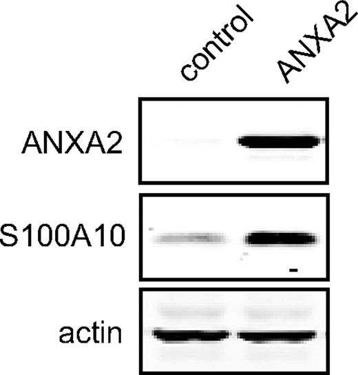
The role of cell surface AIIt in plasminogen binding and activation has also been extensively studied by other laboratories. Nevertheless, the majority of studies that investigate the role of ANXA2 in plasminogen activation neglect the potential role of its binding partner S100A10. For instance, several studies draw conclusions about ANXA2 function using antibodies directed against ANXA2. However, because S100A10 is in close proximity to and interacts with ANXA2 within the heterotetrameric complex, it is unclear from these studies whether the anti-ANXA2 antibodies function by inhibiting ANXA2 or by indirectly altering S100A10 function. The concept of a binding event on one subunit of a protein complex affecting the structure-function of a distinct subunit is not novel and is referred to as allostery.97 Other studies have used RNA interference to deplete ANXA2; however, it is now well established that down-regulation of ANXA2 also results in depletion of S100A10.45,67,80,81 Therefore, loss of a cellular function because of ANXA2 depletion must be interpreted with caution and is merely suggestive of a cellular function regulated by AIIt and not ANXA2. Overexpression of ANXA2 is also another approach used to show increased plasmin activation in cells, but this also up-regulates S100A10 levels in the cells, and this issue is commonly left unaddressed (Figure 4). The same problem arises from attempting to derive conclusions about the physiologic function of ANXA2 with the ANXA2-null mouse model, which, not surprisingly, has been shown to also be essentially void of S100A10 protein.67 Thus, the report of a role for ANXA2 in fibrinolysis, based on fibrin accumulation in the tissues of the ANXA2-null mouse, is not accurate.
Overexpression of ANXA2 in 293T cells up-regulates S100A10 expression. The 293T cells were transfected with either pcDNA3 empty vector (lane 1, control) or pANXA2 vector (lane 2) and incubated at 37°C, 5% CO2 for 72 hours. A total of 20 μg of each protein lysate was subjected to SDS-PAGE, followed by Western blotting with the antibodies indicated.
Overexpression of ANXA2 in 293T cells up-regulates S100A10 expression. The 293T cells were transfected with either pcDNA3 empty vector (lane 1, control) or pANXA2 vector (lane 2) and incubated at 37°C, 5% CO2 for 72 hours. A total of 20 μg of each protein lysate was subjected to SDS-PAGE, followed by Western blotting with the antibodies indicated.
The role of S100A10 in fibrinolysis
It is therefore imperative to use a rigorous approach to address the specific roles of ANXA2 and S100A10 subunits within the AIIt complex when investigating fibrinolysis and other plasminogen-dependent processes. We have proposed that ANXA2 functions to stabilize S100A10 protein levels and to localize S100A10 to the cell surface of endothelial cells, whereas S100A10 is the AIIt subunit that is directly responsible for plasminogen binding and activation during vascular fibrinolysis (Figure 1).29
S100A10 exhibits 3 important properties that directly contribute to its effectiveness as a plasminogen receptor. First, the binding of tPA and plasmin to S100A10 protects these proteins from inactivation by their physiologic inhibitors, PAI-1 and α2-antiplasmin, respectively.36 This is critically important as tPA and plasmin can be rapidly inactivated by these inhibitors in the blood/plasma. Second, S100A10 has also been shown to colocalize plasminogen with the uPA–uPA receptor (uPAR) complex.37,38 We have shown the presence of uPAR in S100A10 immunoprecipitates and have also observed the colocalization of uPAR and S100A10 by immunofluorescence microscopy. Third, plasmin generated by S100A10 participates in the activation of uPA.37 The uPA is secreted by cells in its single-chain pro-enzyme form, pro-uPA. The pro-uPA is rapidly converted to its active 2-chain form by cell-bound plasmin. uPA produced by the action of plasmin on pro-uPA then converts plasminogen to plasmin, participating in an exponential double-reciprocal zymogen activation cycle.98
The role of S100A10 in plasminogen binding, plasmin generation, and protease-directed chemotaxis has been evaluated using cancer cells, macrophages, and endothelial cells (Table 1). Because the truncated form of ANXA2 has been undetectable in these cells, we have ruled out a contribution to these processes by ANXA2. Essentially, these studies have established that S100A10 accounts for the majority of plasminogen binding and plasmin generation in these cells. As a measure of cell surface protease activity, we have used the Boyden chamber invasion assay. In this system, cells are placed on the surface of a Matrigel barrier, and the ability of the cells to proteolyse this barrier and migrate to the opposite side is measured in the presence of a chemotactic agent. This assay measures not only the ability of the cells to generate plasmin but to mobilize plasmin at the cell surface where it is used to proteolyse a pathway through the Matrigel. Because plasmin generated by the cell also activates other proteases released by the cells, the proteolytic activity mobilized by the cells is not only restricted to plasmin but to other plasmin-activated proteases. Results from the Matrigel invasion assay have established that S100A10 regulates not only cell surface plasmin generation but also the plasmin-dependent activation of matrix metalloproteinase-9.39 S100A10 can also regulate other cell surface proteases, such as cathepsin B.99
Percentage of decrease in plasminogen binding, plasmin generation, and chemotaxis resulting from S100A10 depletion
| Cell line . | Plasminogen binding, % . | Plasmin generation, % . | Chemotaxis, % . | Reference . |
|---|---|---|---|---|
| HT1080* (fibrosarcoma) | 28 | 80-95 | 60 | 71 |
| CCL-222* (carcinoma) | 45 | 65 | 38 | 83 |
| LLC* (carcinoma) | ND | 50 | 50 | Unpublished |
| NB4* (APL) | 70 | 70 | 60 | 72 |
| PR9* (histiomonocytic) | 72 | 49 | 55 | 72 |
| Macrophage† | 30 | 45 | 50 | 73 |
| TIME* (endothelial) | 50 | 62 | 74 | 53 |
| MMEC† (endothelial) | 40 | 40 | 55 | 53 |
ND indicates not determined.
S100A10 depletion accomplished by shRNA.
S100A10 depletion was accomplished by gene knockout.
The role of S100A10 in fibrinolysis in vivo
Using the S100A10-null mouse model, we observed enhanced accumulation of fibrin in the tissues compared with WT mouse littermates.41 These results establish that S100A10 plays a major role in fibrinolysis in vivo. In addition, studies with the S100A10-null mice also showed that the fibrin accumulation in the tissues of S100A10-null mice was not the result of increased coagulation because the prothrombin time and activated partial thromboplastin time assays, which directly measure coagulation, were identical in the WT and S100A10-null mice. In another series of experiments, we injected WT and S100A10-null mice with batroxobin and compared the ability of the mice to dissolve the batroxobin-induced blood clots. Batroxobin is a thrombin-like enzyme that rapidly cleaves fibrinogen, resulting in the production of fibrin microclots, which are removed from the vasculature by fibrinolysis. We showed that the S100A10-null mice have dramatically lower rates of fibrinolysis of the batroxabin-induced blood clots in vivo. We also observed that mice lacking S100A10 had a 4-fold reduction in the bleeding time after the tail clip, which was the result of decreased fibrinolysis of the tail clip-induced blood clot. Therefore, a central defect in the S100A10-null mice involves plasmin generation by the endothelium.
The use of Matrigel plugs in mice is a well-established procedure for the measurement of the angiogenic response toward a growth factor stimulus in vivo. We used this methodology to investigate whether S100A10 played a role in angiogenesis. These studies showed that the Matrigel plugs in the S100A10-null mice were poorly vascularized compared with WT mice, indicating that S100A10 is important for angiogenesis. A role for S100A10 in angiogenesis was also supported by the dramatic loss in the ability of S100A10-depleted endothelial cells to migrate through Matrigel barriers. In conclusion, studies with the S100A10-null mouse established an important role for S100A10 in positively regulating fibrinolysis and angiogenesis.
The role of S100A10 in disease
The uPAR is an important regulator of plasmin-dependent extracellular matrix proteolysis. uPAR regulates the generation of plasmin at the extracellular surface of cancer cells by binding uPA and its zymogen form pro-uPA.100 Activated uPA that is bound to uPAR cleaves plasminogen, and the resultant plasmin reciprocally cleaves and activates pro-uPA. This positive-feedback loop is amplified by the localization and concentration of the active and zymogen forms of uPA and plasmin at the cell surface through uPA or pro-uPA binding to uPAR and plasminogen or plasmin binding to plasminogen receptors.101-103 Our demonstration that S100A10 colocalizes with uPAR and contributes to the conversion of pro-uPA to active uPA suggests that S100A10 is an important player in uPA-directed plasmin generation. The activation of proteases at the cell surface of cancer cells plays an important role in the migration of primary tumor cells as well as the establishment of metastatic foci at secondary sites. S100A10 has been shown to play an important role in tumor invasion and metastasis. We have observed that the number of metastatic foci in the lungs of mice decreased 3-fold for the S100A10-depleted HT1080 fibrosarcoma cells and increased 16-fold for the HT1080 cells overexpressing S100A10.37 These results indicate that the ability of these tumor cells to extravasate and metastasize is directly related to extracellular S100A10 expression. Collectively, our results with the HT1080 fibrosarcoma cells establish S100A10 as a potential regulator of tumor cell invasiveness and metastasis.
Recruitment of macrophages in response to an inflammatory stimulus is severely compromised in plasminogen-null mice, indicating that macrophages use the plasminogen activation system for migration to the site of inflammation.104,105 We have reported that the migration of macrophages to an inflammatory stimulus is severely compromised in S100A10-null mice.39 To determine the mechanism for this phenomenon, we isolated macrophages from the S100A10-null mouse and observed that S100A10-null macrophages bound 40% less plasminogen and generated 45% less plasmin compared with WT macrophages. Furthermore, S100A10 deficiency resulted in decreased plasmin generation and decreased matrix metalloproteinase-9 activation in vitro. Taken together, these data highlight the importance of S100A10 in macrophage cell surface plasmin regulation and matrix metalloproteinase-9 activation and, therefore, in the invasive and degradative capabilities of macrophages.
Perhaps one of the most exciting findings was the report that increased S100A10 levels are implicated in the hyperfibrinolytic complications of acute promyelocytic leukemia (APL).40 APL is a special subtype of acute myeloid leukemia whose biologic and clinical features include the presence of the specific t(15;17) chromosomal translocation in leukemic blasts and the frequent existence of severe hemorrhagic diathesis at the time of diagnosis.106-108 The mechanism for the hemorrhagic deaths partially involves hyperfibrinolysis, which occurs because of the excessive production of plasmin at the surface of the promyelocytic leukemic cells.109 We observed that depletion of S100A10 from the APL cell line, NB4, which constitutively expresses the PML-RAR-α oncoprotein, resulted in a 70% loss in plasminogen binding and a 64% loss in plasmin generation by the NB4 cells.40 In addition to measuring plasmin generation, we also examined the ability of NB4 cells to migrate through a fibrin barrier as an independent measure of fibrinolytic activity of the cells. We observed that depletion of S100A10 resulted in 60% fewer NB4 cells migrating through the fibrin barrier. The enhanced fibrinolytic activity of the NB4 cells was reversed by treatment with all-trans retinoic acid, which also causes a loss in cell surface S100A10 levels. Importantly, the induced expression of the PML-RAR-α oncoprotein, the hallmark of APL, increased both cell surface levels of S100A10 and cellular fibrinolytic activity. The enhanced fibrinolytic activity resulting from expression of the PML-RAR-α oncoprotein was inhibited by blocking the expression of S100A10. Overexpression of S100A10 may therefore be responsible for the hemorrhagic complications of APL. These results establish the importance of S100A10 in plasmin generation by promyelocytic leukemia cells and highlights the important role that S100A10 plays in fibrinolysis in general.
Future directions
Studies have identified S100A10 as a key plasminogen receptor and a major regulator of cellular plasmin generation. The accumulation of fibrin in the tissues of the S100A10-null mouse as well as the reduction in fibrinolysis in this mouse model illustrate the importance of S100A10 in fibrinolysis. Two questions remain to be addressed. First, although we have shown that the C-terminal lysine of S100A10 forms the binding site for tPA and plasminogen, it is unclear what other structural determinants are required for tPA or plasminogen binding. If the C-terminal lysine was the sole determinant for tPA or plasminogen binding, then all C-terminal lysine-containing proteins would be tPA and plasminogen receptors; clearly, this is not the case, and other residues must play a role in presenting the C-terminal lysine in the correct 3-dimensional orientation for recognition by the kringle domain(s) of tPA and plasminogen. Second, in addition to S100A10, several other plasminogen receptors have been shown to play an important role in plasmin regulation. These plasminogen receptors include α-enolase,110,111 histone H2B,92 TIP49a,112 and Plg-RTK.113 The challenge of the future will be to identify the biologic processes regulated by this group of plasminogen receptors and to detail if and how these receptors interact with each other. It also remains to be established whether any functional redundancy exists among these or other C-terminal lysine-containing plasminogen receptors.
Acknowledgments
The authors thank Dr P. Svenningsson and Dr P. Greengard for providing S100A10-null mice.
This work was supported by the Canadian Heart and Stroke Foundation of New Brunswick and Nova Scotia, and the Canadian Institutes of Health Research. M.A.S.T. and P.A.M. are trainees of the Beatrice Hunter Cancer Research Institute with funds provided by the Cancer Research Training Program as part of the Terry Fox Strategic Health Research Training Program in Cancer Research at Canadian Institutes of Health Research.
Authorship
Contribution: P.A.M. and D.M.W. wrote the manuscript; P.A.M. and K.D.P. performed experiments; M.A.S.T. designed Figure 1; P.A.M. designed Figure 2; and A.P.S. and V.A.M. edited the manuscript.
Conflict-of-interest disclosure: The authors declare no competing financial interests.
Correspondence: David M. Waisman, Departments of Biochemistry & Molecular Biology and Pathology, Dalhousie University, Halifax, NS B3H 1X5; e-mail: david.waisman@dal.ca.


