Abstract
Fms-like tyrosine kinase-3 (FLT3) inhibitors have been used to overcome the dismal prognosis of acute myeloid leukemia (AML) with FLT3 mutations. Clinical results with FLT3 inhibitor monotherapy have shown that bone marrow responses are commonly less pronounced than peripheral blood responses. We investigated the role of p53 in bone marrow stromal cells in stromal cell-mediated resistance to FLT3 inhibition in FLT3 mutant AML. While the FLT3 inhibitor FI-700 induced apoptosis in FLT3 mutant AML cells, apoptosis induction was diminished under stromal coculture conditions. Protection appeared to be mediated, in part, by CXCL12 (SDF-1)/CXCR4 signaling. The protective effect of stromal cells was significantly reduced by pre-exposure to the HDM2 inhibitor Nutlin-3a. p53 activation by Nutlin-3a was not cytotoxic to stromal cells, but reduced CXCL12 mRNA levels and secretion of CXCL12 partially through p53-mediated HIF-1α down-regulation. Results show that p53 activation in stroma cells blunts stroma cell-mediated resistance to FLT3 inhibition, in part through down-regulation of CXCL12. This is the first report of Nutlin effect on the bone marrow environment. We suggest that combinations of HDM2 antagonists and FLT3 inhibitors may be effective in clinical trials targeting mutant FLT3 leukemias.
Introduction
Activating mutations of the Fms-like tyrosine kinase-3 gene (FLT3) occur in ∼ 30% of patients with acute myeloid leukemia (AML), and are associated with poor prognosis.1-3 FLT3 mutations consist of 2 major types: internal tandem duplication (ITD) of the juxtamembrane domain region (20%-25% of AML patients) and kinase domain (KD) point mutations mostly in codons 835 and 836 (∼ 7% of AML patients). FLT3 mutations result in constitutive activation of the FLT3 receptor kinase in the absence of FLT3 ligand and abnormal activation of downstream signaling pathways, including signal transducer and activator of transcription 5 (STAT5), mitogen-activated protein kinase kinase (Mek)/extracellular signal–regulated kinase (Erk) and phosphatidylinositol-3 kinase (PI3K).4-6 Activation of these downstream effectors up-regulates Mcl-1, Bcl-XL, and survivin, allowing leukemia cells to evade apoptosis.7-11 Mcl-1 is an antiapoptotic Bcl-2 family protein, which plays critical roles in promoting survival of normal and malignant hematopoietic cells. We and others have recently reported that FLT3 inhibition has been shown to reduce Mcl-1 levels and induce Bax activation, resulting in mitochondrial apoptosis in FLT3/ITD AML cells.7,11
FLT3 inhibitors have recently been used clinically to improve the dismal prognosis of AML with FLT3/ITD mutations.12-16 However, while circulating blasts are rapidly eliminated, bone marrow responses are in general less impressive.17,18 One potential explanation for the reduced bone marrow response compared with the striking activity against circulating blast cells may be microenvironmental resistance to FLT3 inhibitors. In this context, the impact of CXCL12 (also known as stromal cell-derived factor-1: SDF-1)/CXCR4 signaling has recently been linked to chemoresistance in FLT3/ITD AML.19-23 Marrow-derived stromal cells constitutively secrete CXCL12, and increased CXCR4 expression has been associated with FLT3/ITD AML.20,21 Overexpression of FLT3/ITD mutants in Ba/F3 cells has been associated with activated CXCL12/CXCR4 signaling.22 Furthermore, we have reported that CXCR4 inhibition increased the sensitivity of FLT3/ITD AML cells to apoptosis induced by the FLT3 inhibitor sorafenib in stromal cocultures,23 and have initiated a clinical trial to validate this concept in patients using sorafenib and the CXCR4 antagonist AMD3100 (NCT00943943).
There is evidence that transformed cells are more sensitive to p53-induced apoptosis than their normal counterparts, leading to the suggestion that activation of p53 may cause tumor-specific cell killing. Small molecule inhibitors have been described that disrupt HDM2–p53 binding, which liberate p53 from its inhibitor and enable p53 activation.24,25 One of these compounds, Nutlin-3, binds HDM2 in the p53-binding pocket, effectively dislodges p53 from HDM2 and induces pronounced p53 response, inhibits growth and induces p53-mediated apoptosis in AML cells.26 An ongoing clinical trial in leukemias with encouraging results seems to provide “proof-of-concept” (NCT00623870).27
Here we investigated if p53 activation of bone marrow stromal cells affects stromal cell-mediated resistance to FLT3 inhibition in FLT3/ITD AML.
Methods
Reagents
The selective FLT3 inhibitor FI-700 and the multi-kinase inhibitor KW-2449 (FLT3, ABL, ABL-T315I and Aurora kinase inhibitor) were kindly provided by Dr Yukimasa Shiotsu (Kyowa Hakko Kirin, Shizuoka, Japan).28,29 The small-molecule antagonists of HDM2, Nutlin-3a and its 150 times less active enantiomer Nutlin-3b (diluted in DMSO, as 10mM stock solution), were kindly provided by Dr Lyubomir T. Vassilev (Hoffmann-La Roche, Nutley, NJ).25 Recombinant human CXCL12/SDF-1α was purchased from R&D Systems. Doxorubicin, AraC and the CXCR4 antagonist AMD3100 were purchased from Sigma-Aldrich.
Cell culture
Human adult bone marrow mesenchymal stromal cells (MSCs) were isolated by bone marrow aspirates from the iliac crest of normal healthy volunteers or AML patients after informed consent, according to institutional guidelines per Declaration of Helsinki. Mononuclear cells were collected by gradient centrifugation and seeded at a density of 1 × 10⁵ cells/cm2 in growth medium containing MEM-α and 20% heat-inactivated FBS and 1% l-glutamine. The nonadherent cells were removed after 2 days. Medium was changed every 3-4 days thereafter. When 70%-80% confluent, adherent cells were trypsinised and expanded for 3-5 weeks. MSCs were checked for positivity of CD105, CD73, and CD90 and the lack of expression of CD45 and CD34.30 The purity of MSC preparation was > 99%.
To study the effect of MSCs on leukemia cells, bone marrow and peripheral blood samples were obtained from AML patients (> 70% blasts) after informed consent, according to institutional guidelines per Declaration of Helsinki. Mononuclear cells were collected by gradient centrifugation and AML cells were cultured with or without a layer of MSCs. MSCs were seeded at 3 × 10⁴ cells/cm2 in 12-well plates in MEM-α medium containing 10% heat-inactivated FBS and 1% l-glutamine (hereafter referred to as “MEM-α medium”) for 72 hours before the addition of AML cells. In some experiments, MSCs were pretreated with 10μM Nutlin-3a or -3b for the last 24 hours of the culture period. The wells were washed 3 times with MEM-α medium, and 4 × 10⁵ AML cells in 1 mL MEM-α medium were added to each well. The cells were then exposed to compounds. Floating and attached AML cells were collected together after attached cells were removed by vigorous pipetting. Conditioned medium was collected from MSC cultures after 24 hours of incubation and filtered through a 0.22-μm filter. Control medium was simultaneously obtained by incubation of MEM-α medium in the absence of cells. MOLM-13 cells were purchased from Deutsche Sammlung von Mikroorganismen und Zellkulturen GmbH (DSMZ), and MV4-11 and HL-60 cells were purchased from American Type Culture Collection. MOLM-13 and MV4-11 cells have wild-type p53, while p53 is disabled by a large deletion of TP53 in HL-60.11,26 MOLM-13 and MV4-11 cells have FLT3/ITD, while HL-60 cells have wild-type FLT3.11 Cell lines were seeded at a density of 2 × 10⁵ cell/mL. Cell viability was evaluated by triplicate counts of trypan blue dye excluding cells.
Transfection of p53 siRNA
MSCs were transfected with small interfering RNA (siRNA) oligonucleotides in 12-well plates using Lipofectamine 2000 according to manufacturer instructions (Invitrogen). To evaluate the transfection efficiency, cells were transfected with the BLOCK-iT Fluorescent Oligo (Invitrogen). Efficiency of transfection was 98%, with > 95% cell viability at 72 hours. Cells were transfected with negative control siRNA (12 935-400; Invitrogen) or with p53 siRNA (12 935-035; Invitrogen). Twenty-four hours after transfection, some cells were subsequently treated with 10μM Nutlin-3a.
Tetracycline-inducible mutant HIF-1α MSCs
A Tet-On advanced inducible gene expression system was used to generate stably transduced normal bone marrow MSCs expressing a degradation-resistant HIF-1α mutant in a tetracycline-inducible manner. In the HIF-1α mutant, the proline residues 402 and 564 within the oxygen-dependent degradation domain of HIF-1α were mutated to alanine and the mutant became insensitive to oxygen-dependent proteasomal degradation. The transduced cells were selected with 2 μg/mL puromycin for 2 weeks. Doxycycline-induced HIF-1α and CopGFP expression was confirmed by immunoblotting and fluorescence microscopy, respectively.
Apoptosis analysis
For the sub-G1 assay, cells were fixed in ice-cold ethanol (70% vol/vol) and stained with propidium iodide solution (25 μg/mL propidium iodide, 180 U/mL RNase, 0.1% Triton X-100, and 30 mg/mL polyethylene glycol in 4mM citrate buffer, pH 7.8; Sigma-Aldrich). The DNA content was determined using a FACSCalibur flow cytometer (Becton Dickinson Immunocytometry Systems). Cells with a hypodiploid DNA content were counted as apoptotic on the basis of DNA fragmentation. Cell debris was defined as events in the lowest 10% range of fluorescence and eliminated from analysis. Annexin V binds specifically to phosphatidylserine, a lipid that is normally on the inside of the cell membrane but is exposed on the cell surface early in the apoptotic process. For annexin V binding studies, cells were washed twice with binding buffer (10mM HEPES, 140mM NaCl, and 5mM CaCl2 at pH 7.4) and incubated with FITC-conjugated annexin V (Roche Diagnostics). Stained cells were analyzed by flow cytometry while membrane integrity was simultaneously assessed by propidium iodide exclusion. All experiments were conducted in triplicate.
Immunophenotype analysis and CXCR4 expression by flow cytometry
Cells were stained with phycoerythrin (PE)-conjugated antibodies against CD34, CD45, CD73, CD90, CD105, and CD184 (CXCR4; BD Pharmingen), or isotype controls. Cells were stained for individual antigens and analyzed by flow cytometry.
Quantitation of intracellular proteins by flow cytometry
Involvement of BAX conformational change was analyzed using an antibody directed against the NH2-terminal region of BAX (YTH-6A7; Trevigen), as previously reported.31 Cellular fixation, permeabilization and staining with primary antibody or an isotypic control were performed using the Dako IntraStain kit (Dako Cytomation), according to manufacturer's instructions. After washing, cells were incubated with Alexa Fluor 488 chicken anti–mouse secondary antibodies (Invitrogen) for 30 minutes at 4°C. Cleaved caspase-3 was labeled with FITC-conjugated anti-active caspase-3 antibody (BD Pharmingen).
Western blot analysis
Equal amounts of protein lysate were separated by SDS-PAGE (12% gel) for 2 hours at 80 V. Proteins were transferred to nitrocellulose membrane, immunoblotted with primary antibodies followed by infrared secondary antibodies (LI-COR Biosciences), and detected by the Odyssey imaging system (LI-COR Biosciences). The following antibodies were used: mouse monoclonal anti-p53 (Santa Cruz Biotechnology); mouse monoclonal anti–phospho-p53 (Ser15; Cell Signaling Technology); mouse monoclonal anti–Mcl-1 (BD Pharmingen); mouse monoclonal anti–Bcl-X (BD Pharmingen); rabbit polyclonal anti-survivin; mouse monoclonal anti–HIF-1α (BD Transduction Laboratories); and mouse monoclonal anti–β-actin (Sigma-Aldrich). An anti–β-actin blot was used as a loading control.
Quantitative real-time PCR
RNA was prepared from cells using a RNeasy Mini Kit (QIAGEN), and first-strand cDNA was generated using random hexamers (SuperScript III First-Strand Synthesis SuperMix; Invitrogen) from 1 μg total RNA. The mRNA expression levels were quantified using TaqMan gene expression assays (CXCL12: Hs00171022_m1, IL-6: Hs00985639_m1, IL-8: Hs00174103_m1, IL-11: Hs00174148_m1, M-CSF: Hs00174164_m1, GM-CSF: Hs00929873_m1, FLT3 ligand: Hs00181740_m1, BAX: Hs00180269_m1, FAS: Hs00163653_m1, BBC3 (PUMA): Hs00248075_m1, CDKN1A (p21): Hs00171132_m1, HDM2: Hs00242813_m1, 18S: Hs99999901_s1, Applied Biosystems) on a 7900HT Fast Real-Time PCR System. The level of expression was calculated based on the PCR cycle number (Ct) at which the exponential increase in fluorescence from the probe exceeds a certain threshold value. For each sample, relative gene expression level was determined by subtracting the Ct value of the housekeeping gene 18S rRNA to the Ct value of the target gene (ΔCt = CtTarget gene – Ct18S rRNA). Relative quantification (fold change) between different samples (eg, treated vs control) was then determined according to the 2–ΔΔCt method (ΔΔCt = ΔCttreated sample – Ctcontrol sample).
ELISA
CXCL12 and FLT3 ligand levels in culture media were quantified using ELISA (Quantikine Human CXCL12/SDF-1α Immunoassay and Quantikine Human Flt-3/Flk-2 Ligand Immunoassay; R&D Systems) according to the manufacturer's protocol.
Cell senescence assay
Senescence-associated β-galactosidase (SA-β-Gal) assays were carried out using a commercial kit (Cell Signaling Technology), according to the manufacturer's instructions.
Statistical analysis
Statistical analysis was performed using the 2-tailed Student t test, or 1-way ANOVA followed by Tukey multiple comparison tests if indicated (GraphPad Prism 5; GraphPad Software). In experiments using primary AML samples, statistical analyses on data from the same samples (all FLT3 mutants or all FLT3 wild types) were performed using the 2-tailed paired Student t tests. Analyses on data from different samples (comparing FLT3 mutants with wild types) were performed using the 2-tailed 2-sample Student t tests. Results were considered statistically significant at P values < .05. Unless otherwise indicated, average values were expressed as mean ± SD. In some cases, average values were expressed as mean ± SEM.
Results
MSCs protect FLT3/ITD AML cells from the cytotoxicity of FLT3 inhibition
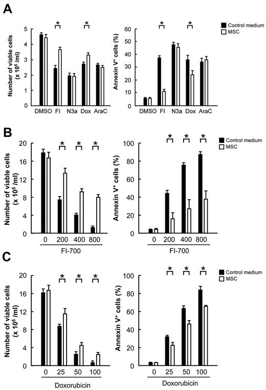
First we examined the effect of coculture with MSCs on the protection of FLT3/ITD AML cells from drug-induced apoptosis. FLT3/ITD-expressing MOLM-13 cells were treated with 800nM FI-700, 2μM Nutlin-3a, 100nM doxorubicin, 200nM AraC or left untreated and cultured in the presence or absence of MSCs for 24 hours, and the numbers of viable cells and annexin V–positive fractions were measured. Doxorubicin and AraC were included in this experiment since they are highly effective chemotherapeutic agents for the therapy of AML and remain the backbone of induction and consolidation regimens.32 Doxorubicin and AraC induce apoptosis in leukemia cells, which is a key determinant of drug response in AML.33-35 As shown in Figure 1A, coculture with normal MSCs protected MOLM-13 cells from FI-700 and doxorubicin-induced apoptosis (P < .05). Evidence for the protective effect was not obtained in Nutlin-3a or AraC, even when cells were exposed for longer time period (48 or 72 hours) or to a range of compound concentration (0.5-10μM Nutlin-3a and 50-1000nM AraC; data not shown). Importantly, the protective effect was more prominent for FI-700 than for doxorubicin (Figure 1B-C). The protective effect of MSC on the survival of MOLM-13 cells treated with FI-700 was well maintained for 72 hours (Figure 1B) and FI-700 itself did not induce apoptosis in MSCs when added alone to the MSC (data not shown). Doxorubicin did not induce apoptosis in MSCs either (data not shown). We investigated if MSCs protect MV4-11 or HL-60 cells from FI-700–induced apoptosis. FLT3/ITD MV4-11 cells were sensitive to FI-700 (4.8 ± 0.2% annexin V–positive cells in untreated cells vs 45.4 ± 1.1% in 400 nM FI-700–treated cells at 24 hours; P < .05), and coculture with normal MSCs partially protected MV4-11 cells from FI-700–induced apoptosis (23.4 ± 2.0% annexin V–positive cells in 400nM FI-700–treated cells at 24 hours; P < .05 vs 400nM FI-700–treated cells in medium). FLT3 wild-type HL-60 cells were resistant to FI-700, irrespective of the presence of MSCs (data not shown). These results suggest that MSCs actively protect FLT3/ITD AML cells from the cytotoxicity of FLT3 inhibition. FI-700 induced G1-phase cell cycle arrest and apoptosis in FLT3/ITD-expressing cells, and increases in annexin V–positive cells were detectable as early as 16 hours after exposure.11 On the other hand, cell viabilities determined by trypan blue dye exclusion were minimally decreased (> 90%) at 24 hours. In the following experiments, we took advantage of the annexin V–binding assay that detects early stage apoptotic cells more efficiently and specifically than the trypan blue dye exclusion assay.
MSCs protect FLT3/ITD AML cells from FI-700- and doxorubicin-induced cell death. (A) FLT3/ITD MOLM-13 cells were treated with 800nM FI-700 (FI), 2μM Nutlin-3a (N3a), 100nM doxorubicin (Dox), 200nM AraC or left untreated and cultured for 24 hours in the presence or absence of MSCs from 3 normal subjects, and the numbers of viable cells and annexin V–positive fractions were measured. (B-C) MOLM-13 cells were treated for 72 hours with the indicated concentrations of FI-700 (B) or doxorubicin (C) in the presence or absence of MSCs, and the numbers of viable cells and annexin V–positive fractions were measured. Asterisk (*) indicates significance at P < .05.
MSCs protect FLT3/ITD AML cells from FI-700- and doxorubicin-induced cell death. (A) FLT3/ITD MOLM-13 cells were treated with 800nM FI-700 (FI), 2μM Nutlin-3a (N3a), 100nM doxorubicin (Dox), 200nM AraC or left untreated and cultured for 24 hours in the presence or absence of MSCs from 3 normal subjects, and the numbers of viable cells and annexin V–positive fractions were measured. (B-C) MOLM-13 cells were treated for 72 hours with the indicated concentrations of FI-700 (B) or doxorubicin (C) in the presence or absence of MSCs, and the numbers of viable cells and annexin V–positive fractions were measured. Asterisk (*) indicates significance at P < .05.
Nutlin-pretreated MSCs exhibit reduced protection of MOLM-13 cells from FI-700– and doxorubicin-induced apoptosis
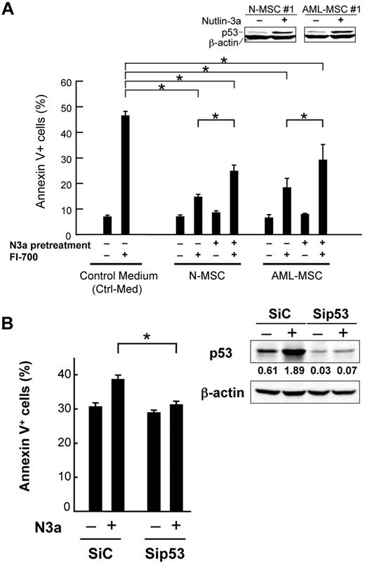
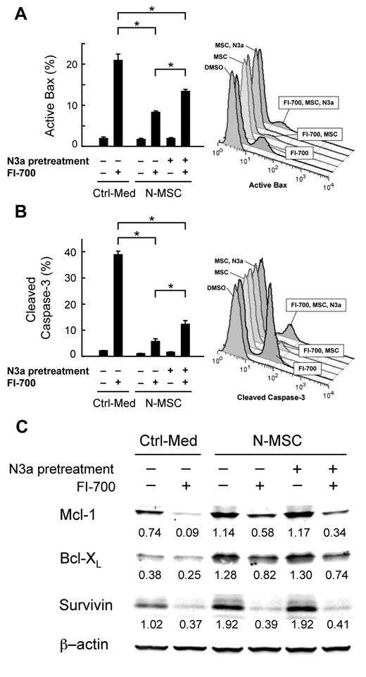
Chemotherapeutic agents for the therapy of AML may activate p53 in normal cells as well as in leukemia cells. The functional consequence of p53 activation in AML/MSC interactions, however, remains unknown. We investigated if p53 activation in MSCs affects their protective effect from FLT3 inhibition-induced apoptosis. Pretreatment of MSCs with 10μM Nutlin-3a for 24 hours protected MOLM-13 cells from FI-700–induced apoptosis significantly less than did the untreated MSCs (P < .05; Figure 2A). Nutlin-3b did not affect the protective effect of MSCs (supplemental Figure 1, available on the Blood Web site; see the Supplemental Materials link at the top of the online article), excluding the possibility of an off-target effect. There was no significant difference between normal MSC and AML-MSC (Figure 2A). To further confirm that the attenuated protection after Nutlin-3a treatment depends on p53 activation in MSCs, p53 levels were reduced in MSCs using siRNA. p53 siRNA led to reduced basal and Nutlin-induced p53 protein expression by > 95% (Figure 2B). As expected, p53 knockdown MSCs were less sensitive to Nutlin-induced de-protection against FI-700–induced apoptosis than control siRNA cells (P < .01; Figure 2B). FI-700 induces apoptosis in FLT3/ITD AML cells through the mitochondrial apoptotic pathway. We therefore investigated if MSC protects MOLM-13 cells from Bax and caspase-3 activation after FI-700 treatment and if Nutlin-3a pretreatment of MSC abrogates the protection. As shown in Figure 3A-B, Nutlin-3a–pretreated MSCs protected MOLM-13 cells from FI-700–induced Bax conformational change and caspase-3 activation significantly less than did the untreated MSCs (P < .05). These findings suggest that p53 activation in MSCs partially abrogates their protective effect from FLT3 inhibition-induced mitochondrial apoptosis. It has been reported that FLT3/ITD up-regulates Mcl-1, Bcl-XL, and survivin to promote leukemia survival.7-11 We investigated if coculture with MSCs results in up-regulation of Mcl-1, Bcl-XL, or survivin in FLT3/ITD-expressing AML cells and if Nutlin-3a pretreatment of MSCs affects their expression. As shown in Figure 3C, MOLM-13 cells expressed higher levels of Mcl-1, Bcl-XL, and survivin under stromal cocultures compared with cells maintained in medium alone, suggesting that MSCs actively support FLT3/ITD-expressing AML cell survival. MSCs prevented the FLT3 inhibitor FI-700 from reducing Mcl-1 and Bcl-XL levels. Importantly, FI-700–treated AML cells expressed lower levels of Mcl-1 and Bcl-XL when cocultured with Nutlin-pretreated MSCs, compared with cocultures with Nutlin-naive MSCs. FI-700 reduced Mcl-1 (but not Bcl-XL) levels to below basal levels in cells under coculture. Decreased Mcl-1 levels were closely associated with increased percentages of cells with Bax activation (Figure 3A), cleaved caspase-3 (Figure 3B) or annexin V–positive cells (Figure 2A), supporting the notion that Mcl-1 is required for the survival of FLT/ITD-expressing AML cells. In contrast to Mcl-1 or Bcl-XL, MSCs did not protect MOLM-13 cells from survivin reduction after FI-700 treatment, suggesting a minor role of survivin in MSC-mediated leukemia cell protection. In FLT3 wild-type HL-60 cells, FI-700 did not significantly induce Bax conformational change, caspase-3 activation or Mcl-1 reduction (data not shown).
p53-activated MSCs exhibit reduced protection of FLT3/ITD AML cells from FI-700–induced apoptosis. (A) MSCs were pretreated with 10μM Nutlin-3a (N3a) for 24 hours. After the wells were washed 3 times with MEM-α medium (control medium: Ctrl-Med), MOLM-13 cells were treated with 800nM FI-700 for 24 hours in the presence or absence of MSCs from 3 normal subjects (N-MSC) or 3 AML patients (AML-MSC). Asterisk (*) indicates significance at P < .05 (1-way ANOVA/Tukey). (B) MSCs were transfected with either control (SiC) or p53 siRNA (Sip53). Twenty-four hours posttransfection, cells were subsequently treated with 10μM Nutlin-3a (N3a). After washing, MOLM-13 cells were treated with 800nM FI-700 for 24 hours. Intensity of the immunoblot signals was quantified and the relative intensity of p53 compared with β-actin was calculated.
p53-activated MSCs exhibit reduced protection of FLT3/ITD AML cells from FI-700–induced apoptosis. (A) MSCs were pretreated with 10μM Nutlin-3a (N3a) for 24 hours. After the wells were washed 3 times with MEM-α medium (control medium: Ctrl-Med), MOLM-13 cells were treated with 800nM FI-700 for 24 hours in the presence or absence of MSCs from 3 normal subjects (N-MSC) or 3 AML patients (AML-MSC). Asterisk (*) indicates significance at P < .05 (1-way ANOVA/Tukey). (B) MSCs were transfected with either control (SiC) or p53 siRNA (Sip53). Twenty-four hours posttransfection, cells were subsequently treated with 10μM Nutlin-3a (N3a). After washing, MOLM-13 cells were treated with 800nM FI-700 for 24 hours. Intensity of the immunoblot signals was quantified and the relative intensity of p53 compared with β-actin was calculated.
Nutlin-pretreated MSCs exhibit reduced protection of FLT3/ITD AML cells from FI-700–induced mitochondrial apoptosis. Normal MSCs (N-MSC) were pretreated with 10μM Nutlin-3a (N3a) for 24 hours. MOLM-13 cells were treated with 800nM FI-700 for 24 hours in the presence or absence of MSCs. Bax conformational change (A) and activation of caspase-3 (B) were analyzed by flow cytometry. Representative flow cytometry results are also shown. Asterisk (*) indicates significance at P < .05 (1-way ANOVA/Tukey). (C) Mcl-1, Bcl-XL and survivin levels were determined by Western blotting. Intensity of the immunoblot signals was quantified and the relative intensity compared with β-actin was calculated.
Nutlin-pretreated MSCs exhibit reduced protection of FLT3/ITD AML cells from FI-700–induced mitochondrial apoptosis. Normal MSCs (N-MSC) were pretreated with 10μM Nutlin-3a (N3a) for 24 hours. MOLM-13 cells were treated with 800nM FI-700 for 24 hours in the presence or absence of MSCs. Bax conformational change (A) and activation of caspase-3 (B) were analyzed by flow cytometry. Representative flow cytometry results are also shown. Asterisk (*) indicates significance at P < .05 (1-way ANOVA/Tukey). (C) Mcl-1, Bcl-XL and survivin levels were determined by Western blotting. Intensity of the immunoblot signals was quantified and the relative intensity compared with β-actin was calculated.
Since MSCs protected MOLM-13 cells from doxorubicin-induced apoptosis, we investigated if Nutlin-induced p53 activation in MSCs reduces their protective effect. Pretreatment of MSCs with 10μM Nutlin-3a for 24 hours protected MOLM-13 cells from doxorubicin-induced apoptosis significantly less than did the untreated MSCs (P < .01; supplemental Figure 2).
MSCs protect MOLM-13 cells by production of soluble factors, regulated by p53 activation
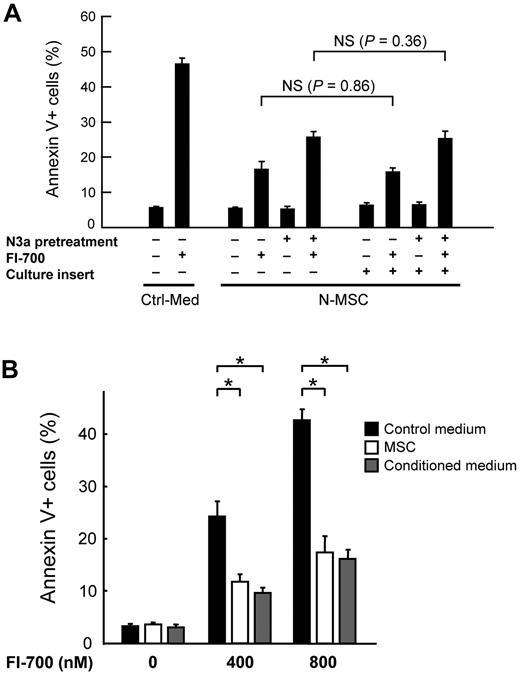
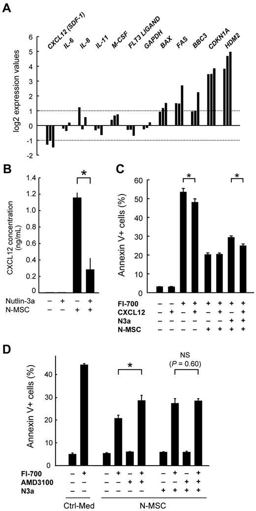
To investigate if the protection is mediated by MSC-leukemia cell contact or soluble MSC-derived factors, we cocultured MOLM-13 cells with normal MSCs in the cell culture inserts that prevented direct contact of the 2 cell populations. As shown in Figure 4A, the presence of culture inserts did not significantly affect the protective effect of Nutlin-pretreated or untreated MSCs, suggesting that soluble MSC-derived factors are important for the protection. The idea that secretable factors are strongly implicated in the protection was supported by the data that stromal cell-conditioned medium efficiently protected MOLM-13 cells from FI-700–induced apoptosis (Figure 4B). It has been reported that MSCs secrete multiple chemokines and cytokines to support hematopoiesis.36 Since p53 functions as a transcription factor, changes in mRNA levels of secretable factors after Nutlin-3a treatment were determined in MSCs from 3 normal subjects. As expected, Nutlin-3a treatment led to a significant induction of p53 target genes including Bax, FAS, BBC3, CDKN1A and HDM2 (Figure 5A). Induction of CDKN1A and HDM2 was more pronounced than that of the proapoptotic genes Bax, FAS or BBC3. Importantly, Nutlin-3a significantly reduced CXCL12 mRNA levels by 57.2 ± 6.7% (P < .01 vs untreated cells). mRNA levels of IL-6, IL-8, IL-11, M-CSF, and FLT3 ligand were similar in untreated and Nutlin-treated MSCs. The mRNA of GM-CSF was not detected in MSCs. Consistent with RQ-PCR, MSCs treated with Nutlin-3a secreted lower levels of CXCL12 than untreated MSCs (P < .01, n = 5; Figure 5B). Mean CXCL12 concentration in the supernatant was reduced by 75.6%. Nutlin-3a treatment affected CXCL12 expression in AML-derived MSCs as well as in normal MSCs (supplemental Figure 3). Nutlin-3a significantly reduced CXCL12 mRNA levels of AML-derived MSCs by 67.7 ± 7.8% (P < .01 vs untreated cells) and the mean CXCL12 concentration in the supernatant by 67.4% (P < .01 vs untreated cells). FI-700 treatment did not significantly affect CXCL12 expression in normal and AML-derived MSCs (data not shown). Recently, it has been reported that FLT3 ligand impedes bioactivity of FLT3 inhibitors in FLT3 mutant AML patients.37 In that publication, plasma FLT3 ligand levels rose to a mean of 1148 pg/mL in relapsed patients and FLT3 ligand levels greater than 1 ng/mL impaired the cytotoxic effects of FLT3 inhibition. We thus investigated FLT3 ligand levels in MSC supernatant (n = 5). FLT3 ligand was not detectable (< 7 pg/mL) in any of the supernatant samples. We next investigated a contribution of the CXCL12/CXCR4 signaling in protecting FLT3/ITD AML cells from FLT3 inhibition-induced apoptosis. The addition of 1 ng/mL CXCL12 to control medium partially protected MOLM-13 cells from FI-700–induced apoptosis (P < .05; Figure 5C), while 200 μM AMD3100, a small nonpeptide CXCR4 inhibitor, blunted the protection conferred by MSCs in MOLM-13 cells (P < .01; Figure 5D), suggesting an involvement of the CXCL12/CXCR4 signaling in the protective mechanisms. CXCL12 partially (46%) rescued MOLM-13 cells from de-protective effect of Nutlin-3a on MSCs (P < .05; Figure 5C). Interestingly, addition of AMD3100 did not sufficiently sensitize MOLM-13 cells to FI-700 when MSCs were pretreated with Nutlin-3a (Figure 5D), supporting the hypothesis that Nutlin-3a abrogates MSC-mediated protection by disturbing the CXCL12/CXCR4 signaling.
MSCs protect MOLM-13 cells by production of soluble factors, regulated by p53 activation. (A) Normal MSCs (N-MSC) were pretreated with 10μM Nutlin-3a (N3a) for 24 hours. MOLM-13 cells were treated with 800nM FI-700 for 24 hours in the presence or absence of normal MSCs. Cells were collected from upper compartment when culture inserts were used. NS indicates not significant. (B) MOLM-13 cells were treated with the indicated concentrations of FI-700 in MEM-α medium (control medium), MSC-conditioned medium (conditioned medium) or with normal MSCs. Asterisk (*) indicates significance at P < .05 (1-way ANOVA/Tukey).
MSCs protect MOLM-13 cells by production of soluble factors, regulated by p53 activation. (A) Normal MSCs (N-MSC) were pretreated with 10μM Nutlin-3a (N3a) for 24 hours. MOLM-13 cells were treated with 800nM FI-700 for 24 hours in the presence or absence of normal MSCs. Cells were collected from upper compartment when culture inserts were used. NS indicates not significant. (B) MOLM-13 cells were treated with the indicated concentrations of FI-700 in MEM-α medium (control medium), MSC-conditioned medium (conditioned medium) or with normal MSCs. Asterisk (*) indicates significance at P < .05 (1-way ANOVA/Tukey).
Nutlin-3a treatment reduces CXCL12 expression in MSCs and may affects CXCL12/CXCR4 signaling in AML cells. (A) MSCs from 3 normal subjects were treated for 24 hours with 10μM Nutlin-3a, and transcripts were quantitated by real-time PCR. Each real-time PCR was performed in duplicate, and the average fold induction relative to untreated cells is shown. (B) MSCs from 5 normal subjects (N-MSC) were cultured for 48 hours in the presence or absence of 10μM Nutlin-3a, and CXCL12 concentrations in the culture medium were determined. Results are expressed as mean ± SEM (C-D) MOLM-13 cells were treated with 800nM FI-700, 1 ng/mL CXCL12 and 200μM AMD3100 for 24 hours in the presence or absence of MSCs, and annexin V–positive fractions were measured. In some cases, MSCs were pretreated with 10μM Nutlin-3a (N3a) for 24 hours. Asterisk (*) indicates significance at P < .05.
Nutlin-3a treatment reduces CXCL12 expression in MSCs and may affects CXCL12/CXCR4 signaling in AML cells. (A) MSCs from 3 normal subjects were treated for 24 hours with 10μM Nutlin-3a, and transcripts were quantitated by real-time PCR. Each real-time PCR was performed in duplicate, and the average fold induction relative to untreated cells is shown. (B) MSCs from 5 normal subjects (N-MSC) were cultured for 48 hours in the presence or absence of 10μM Nutlin-3a, and CXCL12 concentrations in the culture medium were determined. Results are expressed as mean ± SEM (C-D) MOLM-13 cells were treated with 800nM FI-700, 1 ng/mL CXCL12 and 200μM AMD3100 for 24 hours in the presence or absence of MSCs, and annexin V–positive fractions were measured. In some cases, MSCs were pretreated with 10μM Nutlin-3a (N3a) for 24 hours. Asterisk (*) indicates significance at P < .05.
The activation of the p53 pathway, either by strong DNA damage or by oncogenic stress, is known to result in apoptosis or senescence. We investigated if transient activation of p53 by Nutlin-3a induces apoptosis or senescence in normal MSCs (n = 3) or AML-MSCs (n = 3), to exclude the possibility that Nutlin-3a induces apoptosis or senescence thereby reducing CXCL12 expression in MSCs. After primary MSCs were exposed to 10μM Nutlin-3a for 96 hours, the cells were analyzed for sub-G1 apoptotic cell population or stained for senescence-associated β-galactosidase. Nutlin-3a did not increase the percentage of sub-G1 cell population or that of β-galactosidase–positive cells (data not shown), suggesting that MSCs are resistant to transient p53 activation-induced apoptosis and senescence.
Nutlin-pretreated MSCs exhibit reduced protection of primary AML cells from FI-700–induced apoptosis
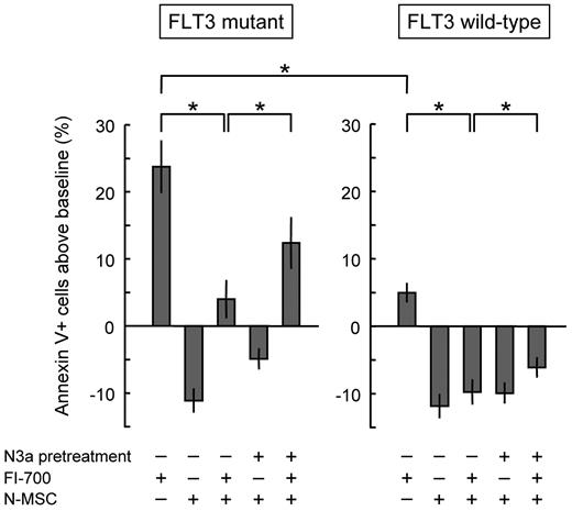
We tested Nutlin-pretreated and untreated MSCs for their ability to protect primary AML cells from spontaneous and FI-700–induced apoptosis. Of 30 AML samples analyzed 15 were positive for mutant FLT3 (13 cases had FLT3/ITD and 2 cases had D835/836 mutation). FLT3 mutant cells were more sensitive to FI-700–induced apoptosis than FLT3 wild-type cells (P = .0001; Figure 6). MSCs protected both FLT3 mutant (P < .0001) and FLT3 wild-type cells (P < .0001) from FI-700–induced apoptosis. Nutlin-3a significantly abrogated MSC-mediated resistance to FI-700 in both FLT3 mutant (P < .0001) and FLT3 wild-type (P < .05) samples. The de-protective effect of Nutlin-3a was higher in FLT3 mutant samples (8.4 ± 1.3%, mean ± SEM) than FLT3 wild-type samples (3.6 ± 1.5%; P < .05). Data suggest hat Nutlin-3a may abrogate the protection conferred by MSCs in primary AML patient samples.
Nutlin-3a abrogates MSC-mediated resistance to FI-700 in primary AML cells. Fifteen FLT3 mutant samples and 15 FLT3 wild-type samples were treated for 72 hours with 800nM FI-700 in the presence or absence of MSCs. Annexin V–positive fractions were determined. MSCs were either Nutlin-3a–pretreated or untreated. In each patient sample, the percentage of annexin V–positive cells in control medium (spontaneous apoptosis) was set as “0” (control) and the extent of apoptosis was expressed as percent changes from this baseline. Results are expressed as mean ± SEM. Asterisk (*) indicates significance at P < .05.
Nutlin-3a abrogates MSC-mediated resistance to FI-700 in primary AML cells. Fifteen FLT3 mutant samples and 15 FLT3 wild-type samples were treated for 72 hours with 800nM FI-700 in the presence or absence of MSCs. Annexin V–positive fractions were determined. MSCs were either Nutlin-3a–pretreated or untreated. In each patient sample, the percentage of annexin V–positive cells in control medium (spontaneous apoptosis) was set as “0” (control) and the extent of apoptosis was expressed as percent changes from this baseline. Results are expressed as mean ± SEM. Asterisk (*) indicates significance at P < .05.
Nutlin-pretreated MSCs exhibit reduced protection of MOLM-13 cells from KW-2449–induced apoptosis
KW-2449 is a multi-kinase inhibitor in clinical development, with a potent activity of FLT3.29 We investigated if Nutlin-3a treatment of MSCs can abrogate MSC-mediated leukemia cell protection against KW-2449. In a manner similar to FI-700, MSCs partially protected MOLM-13 cells from KW-2449–induced apoptosis, which was blunted by Nutlin-3a pretreatment of MSCs (P < .05; supplemental Figure 4).
Nutlin-3a reduces CXCL12 mRNA levels in MSCs partially through the HIF-1α pathway
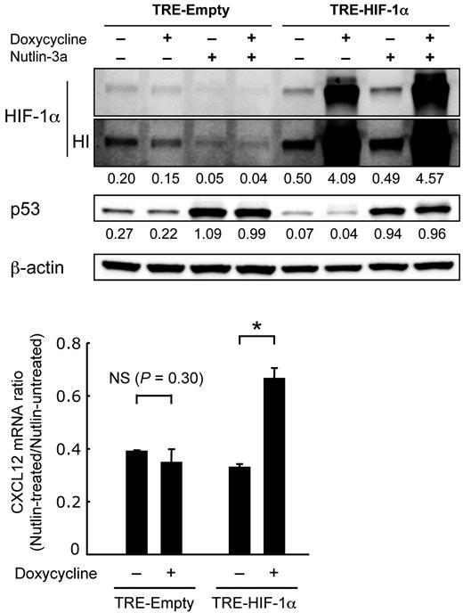
The promoter of CXCL12 does not have p53 binding sites but contains 2 HIF-1α binding sites.38,39 HIF-1α is a critical mediator of MSC responsiveness to bone marrow hypoxia and directly up-regulates CXCL12 expression. On the other hand, p53 has been reported to promote proteasomal degradation of HIF-1α protein.40 To investigate a possible role of HIF-1α degradation in p53-mediated CXCL12 down-regulation, we introduced a doxycycline-inducible HIF-1α P402A/P564A mutant into normal MSCs. In the HIF-1α mutant, the proline residues 402 and 564 within the oxygen-dependent degradation domain of HIF-1α were mutated to alanine and the mutant became insensitive to proteasomal degradation. As shown in Figure 7, Nutlin-3a treatment down-regulated wild-type HIF-1 but not HIF-1α P402A/P564A mutant. Nutlin-3a reduced CXCL12 mRNA levels in the empty vector-transduced MSCs, irrespective of doxycycline administration. Induced HIF-1α mutant significantly abrogated CXCL12 mRNA reduction by Nutlin-3a (P < .01), implying that Nutlin-3a reduces CXCL12 mRNA levels in MSCs partially through down-regulation of HIF-1α.
Nutlin-3a reduces CXCL12 mRNA levels in MSCs partially through the HIF-1α pathway. MSCs were treated with 10μM Nutlin-3a for 24 hours in the presence or absence of 0.1 μg/mL doxycycline. Intensity of the immunoblot signals was quantified and the relative intensity compared with β-actin was calculated. The bar graphs show the ratio of the CXCL12 mRNA level in Nutlin-treated cells compared with that in Nutlin-untreated cells. Asterisk (*) indicates significance at P < .05. HI indicates high intensity image.
Nutlin-3a reduces CXCL12 mRNA levels in MSCs partially through the HIF-1α pathway. MSCs were treated with 10μM Nutlin-3a for 24 hours in the presence or absence of 0.1 μg/mL doxycycline. Intensity of the immunoblot signals was quantified and the relative intensity compared with β-actin was calculated. The bar graphs show the ratio of the CXCL12 mRNA level in Nutlin-treated cells compared with that in Nutlin-untreated cells. Asterisk (*) indicates significance at P < .05. HI indicates high intensity image.
Nutlin-3a more potently induces p53-mediated responses compared with doxorubicin or AraC in MSCs
Doxorubicin and AraC are potential p53 activators and have p53-dependent and p53-independent mechanisms to induce apoptosis in AML cells. To investigate if doxorubicin or AraC efficiently induce p53 and p53-mediated response in MSCs, normal MSCs from 4 normal subject were treated with 20 or 100nM doxorubicin, 1 or 10μM AraC, 10μM Nutlin-3a or left untreated and cultured for 48 hours. These conditions did not affect cell viability of MSCs. The steady-state plasma concentration in patients treated with 60-75 mg/m2 doxorubicin is 15-20nM, and 10μM AraC corresponds to the concentration achieved in serum of patients treated with high-dose AraC (1-3 g/m2/d). As shown in supplemental Figure 5, the amplitude of induction of p53 and its gene targets (FAS, p21, and HDM2) and repression of CXCL12 mRNA by chemotherapeutic agents doxorubicin and AraC was smaller relative to that by Nutlin-3a, suggesting that HDM2 inhibition has high potential to induce p53-mediated responses in MSCs.
Discussion
Our findings confirm that selective FLT3 inhibition may not eradicate FLT3 mutant AML cells protected by stroma and show, for the first time, that p53 activation in stroma cells blunts stroma cell-mediated resistance to a kinase, here a FLT3 inhibitor, in part through regulation of CXCL12. FLT3/ITD AML cells have been reported to express CXCR4 and rely on bone marrow stroma-derived CXCL12 for optimal growth and survival.20-23 TP53 mutations are rare in mutant FLT3 AML and, more importantly, the presence of FLT3/ITD has been associated with high sensitivity to HDM2 inhibitors in AML.41 Furthermore, concomitant Inhibition of HDM2 and mutant FLT3 has been reported to synergistically induce leukemia cell death in FLT3/ITD AML.11 Taken together, pharmacologic p53 activation by HDM2 inhibition is expected to enhance FLT3 inhibition-induced leukemia cell death not only by activation of p53 in mutant FLT3 AML cells but also by depriving those cells of the protective benefits afforded by bone marrow stroma cells.
In the present study, CXCL12 partially rescued FLT3/ITD-expressing AML cells from the de-protective effect of Nutlin-3a on MSCs. The CXCR4 antagonist AMD3100 blunted MSC–mediated protection with the same efficiently as Nutlin-3a. We used CXCL12 at 1 ng/mL, based on the observed CXCL12 concentrations in MSC supernatant that efficiently protected FLT3/ITD-expressing AML cells from FI-700–induced apoptosis. Therefore, CXCL12 should not be the only mediator involved in the attenuation of MSC-mediated leukemia cell protection after Nutlin-3a treatment. Recent studies have shown that the CXCL12/CXCR4 relation is not exclusive.42-46 Macrophage migration inhibitory factor and extracellular ubiquitin have been shown to be CXCR4 ligands that activate CXCR4 signaling, and importantly, the activated CXCR4 signaling can be blocked by the receptor antagonist AMD3100.42-44 On the other hand, it has been reported that CXCL12 binds to and signals through CXCR7.45 AMD3100 also binds to the alternative CXCL12 receptor CXCR7.46 These findings suggest that AMD3100 may influence a broad range of cell-cell communication signaling. Multiple MSC-derived factors should be involved in the attenuation of MSC-mediated protection after Nutlin-3a treatment.
Normal MSC and AML-derived MSC showed similar phenotype in terms of leukemia cell protection from FLT3 inhibition. Both MSCs protect FLT3/ITD AML cells from FI-700–induced apoptosis to a similar extent and importantly, p53 activation by Nutlin resulted in reduced protection. The origin of MSCs in patients with de novo AML is not currently unknown.47 Considering the rarity of TP53 mutations, however, it is anticipated that treatment with HDM2 inhibitors would affect MSCs as well as AML cells even if the MSCs are derived from AML progenitors. The hypothesis that MSC-mediated leukemia cell protection is abrogated by HDM2 inhibition may be applied to conventional chemotherapy to AML, as Nutlin-pretreated MSCs exhibited reduced protection of AML cells from the anthracycline doxorubicin-induced apoptosis. Anthracyclines are commonly used in the treatment of acute leukemias, and remain the backbone of induction and consolidation regimens. Nutlin-3a has been found to synergize with doxorubicin to induce apoptosis in AML,26 further supporting the idea that HDM2 inhibitor could be combined with FLT3 inhibitors and/or anthracyclines in the treatment strategies to overcome resistance. HDM2 inhibitor R7112 (Nutlin-3 analog) is being evaluated in a phase 1 study to determine the safety and dose in leukemia (NCT00623870) and solid tumor (NCT00559533) patients, and has shown to activate p53 signaling, as expected.27
Previously proposed strategies for overcoming stroma-mediated resistance to FTL3 inhibitors include the Smac mimetic LBW242,48 the CXCR4 inhibitor AMD3465,23 and the PIM1 inhibitor K00486.21 The latter 2 agents interfere with the CXCL12/CXCR4 signaling of AML/MSC interactions at the level of CXCR4, as AMD3465 is a competitive CXCR4 antagonist and K00486 treatment induces decreased surface CXCR4 expression in AML cells. Pharmacologic activation of p53 by Nutlin-3a, on the other hand, reduced CXCL12 production in MSCs without affecting CXCR4 expression levels in AML cells. Although p53 is not a universal repressor of CXCL12 expression, it has been reported that p53 can repress the production of CXCL12 by cultured normal human and mouse fibroblasts.49 The promoter of CXCL12 does not contain p53 binding sites38 and previous studies have found that the silencing of p53 does not lead to changes in the CXCL12 promoter activity,50 suggesting that p53 down-regulates CXCL12 via indirect repressive mechanisms. In this context, the CXCL12 promoter contains 2 HIF-1α binding sites.39 HIF-1α is a critical mediator of MSC responsiveness to bone marrow hypoxia and directly up-regulates CXCL12 expression. HDM2 inhibition has been reported to actively down-regulate HIF-1α through p53 activation.51 We found that an induced degradation-resistant HIF-1α mutant significantly abrogated CXCL12 mRNA reduction by Nutlin-3a, suggesting that HIF-1α is actively involved in the Nutlin–mediated down-regulation of CXCL12. As the HIF-1α mutant did not completely abrogate CXCL12 mRNA reduction by Nutlin-3a, there should be additional mechanisms that contribute to the Nutlin–mediated down-regulation of CXCL12. Future studies of unexplored mechanisms of CXCL12 regulation by p53 would lead to important new insights relevant to the cancer microenvironment and may identify new targets for the development of novel therapeutic drugs.
We propose that combinations of HDM2 inhibitors with FLT3 inhibitors and chemotherapy can be investigated in clinical trials targeting FLT3 mutant leukemias, with the dual goal of inducing apoptosis in leukemia cells and, concomitantly, reduce the protective effects exerted by the marrow microenvironment.
The online version of this article contains a data supplement.
The publication costs of this article were defrayed in part by page charge payment. Therefore, and solely to indicate this fact, this article is hereby marked “advertisement” in accordance with 18 USC section 1734.
Acknowledgments
The authors thank Vivian Ruvolo, Duncan Mak, Zhihong Zeng, and Rui-Yu Wang for technical help. The authors also thank Dr Shinako Takada for valuable discussions.
This work was supported in part by grants from National Institutes of Health Lymphoma SPORE (CA136411), P01 “The Therapy of AML” (CA55164), Leukemia SPORE (CA100632), and the Paul and Mary Haas Chair in Genetics (to M.A.).
National Institutes of Health
Authorship
Contribution: K.K. designed and performed the research, analyzed the data, and wrote the paper; T.M., Y.C., R.J., N.S. and E.S. assisted in research; M.K. contributed to discussion; X.H. analyzed the data; and M.A. designed the research, analyzed the data, and edited the paper.
Conflict-of-interest disclosure: The authors declare no competing financial interests.
Correspondence: Michael Andreeff, MD, PhD, Section of Molecular Hematology and Therapy, M. D. Anderson Cancer Center, The University of Texas, 1515 Holcombe Blvd, Unit 448, Houston, TX 77030; e-mail: mandreef@mdanderson.org.

