Abstract
Severe sepsis leads to massive activation of coagulation and complement cascades that could contribute to multiple organ failure and death. To investigate the role of the complement and its crosstalk with the hemostatic system in the pathophysiology and therapeutics of sepsis, we have used a potent inhibitor (compstatin) administered early or late after Escherichia coli challenge in a baboon model of sepsis-induced multiple organ failure. Compstatin infusion inhibited sepsis-induced blood and tissue biomarkers of complement activation, reduced leucopenia and thrombocytopenia, and lowered the accumulation of macrophages and platelets in organs. Compstatin decreased the coagulopathic response by down-regulating tissue factor and PAI-1, diminished global blood coagulation markers (fibrinogen, fibrin-degradation products, APTT), and preserved the endothelial anticoagulant properties. Compstatin treatment also improved cardiac function and the biochemical markers of kidney and liver damage. Histologic analysis of vital organs collected from animals euthanized after 24 hours showed decreased microvascular thrombosis, improved vascular barrier function, and less leukocyte infiltration and cell death, all consistent with attenuated organ injury. We conclude that complement-coagulation interplay contributes to the progression of severe sepsis and blocking the harmful effects of complement activation products, especially during the organ failure stage of severe sepsis is a potentially important therapeutic strategy.
Introduction
Severe sepsis is a multistage, multifactorial, and life-threatening clinical syndrome that arises through the innate response to infection and can appear as a complication in conditions like trauma, cancer, and surgery.1 Despite important strides made in understanding its pathophysiology, the sepsis-related mortality and morbidity rates still remain unacceptably high. Sepsis affects approximately 700 000 people and accounts for approximately 210 000 deaths per year2 in the United States alone. In its most fulminant form, sepsis can produce cardiovascular collapse and death within hours. More common is the development of multiple organ failure (MOF) secondary to hypoperfusion and intravascular thrombosis. The MOF may run a protracted clinical course and eventually proves fatal in 30% to 40% of patients. The mechanisms responsible for the persistent and progressive organ failure are less understood. To examine this problem we have developed nonhuman primate models of Escherichia coli sepsis, which, depending on the bacterial dose, mimic the different pathophysiologic syndromes observed in clinical practice.3 Challenge with 1010 cfu/kg E coli (LD100) results in an explosive inflammatory and coagulopathic response leading to irreversible shock and death. The administration of a lower dose, 109 cfu/kg E coli (LD50), produces transient hypotension followed by MOF, which may progress and prove fatal in approximately 50% of the animals. The pathophysiology of the LD50 model demonstrates a 2-stage response, each stage driven by distinct mechanisms. The first stage is an exacerbated intravascular host defense response to bacterial infection; the second stage is an uncontrolled extravascular host recovery response driven by ischemia-reperfusion (IR) injury, which leads to MOF.3 In the most severe cases death occurs rapidly through septic shock.
Complement is critical for the innate immunity against pathogens but uncontrolled complement activation has been associated with many immuno-inflammatory conditions.4 All 3 complement activation pathways—the classical (CP), the lectin (LP), and the alternative (AP)—converge at C3, which is cleaved by CP-, LP- and AP-generated C3 convertases to C3a and C3b. The anaphylatoxin C3a activates platelets, induces their aggregation, and recruits leukocytes. C3b participates in the formation of C5 convertase, which cleaves C5 to C5a and C5b, the latter becoming part of the terminal C5b-9 complex (TCC).5 Elevated levels of C5a could signal through its receptors C5aR and C5L2, contributing to immune paralysis, multiorgan dysfunction, apoptosis, deterioration of the coagulation/fibrinolytic system and contractile dysfunction of the cardiomyocytes.6 We have previously gestured a biphasic activation of the complement cascade in response to sublethal E coli in baboons with maximum peak of complement activation products occurring during the second stage.7,8 Data suggest that early increase of complement activation during sepsis may relate to bacteria opsonization,7,9 thus being beneficial in the host defense response. In contrast, complement activation during the second stage of sublethal sepsis via either CRP or mannose-binding lectin (MBL) could be a major contributor to IR injury, organ dysfunction, and, finally, death.10 We hypothesized that complement inhibition could constitute a therapeutic target for sepsis-induced MOF.
In this paper we report a detailed pathophysiologic characterization of the organ-specific protective effects of complement inhibition using compstatin, a potent C3 convertase inhibitor, in the 2-stage LD50 model of E coli sepsis in baboons. Our data demonstrate significant protective effects of complement inhibition against IR-caused organ-specific injury and support the use of complement inhibitors as interventional drug during the second stage of severe sepsis. Additional beneficial effects, especially on the blood pressure, were observed by inhibiting complement activation during the first stage.
Methods
Reagents
Antibodies and suppliers used were as follows: rabbit anti–human neutrophil elastase (Calbiochem); monoclonal antibodies (mAbs) anti-MHC-II, anti-CD68, anti-thrombomodulin (TM), anti-GPIIIa and rabbit polyclonal anti–human myeloperoxidase (DakoCytomation); mAb anti–tissue factor (TF, clone TF9-10H10; gift from Dr Jim Morrissey, University of Illinois, Urbana-Champaign); rabbit anti–TFPI IgG (raised and characterized in-house); mAb anti-C3a (clone 4DS17.3; gift from Dr Bo Nilsson, Uppsala University, Uppsala, Sweden); mAb anti-C3b (Clone C3-28; gift from Dr Diana Wouters, Academic Medical Center, Amsterdam, The Netherlands); mAb anti-C5b9 (clone aE11; Diatec); mAb anti-MBL (AbD Serotec); mAb anti-CD55 (Abcam Inc); mAb anti-CD59 (BD Biosciences); mAb anti-PAI1 7F5, (gift from Dr P. DeClerck, University of Leuven, Leuven, Belgium). FITC, Cy3 or Cy5 conjugated donkey anti–mouse or anti–rabbit secondary antibodies were from Jackson ImmunoResearch Laboratories.
The primers were synthesized by Integrated DNA Technologies.
Live E coli organisms (serotype B7-086a:K61; ATCC), stored in the lyophilized state at 4°C after growth in tryptic soybean agar, were reconstituted and used as described.13 To eliminate differences due to E coli strain variations, all animals were infused with E coli from this single isolate.
Experimental procedures
The study protocol received prior approval by the Institutional Animal Care and Use Committees of both Oklahoma Medical Research Foundation and the University of Oklahoma Health Science Center (OUHSC). Papio cyanocephalus baboons were held for 30 days at the OUHSC animal facility. Only healthy, tuberculosis-free animals with hemoglobin greater than 10 g/dL and white blood cell (WBC) count less than 12 000 were included in the study. Animals were infused with 1 × 10⁹ live E coli (LD50 dose) as described.13 The time point at which the infusion was started is further indicated as T0, a time point of n hours thereafter referred to as T + n hours. Compstatin was administered as a 10-mg/kg intravenous bolus followed by 60 μg/kg/min continuous infusion. Three experimental E coli groups were studied: (1) E coli challenge only (n = 4); (2) E coli plus compstatin treatment from T0 to T + 8 (n = 4; prevention regimen); and (3) E coli plus compstatin from T + 5 to T + 11 (n = 4; rescue regimen). In one additional experiment (n = 1), aimed to test the inhibitory properties of compstatin on the generation of complement activation products, administration of compstatin was delayed until T + 11.
The control group comprising 3 animals received saline infusion only. Physiologic data (temperature, mean systemic arterial pressure [MSAP], heart and respiration rate) and blood samples were collected at T0 and T + 1, 2, 4, 6, 8, 12, and 24 hours as described.13 The following assays were performed during the time course of the experiments: complete blood cell count, including hematocrit, platelets, and WBC, coagulation tests (fibrinogen, APTT, PT, FDP, TAT), and organ function biochemical tests including lactate, creatinine, lactate dehydrogenase (LDH), alanine aminotransferase (ALT), aspartate transaminase (AST), and alkaline phosphatase (ALP), as described.13 Animals were killed at T + 24 hours and tissue specimens were removed from the lungs, kidney, adrenals, heart and spleen, snap-frozen in liquid nitrogen and stored at −80°C or fixed for microscopy.14
The complement activation products C3a, C3b, and TCC and complement activity were measured as described.15-18 Soluble thrombomodulin (sTM) was measured as described,13 and P-selectin was measured using a monkey sP-selectin enzyme-linked immunosorbent assay (ELISA; Bender MedSystems). Cytokines were measured using a multiplex assay (Nonhuman primate cytokine/chemokine assay; Bio-Rad Laboratories), which can detect 23 different interleukins, chemokines, and cytokines. The assay was performed according the instructions from the manufacturer.
Morphologic analysis
For immunofluorescence, tissues were fixed in 4% paraformaldehyde, washed with phosphate-buffered saline containing 15% sucrose, embedded in OCT, snap-frozen, and stored at −80°C.
Immunolabeling for PAI-1, TF, TFPI, TM, MBL, C5b9, CD55, CD59, and cell markers (MHC-II and CD68 for dendritic cells and macrophages, myeloperoxidase or elastase for neutrophils, gpIIIa for platelets, CD31 for endothelial cells) was performed as described.19 Briefly, cryosections (approximately 10 μm thick) were incubated with the primary antibodies (see “Reagents”) overnight at 4°C; followed by appropriate detection antibodies coupled to FITC, Cy3 or Cy5 fluorophores and mounted with VectaShield hardset mounting medium (Vector Labs) supplemented with ToPro3 (Invitrogen) as nuclear counterstaining.
As negative control for polyclonal antibody staining, the primary antibodies were replaced with equivalent amounts of rabbit nonimmune serum. mAb anti-digoxigenin (IgG1; Roche Diagnostics), a hapten antigen that occurs only in plants, was used as isotype-matched control for mAb staining.19
The samples were analyzed by confocal laser scanning microscopy using a Nikon C1 scanning head mounted on a Nikon ECLIPSE 2000U inverted microscope, equipped with either a 20× plan achromat objective (NA 0.46, dry) or a 60× apochromat objective (NA 1.2, water immersion). The measurement of fluorescence intensity was done as described.19 In brief, 10 to 15 images (12-bit, 4095 gray levels/pixel) were collected for each experimental condition, and the mean fluorescence intensity (MFI) of the whole image or 15 to 20 regions of interest (ROIs) per image (as specified in the figure legends) was integrated using the EZ-C1 software (Nikon). Image collection parameters (neutral density filters, pinhole, and detector gains) were kept constant during image acquisition to make reliable comparisons between specimens. Histopathologic analysis was done on paraffin sections stained with phosphotungstic acid or hematoxylin and eosin13 by an experienced veterinary pathologist (Dr S. Kosanke, Oklahoma University Health Sciences Center), who was blinded to the experimental conditions.
qRT-PCR–based gene expression analysis
Real-time quantitative reverse transcriptase-polymerase chain reaction (qRT-PCR) was used to determine the relative amount of TF, TFPI-α, TM, PAI-1, CD55, CD59, and β-actin mRNA in the baboons' lung or liver as described.20 Primers were designed using Primer Express software (Applied Biosystems). The sequences of the primers are listed in supplemental Table 1 (available on the Blood Web site; see the Supplemental Materials link at the top of the online article).
Total RNA was extracted using TRIzol (Invitrogen), further purified with the Qiagen DNeasy Tissue kit (Qiagen) and the contaminant genomic DNA was removed with a Qiagen on-column DNase digestion kit. For each sample, 5 μg of total RNA was reverse-transcribed using the SuperScript III first-strand synthesis system for RT-PCR (Invitrogen) with random hexamer primers. Real-time PCR was performed in duplicate with 2 μL of the 50-μl RT reaction products using iTaq SYBR Green Supermix with ROX kit (Bio-Rad) in an ABI Prism 7000 sequence detection system (Applied Biosystems). Relative quantification of gene expression was estimated using ΔΔCT method, following the manufacturer's protocol. The relative expression of target genes was normalized with β-actin mRNA level as housekeeping gene or 18S rRNA.
Statistical analysis
For statistical analyses, we used Prism (GraphPad Software). Values are given as mean plus or minus SEM. The differences between E coli–challenged groups, with/without compstatin treatment, were compared by a 2-tailed, unpaired t test or one-way analysis of variance (ANOVA), followed by single comparison with the E coli challenged group by using Dunnett test. Differences were considered as significant when P < .05. All assays were performed at least in duplicate.
Results
Effect of complement inhibition on systemic and tissue markers of complement activation
Compstatin is a 13-residue cyclic, nonimmunogenic peptide that specifically binds to primate C3,21 hindering the interaction of C3 with the C3 convertase complex21 and thus preventing the proteolytic activation of C3.22 Compstatin inhibits baboon and human complement at approximately equimolar concentrations.22
We found that compstatin infusion rapidly inhibited C3a and C3b generation in septic baboons (supplemental Figure 1). Complement activity and plasma TCC levels were measured to evaluate the effect of compstatin treatment on E coli–induced complement activation. Both preventive (T0 to T + 8) and rescue (T + 5 to T + 11) compstatin treatments inhibited plasma complement activity (Figure 1A) and plasma TCC levels, the rescue regimen averting the late rise observed in the prevention group (Figure 1B).
Complement activity and TCC antigen levels in plasma of baboons treated with compstatin during the first (T0) and second (T + 5) stage of experimental sepsis. Data are presented as mean ± SEM 2-tailed Student t test. **P < .01; ***P < .001.
Complement activity and TCC antigen levels in plasma of baboons treated with compstatin during the first (T0) and second (T + 5) stage of experimental sepsis. Data are presented as mean ± SEM 2-tailed Student t test. **P < .01; ***P < .001.
Immunofluorescence staining of kidney cryosections followed by quantitative confocal microscopy analysis showed that E coli sepsis induced significant levels of C3b and C5b9 staining in peritubular capillaries and glomeruli, and MBL in peritubular capillaries (Figure 2). Compstatin treatment significantly inhibited MBL, C3b and C5b9 deposition in the kidney, suggesting decreased endothelial injury and IR induced nephrotoxicity. Moreover, compstatin treatment protected against shedding of CD55 and CD59 from the endothelial surface (Figure 2) without significantly affecting the mRNA expression of these proteins (not shown). CD55 and CD59 are major negative regulators of complement function, which prevent uncontrolled activation of complement and widespread tissue damage.
Immunofluorescence confocal imaging and quantitative analysis of fluorescence intensity in kidneys stained for several complement pathway proteins. (A) Micrograph panel showing on columns immunostaining for mannose-binding lectin (MBL) C3b, TCC (C5b9), CD55, and CD59 in (rows) healthy controls, septic baboons (E coli), and septic baboons treated with compstatin (CS) during the first (E coli + CS T0) or the second (E coli +CS T + 5) stage. The color of each antigen is shown on the upper row. To facilitate recognition of microscopical structures, green autofluorescence (first column) or nuclear staining channels (columns 2-5) were collected. Magnification bar, 50μm. (B) Scatter-plot representation of mean fluorescence intensity (MFI) of the images collected for the above-mentioned proteins and experimental conditions. MBL: 20 peritubular capillary ROI in 15 images (200 spots); all other images: whole-field MFI of at least 15 images for each experiment. Scatter-plot data are shown as mean ± SEM. One-way ANOVA with Dunnett multicomparison test; ***P < .001 compared with the E coli group.
Immunofluorescence confocal imaging and quantitative analysis of fluorescence intensity in kidneys stained for several complement pathway proteins. (A) Micrograph panel showing on columns immunostaining for mannose-binding lectin (MBL) C3b, TCC (C5b9), CD55, and CD59 in (rows) healthy controls, septic baboons (E coli), and septic baboons treated with compstatin (CS) during the first (E coli + CS T0) or the second (E coli +CS T + 5) stage. The color of each antigen is shown on the upper row. To facilitate recognition of microscopical structures, green autofluorescence (first column) or nuclear staining channels (columns 2-5) were collected. Magnification bar, 50μm. (B) Scatter-plot representation of mean fluorescence intensity (MFI) of the images collected for the above-mentioned proteins and experimental conditions. MBL: 20 peritubular capillary ROI in 15 images (200 spots); all other images: whole-field MFI of at least 15 images for each experiment. Scatter-plot data are shown as mean ± SEM. One-way ANOVA with Dunnett multicomparison test; ***P < .001 compared with the E coli group.
Effects of complement inhibition on hematologic parameters
The influence of compstatin treatment on hematologic responses to E coli challenge is shown in Figure 3. E coli infusion induced a rapid fall in leukocyte count during the first hour and a steady decline of platelets. Compstatin treatment led to a faster WBC recovery (Figure 3A) and lower plasma platelet consumption (Figure 3B) both in the prevention and rescue regimens. The higher WBC and platelet counts in blood correlated with lower accumulation of macrophages (Figure 3C) and platelets in the lung (Figure 3D) and on the surface of the large vessels (Figure 3E), as well as with decreased C5b9 deposition on aggregated platelets detected in these organs (Figure 3D-E).
Effect of compstatin treatment on blood cells. Time course of WBC (A) and platelet (B) counts in the blood of baboons treated with compstatin during the first (T0) and second (T + 5) stage of experimental sepsis. Data are presented as mean ± SEM; 2-tailed Student t test; *P < .05. (C) Immunostaining and quantitation of CD68 positive macrophages in the lung of healthy controls, septic baboons (E coli), and septic baboons treated with compstatin (CS) during the first (E coli + CS T0) or the second (E coli + CS T + 5) stage. Scatter-plot data are shown as mean ± SEM; 1-way ANOVA with Dunnett multicomparison test. ***P < .001 compared with E coli group. (D-E) Immunostaining for C5b9 neo-antigen showing TCC deposition on platelets (D) or vascular endothelium (E) in the lung of septic baboons without (E coli) vs with compstatin (CS) treatment during second stage (E coli + CS T + 5). Note the absence of TCC on gpIIIa-positive platelets in compstatin-treated animals. Magnification bars for all images, 50μm.
Effect of compstatin treatment on blood cells. Time course of WBC (A) and platelet (B) counts in the blood of baboons treated with compstatin during the first (T0) and second (T + 5) stage of experimental sepsis. Data are presented as mean ± SEM; 2-tailed Student t test; *P < .05. (C) Immunostaining and quantitation of CD68 positive macrophages in the lung of healthy controls, septic baboons (E coli), and septic baboons treated with compstatin (CS) during the first (E coli + CS T0) or the second (E coli + CS T + 5) stage. Scatter-plot data are shown as mean ± SEM; 1-way ANOVA with Dunnett multicomparison test. ***P < .001 compared with E coli group. (D-E) Immunostaining for C5b9 neo-antigen showing TCC deposition on platelets (D) or vascular endothelium (E) in the lung of septic baboons without (E coli) vs with compstatin (CS) treatment during second stage (E coli + CS T + 5). Note the absence of TCC on gpIIIa-positive platelets in compstatin-treated animals. Magnification bars for all images, 50μm.
Effects of complement inhibition on plasma and tissue coagulation biomarkers
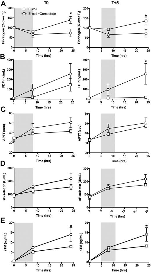
E coli infusion induced a gradual decrease of fibrinogen levels, especially during the first 8 hours after challenge (Figure 4A). Compstatin treatment reduced fibrinogen consumption during this time-frame. Fibrinogen levels fully recovered and overshoot the initial values after 24 hours, compared with nontreated animals (Figure 4A). Consistent with a reduction in the coagulopathic response, FDP levels were significantly lower (Figure 4B) and the APTT was slightly decreased (Figure 4C) in the treated animals. Furthermore, the plasma levels of sP-selectin (Figure 4D) and sTM (Figure 4E) were decreased in compstatin-treated animals, suggesting diminished endothelial cell injury and platelet activation.
Time course of hemostatic parameters. (A, fibrinogen; B, FDP; C, APTT) and plasma levels of soluble P-selectin (D) and soluble thrombomodulin (E) in baboons treated with compstatin during the first (T0) and second (T + 5) stages of experimental sepsis. Data are presented as mean ± SEM; 2-tailed Student t test. *P < .05.
Time course of hemostatic parameters. (A, fibrinogen; B, FDP; C, APTT) and plasma levels of soluble P-selectin (D) and soluble thrombomodulin (E) in baboons treated with compstatin during the first (T0) and second (T + 5) stages of experimental sepsis. Data are presented as mean ± SEM; 2-tailed Student t test. *P < .05.
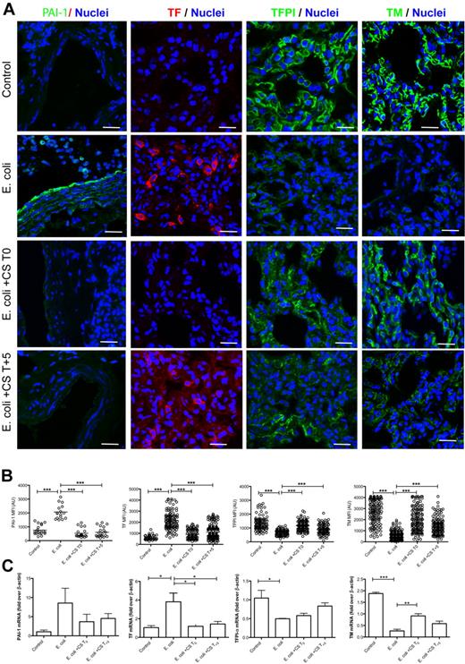
Quantitative immunofluorescence analysis of lung cryosections stained for PAI-1, TF, TFPI and TM (Figure 5A) demonstrated that E coli sepsis markedly increased PAI-1 and TF and decreased TFPI and TM. Both early and late treatments with compstatin decreased E coli induced TF and PAI-1 staining and protected against sepsis-induced down-regulation of TFPI and TM in endothelial cells (Figure 5A). Immunocytochemistry data correlated well with the amount of mRNA transcripts of these proteins (Figure 5B), although, consistent with a small number of animals, the group differences between experimental conditions were not always statistically significant.
Localization and quantitative analysis of hemostatic proteins in the lung. (A) Micrograph panel showing (columns) immunostaining for PAI-1, tissue factor (TF), TFPI and thrombomodulin (TM) in (rows) healthy controls, septic baboons (E coli), and septic baboons treated with compstatin (CS) during the first (E coli + CS T0) or the second (E coli + CS T + 5) stage. The colors of the each antigen and nuclear counterstaining are shown on the top row. ct indicates convolulted tubules; and g, glomerulus. Magnification bars, 50 μm. (B) Scatter-plot representations of MFI of images collected for the above-mentioned proteins and experimental conditions. PAI-1: whole-field MFI of at least 15 images; all other immunostainings: 20 ROI (alveolar capillaries) in 15 images (200 spots). (C) Histogram representation of mRNA expression for PAI-1, TF, TFPI, and TM. Values indicate the mean ± SEM of fold over β-actin housekeeping gene. In panels B and C, data are presented as mean ± SEM. One-way ANOVA with Dunnett multicomparison test. *P < .05; **P < .001; ***P < .001 compared with E coli group.
Localization and quantitative analysis of hemostatic proteins in the lung. (A) Micrograph panel showing (columns) immunostaining for PAI-1, tissue factor (TF), TFPI and thrombomodulin (TM) in (rows) healthy controls, septic baboons (E coli), and septic baboons treated with compstatin (CS) during the first (E coli + CS T0) or the second (E coli + CS T + 5) stage. The colors of the each antigen and nuclear counterstaining are shown on the top row. ct indicates convolulted tubules; and g, glomerulus. Magnification bars, 50 μm. (B) Scatter-plot representations of MFI of images collected for the above-mentioned proteins and experimental conditions. PAI-1: whole-field MFI of at least 15 images; all other immunostainings: 20 ROI (alveolar capillaries) in 15 images (200 spots). (C) Histogram representation of mRNA expression for PAI-1, TF, TFPI, and TM. Values indicate the mean ± SEM of fold over β-actin housekeeping gene. In panels B and C, data are presented as mean ± SEM. One-way ANOVA with Dunnett multicomparison test. *P < .05; **P < .001; ***P < .001 compared with E coli group.
The decrease in TF expression in the lung of compstatin-treated animals correlated with the observed decrease in monocyte/macrophages infiltration. PAI-1 expression in endothelial cells and macrophages was down-regulated in compstatin-treated versus nontreated septic baboons. The changes described in the lung were consistently observed in other organs also. For example, PAI1 mRNA in liver was increased 22-fold (± 9) by E coli sepsis but only 8-fold (± 8) in animals treated with compstatin at T0 and 4-fold (± 0.8) at T + 5 compared with nonchallenged controls (1 ± 0.6; all values are mean ± SEM). Altogether, our data demonstrate that complement inhibition leads to decreased procoagulant activity and a better-preserved endothelial anticoagulant function.
Effects of complement inhibition treatment on plasma cytokines
Nine of the 23 cytokines assayed increase markedly in the E coli sepsis group. The effect of compstatin on the cytokines was only marginal, except for a reduction observed for eotaxin and IL-6 with both the preventive and rescue therapies (data not shown).
Complement inhibition improves blood pressure and organ preservation and function
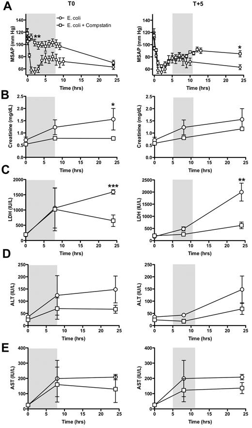
Mean systemic arterial blood pressure decreased markedly in septic animals (Figure 6A). Early compstatin treatment virtually abolished this decrease. Treatment during the second stage led to higher recovery of the blood pressure in the late phase compared with septic untreated baboons, despite the same degree of decrease in the early phase.
Time-course of organ function and biochemical markers in the blood of baboons treated with compstatin during the first (T0) and second (T + 5) stage of experimental sepsis. (A) Mean systemic arterial pressure (MSAP); (B) creatinine; (C) lactate dehydrogenase (LDH); (D) alanine aminotransferase (ALT); (E) aspartate transaminase (AST). Data are presented as mean ± SEM; 2-tailed Student t test. *P < .05; **P < .01; ***P < .001.
Time-course of organ function and biochemical markers in the blood of baboons treated with compstatin during the first (T0) and second (T + 5) stage of experimental sepsis. (A) Mean systemic arterial pressure (MSAP); (B) creatinine; (C) lactate dehydrogenase (LDH); (D) alanine aminotransferase (ALT); (E) aspartate transaminase (AST). Data are presented as mean ± SEM; 2-tailed Student t test. *P < .05; **P < .01; ***P < .001.
To evaluate whether compstatin administration could affect biochemical markers of organ damage, we analyzed 4 markers of tissue injury at T0, T + 8 and T + 24. Time-dependent changes in markers of organ function are presented in Figure 6 B-E. The creatinine, LDH, ALT and AST were increased by E coli sepsis after 8 and 24 hours postchallenge. The magnitude of the response for all 4 markers was lower in the 2 compstatin treatment groups, compared with non treated septic animals (Figure 6 B-E), indicating that complement inhibition attenuates kidney and liver injury. For pathologic examination, all animals were euthanized after 24 hours and tissues were removed for analysis within minutes after death to avoid autolytic postmortem changes. Histologic analysis of organs confirmed that compstatin provided substantial organ-protection. The scoring of the histopathologic lesions of the lung, kidney, liver, adrenals and spleen is shown in Figure 7. Differently from the nontreated group, compstatin-treated animals showed no obvious signs of thrombosis or capillary leak in the lungs, no or less tubular necrosis and glomerular thrombosis in the kidneys, lack of hepatocyte vacuolization and liver degeneration, less leukocyte infiltration in the lung, liver adrenals and spleen, and decreased cell death in adrenals and spleen. Immunostaining for cell specific markers showed a significant decrease in CD68 positive macrophages (Figure 3C) without significant changes in the number of neutrophils (not shown).
Comparison of the histopathologic changes in organs from septic animals with or without compstatin treatment during the first (T0) or second (T + 5) stage. The tissues were collected after euthanasia at T + 24 hours. Evaluations of the parameters were performed in a blinded fashion and graded on a scale from 0 to 4, with 0 being normal and 4 being severe. The histopathologic changes of the tissues collected from the 2 compstatin-treated groups are significantly less severe than those of the E coli challenge group. P < .001, with the exception of the lung congestion. Data are presented as mean ± SEM; 2-tailed Student t test. *P < .05; **P < .01; ***P < .001.
Comparison of the histopathologic changes in organs from septic animals with or without compstatin treatment during the first (T0) or second (T + 5) stage. The tissues were collected after euthanasia at T + 24 hours. Evaluations of the parameters were performed in a blinded fashion and graded on a scale from 0 to 4, with 0 being normal and 4 being severe. The histopathologic changes of the tissues collected from the 2 compstatin-treated groups are significantly less severe than those of the E coli challenge group. P < .001, with the exception of the lung congestion. Data are presented as mean ± SEM; 2-tailed Student t test. *P < .05; **P < .01; ***P < .001.
Discussion
This study demonstrates for the first time that complement inhibition at the C3 convertase level effectively attenuates inflammatory and hemostatic processes, restores systemic blood pressure and improves organ function during E coli sepsis in baboons.
Complement activation is an important innate immune host defense response, supporting leukocyte recruitment to the site of infection, phagocytosis and killing of the bacteria.1
Furthermore, complement activation can be triggered when blood is exposed to damaged vascular tissues, eg, after oxidative stress caused by IR occurring as an aftermath of the inflammatory response to sepsis.10 We hypothesized that the IR-dominated later stage of sepsis could represent a valuable therapeutic window for the use of complement inhibitors to prevent tissue damage and organ failure, and we compared this with a prevention regimen of inhibition.
We have used an E coli intravenous infusion model that exhibits 2 distinct stages of disease progression.3 The first stage is highly coagulopathic and is driven by the inflammatory response to the infused bacteria. The inflammatory mediators up-regulate TF on circulating monocytes and tissue macrophages, which leads to massive intravascular fibrin deposition (disseminated intravascular coagulation [DIC]) and hypoperfusion of vital organs. Under ischemic conditions, hypoxia-inducible genes are up-regulated and reduced oxygen supply leads to increased neutrophil adherence, transmigration, oxidative burst, and enhanced production of reactive oxygen and nitrogen species. These events fuel the second stage by promoting cell and tissue injury and predisposing to organ dysfunction, ultimately leading to organ failure and death in approximately 50% of animals. Histopathologic analysis of organs showed evidence of aberrant tissue repair in the lung, thrombotic angiopathic lesions in the kidney and cell death (apoptosis/necrosis) in kidneys, adrenals and lymphoid organs. We have previously shown that complement activation peaks during both stages of sepsis in baboons,8 although it probably plays distinct pathophysiologic roles in each stage. Interestingly, TCC plasma levels during the IR-driven second stage, when the bacteremia is close to zero, exceeds the activation seen during the highly bacteremic first stage and coincides with the rise of CRP levels. Complement activation and deposition on endothelium during this IR-driven stage can result in a loss of vascular homeostasis, triggering the second round of procoagulant and proinflammatory events observed during the late stages of sepsis in baboons.3
We report here that compstatin, a novel C3 convertase inhibitor efficiently inhibits sepsis-induced complement activation and deposition in organs of MBL, C3b, and C5b9 when administered during both the early and the late stages of sepsis. In kidneys, lower MBL binding on microvasculature and the decreased TCC deposition in glomerular and peritubular capillaries reflect the protective effects of the treatment against endothelial injury and IR-induced nephrotoxicity. In addition, compstatin treatment protected against cell-surface shedding of CD55 and CD59, 2 glycosyl phosphatidyl inositol-anchored negative regulators of complement activation. CD55, also known as the decay-accelerating factor binds to locally generated C4b and C3b accelerating their decay. CD59 regulates the final step of the complement cascade by inhibiting the formation of TCC on cell membranes.
Notably, we observed that complement inhibition partially reversed sepsis-induced leukopenia, thrombocytopenia, DIC and inflammatory responses and prevented sepsis-induced endothelial dysfunction.
It is well established that complement activation, directly or indirectly, promotes blood coagulation and thrombosis (for a review see Markiewski et al23 ). The direct effects include its ability to induce exposure of procoagulant lipids,24,25 activate platelets,26,27 increase TF expression in various cell types,28 induce the release of microparticles and inhibit fibrinolysis by up-regulation of PAI1.29 Our data demonstrate that complement inhibition decreases fibrin deposition by regulating both the coagulation and fibrinolytic pathways via TF and PAI-1 expression. In addition, complement inhibition could indirectly decrease the procoagulant response by suppressing platelets aggregation, endothelial cell dysfunction, leukocyte infiltration and the release of inflammatory cytokines and extracellular proteases involved in the down-regulation or degradation of 2 major anticoagulant pathways, namely TM-protein C (PC) and TFPI, known to attenuate the amplification of the inflammatory response in baboons.30 We have previously reported that endothelial cell TFPI is decreased in septic baboons, partly due to plasmin-dependent proteolysis.14 Similarly, TM is strongly decreased during both early and late stages of sepsis.20,30 We report here that both TM and TFPI proteins and mRNA were maintained at higher levels on the endothelial surface while less sTM and sP-selectin are released in the plasma of compstatin-treated animals, suggesting that complement inhibition protects the anticoagulant function of the endothelium in septic animals. Higher TM levels could promote anti-inflammatory and organ-protective effects via activated PC generation and/or endothelial PC receptor/protease activated receptors signaling.31 On the other side, TM can also provide additional protection by acting as a direct negative regulator of the complement system enhancing complement factor I–mediated inactivation of C3b in the presence of either complement factor H or C4bBP.32 Moreover, TM binds thrombin, thereby preventing it from activating C5,33 and promotes the generation of carboxypeptidase B (TAFIa), which inactivates C3a and C5a.34
Unexpectedly, compstatin treatment reduced the number of macrophages but not of neutrophils accumulated in the lung. This apparent disconnect can be explained in view of the temporal sequence of events after bacterial challenge. Our previous time-course analysis of gene expression in the lung demonstrated that acute inflammation, including neutrophil-specific genes are strongly increased during first 6 hours, whereas at 24 hours predominate macrophage and fibroblast specific genes involved in the resolution of inflammation (clearance of cell debris, extracellular matrix deposition, tissue regeneration, and functional recovery).20 In the experiments reported here animals were killed 24 hours after challenge, thus the tissue composition reflects the healing stage. Because less macrophages indicate less tissue damage, one could speculate that decreased macrophage accumulation in the compstatin-treated group may reflect decreased neutrophil adhesion and subsequent oxidative stress during the early time points (1-8 hours).
Complement inhibition decreased platelet adhesion and accumulation into organs and protected against sepsis-induced thrombocytopenia. DIC and thrombocytopenia have high sensitivity and specificity for predicting MOF,35 and, consistent with our findings, they are likely direct contributors to organ failure. We have detected a decreased C5b9 staining of platelets in compstatin-treated groups compared with nontreated controls supporting the involvement of complement in platelet deposition in tissues and subsequent thrombocytopenia. It is known that C3a induces platelet activation and aggregation27 and that C3 could bind to the platelet surface without36 or with C5b9 formation.26
Inhibition of complement activation by a potent inhibitor of C3 activation, a key event common to all 3 complement activation pathways, had organ-protective effects in both early and late treatment regimens. The protective effect of the complement inhibitor when administered during the IR-driven, sterile second stage suggests that complement activation during this time-frame contributes to disease progression toward organ failure and death. This supports the idea that efficient blocking of complement activation during the second stage could be a better option than during the early bacteremic stage. Nevertheless, blocking of complement at the early phase virtually completely abolished the fall in systemic blood pressure, indicating that complement activation is responsible for one of the most important physiologic disturbances during early sepsis. Thus, inhibition of the early stage could have some beneficial effects as well. The fact that late intervention with compstatin (5 hours after challenge) still provides organ protection could be therapeutically important, as most of the septic patients receive medical attention after the debut of the disease. These findings are particularly notable as not many therapeutic agents evaluated by us show organ protective effects when administered so late. For comparison, our gold-standard agent, activated Protein C (drotrecogin alfa [Xigris, Eli Lilly and Company]) has therapeutic efficiency in baboons only when administered 1 to 2 hours after challenge.
Inhibition of complement did not affect the release of cytokines, except for IL-6 and the chemotaxin eotaxin. This is not surprising, because the generation of cytokines by E coli is mainly CD14-driven, as recently documented in a porcine model of E coli sepsis37 and in an ex vivo human model of Gram-negative sepsis.38 This is in agreement with a hypothesis that we have recently put forward,39 that to inhibit innate immunity and thus reduce the adverse effects of this system in sepsis, combined upstream inhibition of complement and CD14 might be the strategy of choice.
We conclude that blocking the harmful effects of complement activation products during the second (organ failure) stage of severe sepsis is a potentially important therapeutic strategy. Because the pathophysiologic events of the IR-driven stage occur after the bacteria were already cleared, blockade of complement activity should not interfere with the beneficial effects of complement activation. Our data suggest that complement inhibition, although efficiently attenuating hypotension when given early, is still effective even when given during the second stage of progressive organ failure. Thus complement inhibition during the second stage could be a valuable approach for combination therapies with inhibitors of upstream inducers of inflammation, such as anti-CD14 antibodies40 or drotrecogin alfa, which is particularly potent during the early shock/DIC response to severe sepsis.
The online version of this article contains a data supplement.
The publication costs of this article were defrayed in part by page charge payment. Therefore, and solely to indicate this fact, this article is hereby marked “advertisement” in accordance with 18 USC section 1734.
Acknowledgments
We thank Dr Charles Esmon (Oklahoma Medical Research Foundation) for sTM ELISA reagents and critical reading of the manuscript, Dr James H. Morrissey (University of Illinois, Urbana-Champaign) for the mAb anti-TF, Dr Paul DeClerck (University of Leuven, Belgium) for the mAb anti-PAI-1, Dr Bo Nilsson (Uppsala University, Sweden) for the mAb anti C3a (clone 4DS17.3) and Dr Diana Wouters (Academic Medical Center, Amsterdam, The Netherlands) for the mAb anti-C3b (Clone C3-28) and Drs Bo Nilsson, Markus Huber-Lang, and Peter Ward for helpful discussions.
This work was supported by grants from the National Institutes of Health (GM037704-19 and 1RC1GM09739-01 to F.L.; AI068730 and GM062134 to J.D.L.).
This article is dedicated to Fletcher B. Taylor on the occasion of his 80th birthday.
National Institutes of Health
Authorship
Contribution: G.P., R.S.-M., L.I., and N.I.P. performed the animal experimentation; R.S.-M. performed immunofluorescence analysis, H.Z. performed mRNA analysis, C.L. performed experiments, analyzed results and contributed to paper writing; T.E.M. performed TCC and cytokine assays and analyzed results; G.S. performed complement assays; P.M. synthesized compstatin, F.B.T. and G.K. designed research; J.D.L. and F.L. conceived and supervised the project; and F.L. wrote the paper.
Conflict-of-interest disclosure: J.D.L., F.L., F.B.T., and G.K. are coinventors on a patent application to use complement inhibitors to prevent organ failure in sepsis. The remaining authors declare no competing financial interests.
Correspondence: Florea Lupu, Cardiovascular Biology Research Program, Oklahoma Medical Research Foundation, 825 NE 13th Street, Oklahoma City, OK 73 104; E-mail: florea-lupu@omrf.org; Phone: (405) 271 7462; Fax: (405) 271 7417.
References
Author notes
R.S.-M. and H.Z. contributed equally to this study.


This feature is available to Subscribers Only
Sign In or Create an Account Close Modal