Abstract
Graft-versus-host disease (GVHD) is the major complication after allogeneic bone marrow transplantation and is characterized by the overproduction of proinflammatory cytokines. In this study, we have identified interleukin-6 (IL-6) as a critical inflammatory cytokine that alters the balance between the effector and regulatory arms of the immune system and drives a proinflammatory phenotype that is a defining characteristic of GVHD. Our results demonstrate that inhibition of the IL-6 signaling pathway by way of antibody-mediated blockade of the IL-6 receptor (IL-6R) markedly reduces pathologic damage attributable to GVHD. This is accompanied by a significant increase in the absolute number of regulatory T cells (Tregs) that is due to augmentation of thymic-dependent and thymic-independent Treg production. Correspondingly, there is a significant reduction in the number of T helper 1 and T helper 17 cells in GVHD target organs, demonstrating that blockade of IL-6 signaling decreases the ratio of proinflammatory T cells to Tregs. These studies demonstrate that antibody blockade of the IL-6R serves to recalibrate the effector and regulatory arms of the immune system and represents a novel, potentially clinically translatable, strategy for the attenuation of GVHD.
Introduction
Graft-versus-host disease (GVHD) is the major complication associated with allogeneic stem cell transplantation. A prominent characteristic of GVHD is the presence of a proinflammatory milieu that is attributable to conditioning regimen–induced host tissue damage as well as secretion of inflammatory cytokines (eg, interleukin-1 [IL-1], tumor necrosis alpha-α [TNF-α], interferon-γ [IFN-γ], interleukin-6 [IL-6]) by alloactivated donor T cells and other effector cell populations.1-3 These cytokines perpetuate GVHD through direct cytotoxic effects on host tissues,4-6 activation and/or priming of immune effector cells,7 and differentiation of proinflammatory T-cell populations (ie, T helper [TH] 1 and TH17 cells) from naive T-cell precursors.8,9 This inflammatory environment is also promoted by the absence of an effective regulatory T-cell (Treg) response as both a relative and an absolute decline of Tregs in the peripheral blood and target tissues has been demonstrated in a majority of studies.8,10-12 The strong association between a proinflammatory milieu and the absence of an effective counterregulatory response suggests that the inflammatory environment prevents and/or inhibits Treg reconstitution during GVHD. How this occurs, however, is not well understood.
IL-6 is a pleiotrophic cytokine that is produced by a variety of cell types, including T cells, B cells, fibroblasts, endothelial cells, monocytes, and keratinocytes.13 IL-6 is of particular interest with respect to GVHD biology since it occupies a unique position at the crossroads where the fate of naive T cells to become either regulatory cells or proinflammatory T cells is determined. In the presence of IL-6 and transforming growth factor-β (TGF-β), naive T cells differentiate into TH17 cells, whereas in its absence these same cells are induced to become Tregs.14,15 Furthermore, IL-6 produced by dendritic cells after activation through Toll-like receptors is able to inhibit the suppressive function of natural Tregs.16,17 Thus, IL-6 appears to have a pivotal role in directing the immune response toward an inflammatory phenotype and away from a regulatory response. The potential importance of IL-6 in GVHD is also supported by clinical studies that have shown that patients with elevated plasma levels of IL-6,18,19 as well as those with a recipient or donor IL-6 genotype that results in increased IL-6 production,20,21 have an increased incidence and severity of GVHD.
Signaling through IL-6 occurs by binding to a low-affinity IL-6 receptor (IL-6R); thus, together IL-6 and IL-6R induce homodimerization of gp130 and subsequent transduction of the intracellular signal.22 This membrane-bound IL-6R, however, is expressed only on hepatocytes and hematopoietic cells. Notably, the IL-6R can also be shed from the membrane generating a soluble form of the receptor that can complex with IL-6 and induce an intracellular response in cells that lack the membrane-bound IL-6R through a process called transsignaling.23,24 Interference with the actions of IL-6 by administration of an IL-6R antibody that prevents binding of the cytokine to its receptor has been shown effective in the treatment of a variety of inflammatory disease such as rheumatoid arthritis,25,26 amyloidosis,27 and colitis.28 Whether inhibiting the actions of IL-6 affects the severity of GVHD or alters the proinflammatory milieu, however, has not been studied. In this report, we examined the effect that blockade of IL-6 signaling had on the pathophysiology of GVHD and on the ability of the host to reconstitute an effective regulatory T-cell response.
Methods
Mice
C57BL/6 (B6; H-2b), Balb/c (H-2d), B6(C)-H2-Ab1bm12/KhEgl (bm12; H-2b), B6.129S7-Rag-1 (B6 Rag-1), and IL-6–deficient (IL-6−/−; B6 background) mice were bred in the Animal Resource Center (ARC) at the Medical College of Wisconsin (MCW) or purchased from The Jackson Laboratory. Thymectomized Balb/c mice were purchased from The Jackson Laboratory. Foxp3EGFP mice (backcrossed to the B6 background for 6 generations) in which the foxp3 gene is coupled to the enhanced green fluorescent protein (EGFP) were obtained from Dr Calvin Williams (Medical College of Wisconsin) and have been previously described.29 All animals were housed in the Association for Assessment and Accreditation of Laboratory Animal Care (AAALAC)–accredited ARC of the MCW. Experiments were all carried out under protocols approved by the MCW Institutional Animal Care and Use Committee. Mice received regular mouse chow and acidified tap water ad libitum.
Bone marrow transplantation
Bone marrow (BM) was flushed from donor femurs and tibias with Dulbecco modified media (Gibco-BRL) and passed through sterile mesh filters to obtain single-cell suspensions. BM was T-cell depleted in vitro with anti-Thy1.2 monoclonal antibody plus low-toxicity rabbit complement (C-6 Diagnostics). The hybridoma for 30-H12 (anti-Thy1.2, rat IgG2b) antibody was purchased from the ATCC. Host mice were conditioned with total body irradiation administered as a single exposure at a dose rate of 82 cGy using a Shepherd Mark I Cesium Irradiator (J. L. Shepherd and Associates). Irradiated recipients received a single intravenous injection in the lateral tail vein of BM with or without added spleen cells. In some experiments, mice received a transplant of purified splenic CD4+ T cells that were isolated by positive selection using the magnetic-activated cell sorting microbead cell separation system (Miltenyi Biotec).
Cell sorting and flow cytometry
Spleen and peripheral lymph node cells were collected from Foxp3EGFP mice and sorted on a FACSVantage with a DIVA option (Becton Dickinson) or a FACSAria. Spleen cells from transplant recipients were labeled with monoclonal antibodies (mAbs) conjugated to fluorescein isothiocyanate, phycoerythrin (PE), or PE-Cy5.5 that were obtained from BD Biosciences. Cells were analyzed on a FACSCalibur flow cytometer with CellQuest software (Becton Dickinson). Data were analyzed using FlowJo software (TreeStar).
Reagents
Anti–IL-6R antibody (MR-16-1) is a rat IgG antibody that has been previously described.30 Animals received a loading dose of 2 mg intravenously on the day of transplantation and then were treated with 0.5 mg weekly by intraperitoneal injection. Antibody was resuspended in phosphate-buffered saline before injection. Rat IgG (Jackson ImmunoResearch Laboratories) was used as a control and administered at the same dose and schedule as MR-16-1.
Histologic analysis
Representative samples of liver, colon, and lung were obtained from transplant recipients and fixed in 10% neutral-buffered formalin. Samples were then embedded in paraffin, cut into 5-μm-thick sections, and stained with hematoxylin and eosin. A semiquantitative scoring system was used to account for histologic changes in the colon, liver, and lung as previously described.8,31 All slides were coded and read in a blinded fashion. Images were visualized with an Olympus BX45 microscope. Image acquisition was performed with an Olympus DP70 digital camera and software package.
Cytokine analysis
Serum was collected from mice by retro-orbital bleeds and analyzed on a Bioplex System (Bio-Rad Laboratories) according to the manufacturer's instructions. Soluble IL-6R was measured using a specific sandwich enzyme-linked immunosorbent assay (R&D Systems). Concentrations of all other proinflammatory cytokines (IL-1β, TNF-α, IL-6, IL-17, G-CSF, and IFN-γ) in serum were measured using the multiplex cytokine Bio-Rad assay system (Bio-Rad). All samples were run in duplicate.
Cell isolation
To isolate lamina propria lymphocytes, pooled colons were incubated in Hanks balanced salt solution buffer (Gibco-BRL) supplemented with 2% fetal bovine serum, EDTA (0.05 mM), and 15 μg/mL dithiothreitol (Invitrogen) at 37°C for 30 minutes and subsequently digested in a solution of collagenase D (0.15 mg/mL; Roche Diagnostics) in Dulbecco modified Eagle media with 2% fetal bovine serum for 75 minutes at 37°C. The resulting cell suspension was then layered on a 44%/67% Percoll gradient (Sigma-Aldrich). Pooled liver and lung lymphocytes were isolated by collagenase D digestion followed by layering on a Percoll gradient.
Intracellular cytokine staining
Lymphocytes isolated from spleen, liver, and lung were stimulated with 50 ng/mL phorbol 12-myristate 13-acetate (Sigma-Aldrich) and 750 ng/mL ionomycin (Calbiochem) for 2.5 hours and then incubated with GolgiStop (BD Pharmingen) for an additional 2.5 hours. Cells were surface stained with PE Cy5.5 anti-CD4 and then intracellularly stained with PE-labeled antibody to IL-17 and fluorescein isothiocyanate–labeled antibody to IFN-γ.
Real-time q-PCR
Liver, spleen, and colon samples were harvested and immediately snap frozen in liquid nitrogen for RNA extraction. Total RNA was extracted from frozen samples using TRIzol reagent (Gibco-BRL). Real-time quantitative polymerase chain reaction (q-PCR) was performed using Rotor-Gene 3000 (Corbett Research), QuantumRNA 18S Internal Standards (Ambion), IL-6 primers (5′-TCCAATGCTCTCCTAACAGATAAG-3′, 3′-CAAGATGAATTGGATGGTCTTG-5′),32 IL-6R (5′-CCTGTGTGGGGTTCCAGAGGAT-3′, 3′-CTGCCAGTATTCTCAGCAGCTG-5′),33 and QuantiTect SYBR Green PCR Master Mix (QIAGEN) according to the manufacturers' instructions. Synthesis of first-strand cDNA from 1 μg RNA per animal was accomplished with random primers and Superscript II (Invitrogen) according to the manufacturer's instructions. Primers were purchased from Integrated DNA Technologies. Specificity for all q-PCR reactions was verified by melting curve analysis. Data were analyzed with the Rotor-Gene 3000 software using the cycle threshold for quantification. Relative gene expression data (fold change) between samples was accomplished using the mathematic model described by Pfaffl.34
Statistics
Group comparisons of spleen and T-cell populations, cytokine levels, pathology scores, mean weights, and gene expression data were performed using the Mann-Whitney U test. A P value of .05 or less was deemed significant in all experiments.
Results
IL-6 and IL-6R levels are significantly increased during GVHD
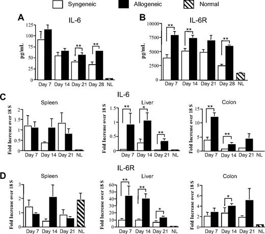
In initial studies, we examined the temporal kinetics of IL-6 and sIL-6R production in the sera of mice undergoing syngeneic and allogeneic bone marrow transplantation (BMT). Lethally irradiated Balb/c mice received a transplant of either Balb/c BM and spleen cells (0.4-0.5 × 10⁶; syngeneic) or B6 BM and an equivalent number of B6 spleen cells (allogeneic) to induce GVHD. Cohorts of mice were killed at weekly intervals and IL-6 levels were quantitated. These studies revealed that IL-6 was substantially increased in the sera of both syngeneic and allogeneic transplant recipients compared with normal animals that did not undergo transplantation at all time points (Figure 1A). Within the first 2 weeks after BMT, no difference was observed in IL-6 levels between recipients of syngeneic versus allogeneic marrow grafts. However, whereas levels of IL-6 progressively declined in syngeneic recipients, levels remained consistently elevated in allogeneic recipients resulting in significantly higher values 21 and 28 days after BMT. Given the restricted expression of the membrane-bound IL-6R and the importance of trans-signaling in inflammatory responses,24 we also examined sIL-6R levels in syngeneic and allogeneic marrow transplant recipients. Quantitation of sIL-6R in serum revealed that levels were significantly increased (2- to 3-fold) in recipients of allogeneic versus syngeneic grafts at nearly all time points (Figure 1B).
IL-6 and soluble IL-6R levels are increased during GVHD. (A-B) Lethally irradiated (900 cGy) Balb/c mice received a transplant of Balb/c BM (10 × 106) and spleen cells (0.4-0.5 × 10⁶, syngeneic) or B6 BM (10 × 106) and an equivalent number of B6 spleen cells (allogeneic). Cohorts of animals (n = 5-9/group) were killed weekly and serum was analyzed for IL-6 and soluble IL-6R levels using either Bioplex or enzyme-linked immunosorbent assay as described in “Cytokine analysis.” Values for normal control mice that did not undergo transplantation (n = 5) are depicted. Data are derived from 2 independent experiments. (C-D) RNA was extracted from spleen, liver, and colon tissues obtained from recipients of syngeneic or allogeneic marrow grafts (n = 7-8 mice per tissue) at the indicated time points, and gene expression of IL-6 (C) and total IL-6R (D) was analyzed by real-time q-PCR as described in “Real-time q-PCR.” Values for normal control mice that did not undergo transplantation (n = 5-6 mice/tissue) are depicted. Data are derived from 2 independent experiments and are presented as the mean ± SEM. (Statistics: *P ≤ .05, **P < .01.)
IL-6 and soluble IL-6R levels are increased during GVHD. (A-B) Lethally irradiated (900 cGy) Balb/c mice received a transplant of Balb/c BM (10 × 106) and spleen cells (0.4-0.5 × 10⁶, syngeneic) or B6 BM (10 × 106) and an equivalent number of B6 spleen cells (allogeneic). Cohorts of animals (n = 5-9/group) were killed weekly and serum was analyzed for IL-6 and soluble IL-6R levels using either Bioplex or enzyme-linked immunosorbent assay as described in “Cytokine analysis.” Values for normal control mice that did not undergo transplantation (n = 5) are depicted. Data are derived from 2 independent experiments. (C-D) RNA was extracted from spleen, liver, and colon tissues obtained from recipients of syngeneic or allogeneic marrow grafts (n = 7-8 mice per tissue) at the indicated time points, and gene expression of IL-6 (C) and total IL-6R (D) was analyzed by real-time q-PCR as described in “Real-time q-PCR.” Values for normal control mice that did not undergo transplantation (n = 5-6 mice/tissue) are depicted. Data are derived from 2 independent experiments and are presented as the mean ± SEM. (Statistics: *P ≤ .05, **P < .01.)
To determine whether similar findings were present in GVHD target organs, gene expression studies were performed to quantitate both IL-6 and total (ie, membrane and soluble) IL-6R mRNA levels. We observed that IL-6 mRNA levels were significantly increased in the colon and liver of mice with GVHD compared with syngeneic control animals (Figure 1C). This was in contrast to the spleen where no differences were observed at any of the time points examined. Notably, IL-6 mRNA levels were highest in the colon, consistent with a prior report demonstrating markedly elevated levels of this cytokine in the colon microenvironment.31 Gene expression analysis of the IL-6R mRNA levels also demonstrated significant increases in the colon and liver at several time points in recipients of allogeneic marrow grafts, but no difference in the spleen (Figure 1D). Whereas IL-6 mRNA levels were highest in the colon, IL-6R mRNA levels were most significantly increased in the liver where they were approximately 10-fold higher than either the spleen or colon. Collectively, these results demonstrate that there is a marked increase in both IL-6 and IL-6R levels during the early stages of GVHD.
Absence of either recipient or donor-derived IL-6 is insufficient to protect animals from GVHD
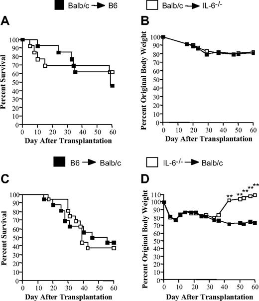
Given the increase in IL-6 production observed in both the sera and specific GVHD target organs, we conducted experiments to determine the relative importance of recipient versus donor IL-6 production in GVHD pathophysiology. Cohorts of lethally irradiated wild-type B6 or IL-6−/− mice received a transplant of Balb/c BM and 15 × 106 Balb/c spleen cells to induce GVHD. No difference in overall survival was observed between these 2 groups of animals (46% versus 62% at day 60, P = .67; Figure 2A). Moreover, serial weight curves demonstrated a similar pattern of weight loss indicating that absence of recipient-derived IL-6 had no protective effect on GVHD (Figure 2B). Reciprocal experiments were then performed to define the role of donor IL-6 production by transplantation of B6 or IL-6−/− BM and 0.5 to 0.7 × 10⁶ splenocytes into lethally irradiated Balb/c animals. There was again no difference in overall survival between these 2 groups (44% vs 38%, P = .85) (Figure 2C). Serial weight curves demonstrated no difference in weight loss over the first 5 weeks; however, surviving mice that received a transplant of IL-6−/− marrow grafts did have enhanced weight gain thereafter. This resulted in a statistically significant difference compared with similar time points in recipients of B6 marrow grafts (Figure 2D). However, no difference in overall GVHD pathology scores was observed in surviving animals that received a transplant of wild-type versus IL-6−/− marrow grafts (mean score, 7.6 ± 1.9 vs 6.7 ± 0.9; P = .93).
Absence of either recipient or donor-derived IL-6 is insufficient to protect mice from lethal GVHD. (A-B) Lethally irradiated (1000 cGy) B6 (n = 13) or IL-6−/− (n = 13) animals received a transplant of 10 × 106 BM and 15 × 106 spleen cells from Balb/c mice. Overall survival (A) and the percentage of original body weight over time (B) are depicted. Data are cumulative results from 4 independent experiments. (C-D) Lethally irradiated (900 cGy) Balb/c mice received a transplant of 10 × 106 BM and 0.5 to 0.7 × 10⁶ spleen cells from either B6 (n = 16) or IL-6−/− (n = 16) animals. Overall survival (C) and the percentage of original body weight over time (D) are depicted. Data are cumulative results from 4 independent experiments. (Statistics: **P < .01.)
Absence of either recipient or donor-derived IL-6 is insufficient to protect mice from lethal GVHD. (A-B) Lethally irradiated (1000 cGy) B6 (n = 13) or IL-6−/− (n = 13) animals received a transplant of 10 × 106 BM and 15 × 106 spleen cells from Balb/c mice. Overall survival (A) and the percentage of original body weight over time (B) are depicted. Data are cumulative results from 4 independent experiments. (C-D) Lethally irradiated (900 cGy) Balb/c mice received a transplant of 10 × 106 BM and 0.5 to 0.7 × 10⁶ spleen cells from either B6 (n = 16) or IL-6−/− (n = 16) animals. Overall survival (C) and the percentage of original body weight over time (D) are depicted. Data are cumulative results from 4 independent experiments. (Statistics: **P < .01.)
Blockade of IL-6 signaling attenuates GVHD severity and effects a significant increase in Treg reconstitution
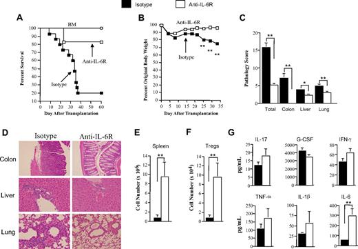
We then conducted studies to determine whether more complete blockade of IL-6 signaling would result in greater protection of GVHD target organs from pathologic damage. To address this question, we used a monoclonal antibody that binds to both the membrane and soluble components of the IL-6R so that IL-6 signaling could be more effectively inhibited in vivo. Lethally irradiated Balb/c mice received a transplant of TCD B6 BM and spleen cells adjusted to yield a T-cell dose of 0.7 × 10⁶ αβ T cells to induce lethal GVHD. Cohorts of mice were then treated weekly for 4 weeks with either anti–IL-6R or rat IgG isotype control antibody. Animals treated with anti–IL-6R antibody had significantly increased survival compared with control antibody–treated mice (83% versus 20%, P = .003) (Figure 3A). To understand the mechanism by which these mice were protected from GVHD lethality, we repeated these studies with a reduced T-cell dose so that mice in all groups would survive and be able to be examined for immunologic and pathologic analysis. Moreover, since IL-6 has also been shown to play a pivotal role in directing naive T-cell differentiation toward TH17 cells and away from Tregs,14,15 we examined this question using donor Foxp3EGFP mice so that Tregs could be definitively identified in recipient animals. In these studies, mice that received anti–IL-6R antibody had significantly less weight loss beginning approximately 3 weeks after transplantation (Figure 3B). Histologic examination of GVHD target organs 5 weeks after transplantation revealed a significant reduction in pathologic damage in the colon, liver, and lung (Figure 3C-D) compared with control antibody–treated mice, indicating that blockade of IL-6 signaling markedly attenuated GVHD severity in all target organs. This was accompanied by an increase in spleen cellularity (Figure 3E) and, most notably, a commensurate 12-fold increase in the number of Tregs (9.5 ± 3.1 × 10⁴) compared with mice administered the isotype antibody (0.78 ± 0.61 × 10⁴, P = .001; Figure 3F). Examination of serum proinflammatory cytokines 1 week after BMT revealed a significant increase in IL-6 levels in anti–IL-6R antibody–treated animals, but no difference in IFN-γ, IL-1β, TNF-α, G-CSF, or IL-17 compared with control mice (Figure 3G). The increase in serum IL-6 levels after anti–IL-6R antibody administration has been previously reported and is postulated to be due to a decrease in clearance as a consequence of circulating IL-6 being unable to bind to blocked IL-6 receptors.35,36
Antibody blockade of the IL-6R significantly attenuates the severity of GVHD. (A) Lethally irradiated (900 cGy) Balb/c mice received a transplant of T cell–depleted (TCD) B6 BM alone (10 × 106; ○, n = 9) or together with B6 spleen cells adjusted to yield a T-cell dose of 0.7 × 10⁶ αβ T cells. Cohorts of mice that received adjunctive spleen cells were then treated with either rat IgG isotype control (n = 15) or anti–IL-6R (MR-16-1) antibody (n = 12) once weekly for 4 weeks beginning on the day of transplantation. Overall survival is depicted. Data are cumulative results from 3 independent experiments. (B) Lethally irradiated Balb/c mice received a transplant of TCD Foxp3EGFP BM (10 × 106) and 0.4 to 0.5 × 10⁶ Foxp3EGFP spleen cells. Cohorts of mice were then treated with either rat IgG isotype control (n = 12) or anti–IL-6R (MR-16-1) antibody (n = 12) once weekly for 4 weeks beginning on the day of transplantation. Mice from both groups were killed 34 to 37 days after BMT. (B) The percentage of original body weight over time in mice from both groups is depicted. (C) Pathological damage in the colon, liver, and lung using a semiquantitative scoring system as detailed in “Histologic analysis.” (D) Histology of colon, liver, and lung from representative recipients treated with either isotype or anti–IL-6R antibody. In isotype control animals, colon shows extensive inflammation in the lamina propria, goblet cell depletion, and crypt cell destruction; liver reveals portal triad inflammation with mononuclear cells and endothelialitis; and lung demonstrates perivascular and peribronchial cuffing with mononuclear cells. In anti–IL-6R antibody–treated mice, colon has normal-appearing mucosa with no attendant inflammation, liver has reduced portal triad inflammation, and lung demonstrates a similar reduction in perivascular and peribronchial cuffing. (E) Total spleen cellularity and (F) absolute number of splenic Tregs (CD4+ EGFP+) are shown. (G) Lethally irradiated (900 cGy) Balb/c mice received a transplant of TCD B6 BM plus 0.4 × 10⁶ B6 spleen cells. Cohorts of animals that underwent transplantation were then treated with either isotype control (n = 12) or anti–IL-6R antibody (n = 12) on the day of transplantation. Mice were bled 6 days after transplantation and serum was assayed for proinflammatory cytokines. Data are presented as the mean (± SEM) and are the cumulative results from 3 independent experiments. (Statistics: *P ≤ .05, **P < .01.)
Antibody blockade of the IL-6R significantly attenuates the severity of GVHD. (A) Lethally irradiated (900 cGy) Balb/c mice received a transplant of T cell–depleted (TCD) B6 BM alone (10 × 106; ○, n = 9) or together with B6 spleen cells adjusted to yield a T-cell dose of 0.7 × 10⁶ αβ T cells. Cohorts of mice that received adjunctive spleen cells were then treated with either rat IgG isotype control (n = 15) or anti–IL-6R (MR-16-1) antibody (n = 12) once weekly for 4 weeks beginning on the day of transplantation. Overall survival is depicted. Data are cumulative results from 3 independent experiments. (B) Lethally irradiated Balb/c mice received a transplant of TCD Foxp3EGFP BM (10 × 106) and 0.4 to 0.5 × 10⁶ Foxp3EGFP spleen cells. Cohorts of mice were then treated with either rat IgG isotype control (n = 12) or anti–IL-6R (MR-16-1) antibody (n = 12) once weekly for 4 weeks beginning on the day of transplantation. Mice from both groups were killed 34 to 37 days after BMT. (B) The percentage of original body weight over time in mice from both groups is depicted. (C) Pathological damage in the colon, liver, and lung using a semiquantitative scoring system as detailed in “Histologic analysis.” (D) Histology of colon, liver, and lung from representative recipients treated with either isotype or anti–IL-6R antibody. In isotype control animals, colon shows extensive inflammation in the lamina propria, goblet cell depletion, and crypt cell destruction; liver reveals portal triad inflammation with mononuclear cells and endothelialitis; and lung demonstrates perivascular and peribronchial cuffing with mononuclear cells. In anti–IL-6R antibody–treated mice, colon has normal-appearing mucosa with no attendant inflammation, liver has reduced portal triad inflammation, and lung demonstrates a similar reduction in perivascular and peribronchial cuffing. (E) Total spleen cellularity and (F) absolute number of splenic Tregs (CD4+ EGFP+) are shown. (G) Lethally irradiated (900 cGy) Balb/c mice received a transplant of TCD B6 BM plus 0.4 × 10⁶ B6 spleen cells. Cohorts of animals that underwent transplantation were then treated with either isotype control (n = 12) or anti–IL-6R antibody (n = 12) on the day of transplantation. Mice were bled 6 days after transplantation and serum was assayed for proinflammatory cytokines. Data are presented as the mean (± SEM) and are the cumulative results from 3 independent experiments. (Statistics: *P ≤ .05, **P < .01.)
Attenuation of GVHD by blockade of IL-6 signaling is not dependent upon an intact thymus
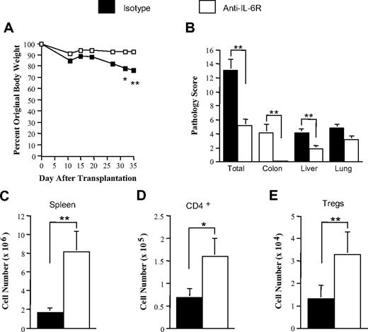
Immune reconstitution is a particular problem in older transplant recipients due, in part, to the involution of the thymus that limits the generation new T cells, including Tregs, and thereby constrains T-cell repertoire complexity.37-39 Thus, we examined the effect of antibody blockade on GVHD protection under conditions where thymic function was absent. Lethally irradiated thymectomized Balb/c mice received a transplant of B6 Foxp3EGFP BM and spleen cells and then administered either anti–IL-6R or isotype control antibodies. Mice treated with anti–IL-6R antibody had significantly less GVHD-associated weight loss (Figure 4A) and there was also reduced overall pathologic damage with specific reductions in the colon and liver (Figure 4B). Coincident with the attenuation in organ pathology, there was a commensurate increase in splenic cellularity and total splenic CD4+ T cells (Figure 4C-D). Notably, animals treated with anti–IL-6R antibody also had a significant increase in the absolute number of splenic Tregs (3.3 ± 1.0 × 10⁴ versus 1.3 ± 0.6 × 10⁴, P = .012; Figure 4E), indicating that the augmentation in Treg numbers occurring as a consequence of IL-6 signaling blockade was not dependent upon an intact thymus.
Attenuation of GVHD by blockade of IL-6 signaling does not require an intact thymus. Lethally irradiated (900 cGy) thymectomized Balb/c mice received a transplant of Foxp3EGFP BM (10 × 106) and 0.4 × 10⁶ Foxp3EGFP spleen cells. Cohorts of mice were then treated with either rat IgG isotype control (n = 12) or anti–IL-6R antibody (n = 12) once weekly for 4 weeks beginning on the day of transplantation. Animals were then killed 46 to 48 days after transplantation. (A) The percentage of original body weight of mice over time from both groups is depicted. (B) Pathological damage in the colon, liver, and lung using a semiquantitative scoring system as detailed in “Histologic analysis.” (C) Total spleen cellularity, (D) absolute number of splenic CD4+ T cells, and (E) absolute number of splenic Tregs are depicted. Data are presented as the mean (± SEM) and are the cumulative results from 3 independent experiments. (Statistics: *P ≤ .05, **P < .01.)
Attenuation of GVHD by blockade of IL-6 signaling does not require an intact thymus. Lethally irradiated (900 cGy) thymectomized Balb/c mice received a transplant of Foxp3EGFP BM (10 × 106) and 0.4 × 10⁶ Foxp3EGFP spleen cells. Cohorts of mice were then treated with either rat IgG isotype control (n = 12) or anti–IL-6R antibody (n = 12) once weekly for 4 weeks beginning on the day of transplantation. Animals were then killed 46 to 48 days after transplantation. (A) The percentage of original body weight of mice over time from both groups is depicted. (B) Pathological damage in the colon, liver, and lung using a semiquantitative scoring system as detailed in “Histologic analysis.” (C) Total spleen cellularity, (D) absolute number of splenic CD4+ T cells, and (E) absolute number of splenic Tregs are depicted. Data are presented as the mean (± SEM) and are the cumulative results from 3 independent experiments. (Statistics: *P ≤ .05, **P < .01.)
In vivo induction of CD4+ foxp3+ T cells from CD4+ foxp3− T cells is negligible during acute GVHD
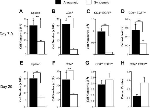
The increased number of Tregs that we observed in thymectomized mice led us to investigate whether treatment with anti–IL-6R antibody enhanced the peripheral conversion of CD4+ foxp3+ T cells from conventional CD4+ foxp3− T cells. To address this question, we first performed studies to determine the extent to which peripheral Treg conversion occurred during the course of acute GVHD. Experiments were conducted using a CD4+ T cell–dependent murine model in which lethally irradiated bm12 (allogeneic) or B6 (syngeneic) mice received a transplant of B6 Rag-1 BM (5-10 × 106) and equivalent numbers of sorted CD4+ EGFP-foxp3− T cells (0.1-0.6 × 10⁶) so that no natural Tregs were administered in the marrow graft inoculum and induced Tregs (iTregs) could be identified by their expression of EGFP. Cohorts of mice from each group were then killed at 1 and 3 weeks after transplantation, and the absolute number and/or percentage of iTregs in the spleen, liver, and colon was quantitated. Overall spleen cell numbers and total CD4+ T cells were increased in GVHD animals at both 1 and 3 weeks after transplantation due, in part, to the expansion of alloreactive CD4+ T cells (Figure 5A-B,E-F). The number of iTregs was also marginally increased at 1 week (Figure 5C), but by 3 weeks there was no difference compared with syngeneic controls (Figure 5G). Similarly, although the mean percentage of CD4+ T cells that were EGFP-foxp3+ was also higher 7 days after transplantation in GVHD mice (Figure 5D), no difference compared with syngeneic animals was observed by week 3 (Figure 5H). The most important aspect of these studies, however, was that the percentage of iTregs relative to total CD4+ T-cell numbers was very low and averaged less than 1% at both time points. Examination of the colon and liver from these same animals also demonstrated that the mean percentage of CD4+ EGFP+ T cells in allogeneic recipients (n = 3-4 mice/group) was 0.48% and 0.23%, respectively, at 1 week and 0.36% and 0.08%, respectively, at 3 weeks (data not shown). Collectively, these data demonstrated that the induction of CD4+ foxp3+ T cells from conventional CD4+ foxp3− T cells was negligible during acute GVHD.
In vivo conversion of CD4+ foxp3− T cells to CD4+ foxp3+ T cells is negligible during acute GVHD. Lethally irradiated bm12 (1000 cGy; n = 8-11/group) or B6 (1000 cGy; n = 8-12/group) mice received a transplant of B6 Rag-1 BM (5 × 10⁶) and sorted CD4+ EGFP-foxp3− T cells (0.6 × 10⁶). Cohorts of mice were killed on either days 7 to 9 or day 20 after transplantation. Spleen cellularity (A,E), absolute number of splenic CD4+ T cells (B,F), percentage of CD4+ EGFP+ T cells in the spleen (C,G), and absolute number of CD4+ EGFP+ T cells in the spleen (D,H) are depicted. Data are presented as the mean (± SEM) and are the cumulative results from 2 to 3 independent experiments. (Statistics: **P < .01.)
In vivo conversion of CD4+ foxp3− T cells to CD4+ foxp3+ T cells is negligible during acute GVHD. Lethally irradiated bm12 (1000 cGy; n = 8-11/group) or B6 (1000 cGy; n = 8-12/group) mice received a transplant of B6 Rag-1 BM (5 × 10⁶) and sorted CD4+ EGFP-foxp3− T cells (0.6 × 10⁶). Cohorts of mice were killed on either days 7 to 9 or day 20 after transplantation. Spleen cellularity (A,E), absolute number of splenic CD4+ T cells (B,F), percentage of CD4+ EGFP+ T cells in the spleen (C,G), and absolute number of CD4+ EGFP+ T cells in the spleen (D,H) are depicted. Data are presented as the mean (± SEM) and are the cumulative results from 2 to 3 independent experiments. (Statistics: **P < .01.)
Antibody blockade of the IL-6R augments conversion of CD4+ foxp3− to CD4+ foxp3+ T cells in the periphery
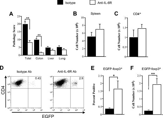
Experiments were then conducted to determine whether anti–IL-6R antibody administration enhanced the conversion of CD4+ foxp3+ T cells from CD4+ foxp3− T cells. To address this question, lethally irradiated Balb/c mice received a transplant of B6 Rag-1 BM cells and sorted CD4+ EGFP− T cells, and then treated with either anti–IL-6R or control antibody. Anti–IL-6R antibody–treated animals had a significant overall reduction in pathologic damage compared with control-treated mice (Figure 6A). Protection, however, was seen only in the colon, whereas there was no statistically significant decrease in pathology scores in either the liver or lung. Although there was no difference in spleen size or total number of CD4+ T cells between groups of mice (Figure 6B-C), we did observe that anti–IL-6R antibody administration significantly augmented the peripheral conversion of CD4+ foxp3− to CD4+ foxp3+ T cells. More specifically, there was a substantial increase in the percentage of iTregs (Figure 6D-E) as well as an 8-fold increase in the absolute number of these cells in the spleen in anti–IL-6R versus isotype antibody–treated mice (1.93 ± 0.37 × 10⁴ versus 0.24 ± 0.05 × 10⁴, P = .002) (Figure 6F). Thus, blockade of IL-6 signaling enhanced the conversion of CD4+ foxp3+ T cells from CD4+ foxp3− T cells without resulting in any statistically significant increase in the absolute number of CD4+ T cells.
Antibody blockade of the IL-6R augments conversion of CD4+ foxp3− to CD4+ foxp3+ Tregs. Lethally irradiated (900 cGy) Balb/c mice received a transplant of B6 Rag-1 BM (5 × 10⁶) and sorted CD4+ EGFP-foxp3− T cells (0.2 × 10⁶). Cohorts of mice were then administered rat IgG isotype control (n = 9) or anti–IL-6R antibody (n = 12) once weekly for 4 weeks as described in “Methods.” Mice in both groups were killed 26 to 36 days after transplantation. (A) Pathological damage in the colon, liver, and lung using a semiquantitative scoring system as detailed in “Histologic analysis.” (B) Total spleen cellularity and (C) absolute number of splenic CD4+ T cells are depicted. (D) Representative dot plot showing percentage of EGFP-foxp3+ iTregs in the gated CD4+ T-cell population from transplant recipients treated with either isotype control or anti–IL-6R antibody. (E) Percentage and (F) absolute number of iTregs in the spleen of animals administered control or anti–IL-6R antibody. Data are presented as the mean (± SEM) and are the cumulative results from 3 independent experiments. (Statistics: *P ≤ .05, **P < .01.)
Antibody blockade of the IL-6R augments conversion of CD4+ foxp3− to CD4+ foxp3+ Tregs. Lethally irradiated (900 cGy) Balb/c mice received a transplant of B6 Rag-1 BM (5 × 10⁶) and sorted CD4+ EGFP-foxp3− T cells (0.2 × 10⁶). Cohorts of mice were then administered rat IgG isotype control (n = 9) or anti–IL-6R antibody (n = 12) once weekly for 4 weeks as described in “Methods.” Mice in both groups were killed 26 to 36 days after transplantation. (A) Pathological damage in the colon, liver, and lung using a semiquantitative scoring system as detailed in “Histologic analysis.” (B) Total spleen cellularity and (C) absolute number of splenic CD4+ T cells are depicted. (D) Representative dot plot showing percentage of EGFP-foxp3+ iTregs in the gated CD4+ T-cell population from transplant recipients treated with either isotype control or anti–IL-6R antibody. (E) Percentage and (F) absolute number of iTregs in the spleen of animals administered control or anti–IL-6R antibody. Data are presented as the mean (± SEM) and are the cumulative results from 3 independent experiments. (Statistics: *P ≤ .05, **P < .01.)
Anti–IL-6R antibody–induced increase in Tregs is associated with a significant reduction in proinflammatory TH1 and TH17 cells
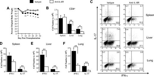
IL-6, in combination with TGF-β, has been shown to play a critical role in the differentiation of TH17 cells from naive T-cell precursors.14,15 Moreover, recent studies suggest that TH17 cells may contribute to the pathophysiology of GVHD,9,40 although their specific role relative to TH1 cells is still not well defined. We therefore conducted studies to determine whether blockade of IL-6 signaling affected the generation of TH17 and TH1 cells during GVHD. To address this question, lethally irradiated Balb/c mice received a transplant of wild-type B6 BM and purified B6 CD4+ T cells and then were treated with either control or anti–IL-6R antibody. Purified CD4+ T cells, which alone are able to induce GVHD in this strain combination, were used to maximize the ability to detect expansion of TH1 and TH17 cells in the tissues of recipient mice. As we previously observed, blockade of IL-6 signaling resulted in improved overall weight curves (Figure 7A) in anti–IL-6R antibody–treated mice. There was also a significant reduction in the total number of CD4+ T cells in the liver and lung in these same animals (Figure 7B). This was accompanied by a relative reduction in the percentage of CD4+ T cells secreting either IL-17 or IFN-γ with the exception of the liver where similar percentages of CD4+ IFN-γ+ cells were observed (Figure 7C). Coincident with the reduction in GVHD-associated weight loss, we also observed a significant decrease in the absolute number of CD4+IL-17+ T cells in spleen, liver, and lung (Figure 7D-F). CD4+ T cells secreting IFN-γ+ were also reduced in the liver and lung, although there was no significant difference in the absolute numbers of these cells in the spleen. Overall, these studies demonstrated that blockade of IL-6 signaling significantly decreased the number of proinflammatory CD4+ T cells.
Blockade of IL-6 signaling results in a significant reduction in proinflammatory TH1 and TH17 cells. Lethally irradiated (900 cGy) Balb/c mice received a transplant of B6 BM (5 × 10⁶) and purified B6 CD4+ T cells (0.3 × 10⁶). Cohorts of mice were then administered rat IgG isotype control (n = 8) or anti–IL-6R antibody (n = 8) for 4 weekly injections as described in “Reagents.” Mice in both groups were killed 28 to 29 days after transplantation. (A) The percentage of original body weight of mice over time from both groups is depicted. (B) Absolute number of CD4+ T cells in the spleen, liver, and lung of animals treated with either isotype control or anti–IL-6R antibody. (C) Representative dot plot depicting the percentage of IL-17– and/or IFN-γ–secreting cells within the gated CD4+ T-cell population. (D-F) Absolute number of CD4+ IFN-γ+ or CD4+ IL-17+ T cells present in the spleen, liver, or lung of animals treated with either isotype control or anti–IL-6R antibody. Data are presented as the mean (± SEM) and are the cumulative results from 2 independent experiments. (Statistics: *P ≤ .05, **P < .01.)
Blockade of IL-6 signaling results in a significant reduction in proinflammatory TH1 and TH17 cells. Lethally irradiated (900 cGy) Balb/c mice received a transplant of B6 BM (5 × 10⁶) and purified B6 CD4+ T cells (0.3 × 10⁶). Cohorts of mice were then administered rat IgG isotype control (n = 8) or anti–IL-6R antibody (n = 8) for 4 weekly injections as described in “Reagents.” Mice in both groups were killed 28 to 29 days after transplantation. (A) The percentage of original body weight of mice over time from both groups is depicted. (B) Absolute number of CD4+ T cells in the spleen, liver, and lung of animals treated with either isotype control or anti–IL-6R antibody. (C) Representative dot plot depicting the percentage of IL-17– and/or IFN-γ–secreting cells within the gated CD4+ T-cell population. (D-F) Absolute number of CD4+ IFN-γ+ or CD4+ IL-17+ T cells present in the spleen, liver, or lung of animals treated with either isotype control or anti–IL-6R antibody. Data are presented as the mean (± SEM) and are the cumulative results from 2 independent experiments. (Statistics: *P ≤ .05, **P < .01.)
Discussion
The reestablishment of effective regulation of alloreactive donor T cell–mediated inflammation is a major goal in the treatment of GVHD. A preponderance of prior studies, however, supports the premise that the reconstitution of Tregs, which plays a pivotal role in regulating T cell–mediated alloresponses, is severely impaired in both acute and chronic GVHD.8,10-12 In fact, the progressive reduction in Treg numbers, which begins during acute GVHD, is a major factor responsible for the development of autoimmunity, which is a defining characteristic of chronic GVHD.8 Why these cells fail to effectively reconstitute during GVHD has not been well understood. The current report now establishes that IL-6 is a critical cytokine that inhibits the reconstitution of Tregs after transplantation during GVHD. Furthermore, these studies reveal that both IL-6 and IL-6R levels are significantly increased in the sera and in specific target organs during the course of GVHD and that targeting of the IL-6/IL-6R complex by way of antibody blockade results in a reduction in GVHD severity.
The most significant observation was the finding that blockade of IL-6 signaling results in a marked increase in the absolute number of Tregs. Our data indicate that this occurs in both thymic-dependent and thymic-independent manners. With respect to the former, we observed that total Treg numbers in the spleen were 3-fold higher in anti–IL-6R antibody–treated recipients with intact thymi as opposed to thymectomized mice (Figure 3E vs 4E). The most plausible explanation for this increase was that blockade of IL-6 signaling enhanced the de novo generation of Tregs in the thymus of these animals. Treatment with anti–IL-6R as opposed to control antibody, however, also augmented absolute Treg numbers in thymectomized mice indicating that de novo production of natural thymic-derived Tregs was not an absolute requirement for GVHD protection to be observed. The fact that blockade of the IL-6R increased the number of Tregs in thymectomized recipients is an important finding that has potential clinical implications. Reconstitution of effective T-cell immunity is a particular problem in older transplant recipients who comprise the majority of patients who requires allogeneic stem cell transplantations. These patients are also at increased risk of developing GVHD due, in part, to impaired thymic function occurring as a consequence of direct thymic damage from the conditioning regimen and/or age-related involution,37-39 both of which serve to limit the generation of new Tregs. Although the increase in Treg numbers in anti–IL-6R antibody–treated mice was approximately 3-fold less than in thymus-intact animals, this was still sufficient to significantly attenuate GVHD-associated pathologic damage. Thus, these results suggest that blockade of IL-6 signaling may be a strategy to augment the establishment of peripheral tolerance in older patients with more limited ability to generate Tregs in the thymus.
Regulatory T cells defined by their expression of foxp3 have been classified into 2 distinct subsets that have different developmental pathways. The first, termed “natural” Tregs, represents 5% to 10% of CD4+ T cells, develops in the thymus,41 and requires costimulation through the CD28/B7 pathway to maintain their survival.42 Natural Tregs are most commonly characterized by the constitutive expression of CD25, CTLA-4, CD134, CD103, and glucocorticoid-induced tumor necrosis factor receptor, which are all markers of cell activation and reflect the continuous exposure of these cells to self-antigens in the periphery.42-44 The second subset, termed induced regulatory T cells (iTregs), is able to up-regulate foxp3 after activation in the periphery, has variable CD25 expression and function in a cytokine-dependent fashion (eg, IL-10).45 TGF-β has been shown to be a critical cytokine that promotes the conversion of conventional T cells into iTregs after ligation through the TCR along with appropriate costimulation.45,46 Prior studies have shown that IL-6 deleteriously affects the generation of iTregs induced by TGF-β by blocking foxp3 expression.47 This has been shown to occur by up-regulation of the TGF-β inhibitor SMAD7.46 Although several reports have examined the fate of natural Tregs during the course of GVHD,48-51 the role of iTregs in this disease has not been critically studied. In the current report, we observed that conversion of CD4+ foxp3− T cells to CD4+ foxp3+ T cells in the spleen or selected target organs during GVHD was very limited and insufficient to prevent pathologic damage. Administration of anti–IL-6R antibody, however, resulted in a significant increase in both the percentage and absolute number of these cells and this was associated with a marked reduction in overall GVHD. Notably, GVHD protection was observed only in the colon as there was no difference in pathology scores in either the lung or liver. This observation suggests that peripheral Treg conversion may be of particular importance in regulating alloimmune reactions in the gut and that IL-6 has a pivotal role in inhibiting this event. The elevated IL-6 mRNA levels in the colon (Figure 1C) are consistent with this interpretation. Moreover, an important role for IL-6 in the colon microenvironment is supported by recent studies demonstrating that IL-6 levels are significantly increased in this organ relative to other proinflammatory cytokines such as TNF-α, IFN-γ, and IL-1β.31
IL-6 has been shown to play a pivotal role in driving the differentiation of naive T cells to become TH17 cells and in inhibiting the generation of CD4+ foxp3+ from CD4+ foxp3− T cells. We therefore examined whether anti–IL-6R antibody administration affected the number of IL-17–secreting CD4+ T cells, which have been shown to be increased during GVHD.8 Our studies demonstrated that blockade of IL-6 signaling was associated with a significant reduction in these cells in the spleen, liver, and lung. Thus, blockade of IL-6 signaling resulted in increased conversion in the periphery along with a corresponding decrease in TH17 cells. The inverse reciprocality in the numbers of these 2 T-cell populations would suggest that blockade of IL-6 signaling was able to divert the differentiation of naive T cells away from the TH17 cell lineage. We cannot exclude, however, that recently described IL-6–dependent plasticity that allows for the reprogramming of committed cells from regulatory to effector phenotypes might not also have been operative under these conditions.52 Notably, we observed that there was a significant reduction in the absolute number of CD4+ IFN-γ+ T cells as well in GVHD target organs. A similar observation has been made by Serada et al in a model of experimental allergic encephalomyelitis.53 A potential explanation for this observation is that the increase in Tregs as a consequence of anti–IL-6R antibody administration could have inhibited the induction of Th1 cells in both scenarios. This interpretation is consistent with data demonstrating that there is an increase in inducible Tregs and a decline in TH1 and TH17 cells in the absence of IL-6.54
The results of this study also have clinical implications with respect to the use of Tregs to prevent GVHD in allogeneic stem cell transplant recipients. Current approaches are now focused on the ex vivo expansion of these cells so that they can be adoptively transferred into recipients after transplantation. However, there are several challenges with trying to implement this approach. First, defining the optimal regulatory T-cell population is controversial and is constrained by the need to identify these cells based on CD25 expression which is also an activation marker. Although expansion strategies have incorporated agents such as rapamycin to eliminate non-Treg CD4+ T-cell populations,55,56 this approach does not completely remove them and thereby raises concerns that transplantation of an impure Treg population could potentially exacerbate GVHD toxicity. Second, Tregs that have the highest CD25 expression and thus the most suppressive capability in vitro are typically those that expand less well ex vivo.57 Consequently, achieving sufficiently large numbers of these cells for transplantation may be problematic. Our approach that uses administration of an anti–IL-6R antibody may be a way to circumvent these obstacles by augmenting the in vivo expansion of these cells. It should also be pointed out that blockade of IL-6/IL-6R interactions is currently a clinically feasible option. Tocilizumab is a humanized anti–IL-6R antibody that has been administered to patients with rheumatoid and juvenile arthritis and found to significantly ameliorate joint inflammation without any serious toxicity.58,59 Thus, formal testing of this premise is now a viable clinical option.
In summary, these studies demonstrate that the increased production of IL-6 during GVHD deleteriously affects the reconstitution of regulatory T cells and thereby adversely impacts the development of peripheral tolerance during this disease. Furthermore, IL-6 drives a proinflammatory phenotype that is characterized by increased numbers of TH1 and TH17 cells in GVHD target organs that contribute to this inflammatory environment. Blockade of IL-6 signaling, on the other hand, significantly reduces GVHD-associated pathologic damage by increasing Treg numbers and decreasing the absolute number of proinflammatory T cells. This serves to recalibrate the effector and regulatory arms of the immune system and thereby mitigate the severity of GVHD.
The publication costs of this article were defrayed in part by page charge payment. Therefore, and solely to indicate this fact, this article is hereby marked “advertisement” in accordance with 18 USC section 1734.
Acknowledgments
This research was supported by grants from the National Institutes of Health (HL064603, HL081650, and AI078713) and by an award from the Midwest Athletes Against Childhood Cancer Fund.
National Institutes of Health
Authorship
Contribution: X.C. designed and performed research, analyzed data, and wrote the paper; R.D. performed research, analyzed data, and wrote the paper; R.K. performed all pathologic analyses; A.B. performed research; M.J.H. assisted with real-time quantitative PCR studies; M.M. provided vital reagents; and W.R.D. designed and supervised research, analyzed data, and wrote the paper.
Conflict-of-interest disclosure: The authors declare no competing financial interests.
Correspondence: William R. Drobyski, Bone Marrow Transplant Program, 9200 West Wisconsin Ave, Milwaukee, WI 53226; e-mail: wdrobysk@mcw.edu.
References
Author notes
*X.C. and R.D. contributed equally to this work and share first authorship.

This feature is available to Subscribers Only
Sign In or Create an Account Close Modal