Abstract
Aberrant overexpression of the miR-17-92 polycistron is strongly associated with B-cell lymphomagenesis. Recent studies have shown that miR-17-92 down-regulates the proapoptotic protein Bim, leading to overexpression of Bcl2, which likely plays a key role in lymphomagenesis. However, the fact that Jeko-1 cells derived from mantle cell lymphoma exhibit both homozygous deletion of BIM and overexpression of miR-17-92 suggests other targets are also involved in B-cell lymphomagenesis. To identify essential target(s) of miR-17-92 in lymphomagenesis, we first transfected miR-17-92 into 2 genetically distinct B-cell lymphoma cell lines: Raji, which overexpress c-Myc, and SUDHL4, which overexpress Bcl2. Raji transfected with miR-17-19b-1 exhibited down-regulated expression of Bim and a slight up-regulation in Bcl2 expression. On the other hand, SUDHL4 transfectants showed aggressive cell growth reflecting facilitated cell cycle progression at the G1 to S transition and decreased expression of CDKN1A mRNA and p21 protein (CDKN1A/p21) that was independent of p53 expression. Conversely, transfection of antisense oligonucleotides against miR-17 and miR-20a into Jeko-1 led to up-regulation of CDKN1A/p21, resulting in decreased cell growth with G1 to S arrest. Thus, CDKN1A/p21 appears to be an essential target of miR-17-92 during B-cell lymphomagenesis, which suggests the miR-17-92 polycistron has distinct targets in different B-cell lymphoma subtypes.
Introduction
MicroRNAs (miRNAs) are small (20-24 nt's), noncoding RNAs that function as key regulators of gene expression. By pairing with the transcripts of protein-coding genes, they mediate cleavage of the targeted mRNAs or repression of their productive translation.1-3 Notably, miRNAs exhibit dynamic temporal and spatial expression patterns, disruption of which may be associated with tumorigenesis.4-7
The C13orf25/miR-17-92 cluster encodes 6 miRNAs (miR-17, miR-18a, miR-19a, miR-20a, miR-19b-1, and miR-92). The miR-17-92 cluster is known to be overexpressed in a variety of B-cell lymphomas, including diffuse large B-cell lymphoma (DLBCL), Burkitt lymphoma, follicular lymphoma, and mantle cell lymphoma,5,8-13 all of which arise through various primary and additional genetic changes, including genomic translocations, losses, and amplifications.8-13 Recurrent overexpression of miR-17-92 in B-cell lymphomas suggests the polycistron possesses tumorigenic potential. Consistent with that idea, He et al demonstrated that forced expression of the miR-17-19b-1 (a miR-17-92 variant) in the Eu-myc transgenic mouse model of B-cell lymphoma accelerated disease onset and progression.5 This suggests dysregulation of the miR-17-92 polycistron contributes to lymphomagenesis by repressing tumor suppressor gene(s). The most likely target of miR-17-92 in lymphomagenesis is the proapoptotic protein Bim,14 which is known to be a tumor suppressor.15 Recently, Xiao et al generated a mouse line that selectively overexpressed miR-17-92 in their lymphocytes.16 As a result, these mice developed lymphoproliferative disease and died prematurely. Following activation, B and T cells from these transgenic mice showed increased proliferation and survival as a result of dysregulation of the proapoptotic gene Bcl2l11/Bim. In addition, Ventura et al established a strain of miR-17-92 knockout mice,17 which enabled the researchers to demonstrate that the miR-17-92 cluster is essential for the survival signal that mediates progression from the pro-B- to the pre-B-cell stage. The absence of miR-17-92 led to elevated Bim protein levels and inhibition of B-cell development. Finally, Koralov et al ablated Dicer in early B-cell progenitors by conditionally deleting dcr-1 in the B-cell lineage of mice. The resulting phenotype was similar to that seen with miR-17-92 deletion, which was characterized by a developmental block at the transition from the pro-B- to the pre-B-cell stage. The researchers also found that cells lacking Dicer showed increased Bim transcription and Bim protein production.18 All of these reports suggest that down-regulation of Bim mRNA and protein induced by miR-17-92 overexpression could contribute lymphomagenesis via antiapoptotic activity. Nonetheless, Bim is an unlikely target of miR-17-92 in some aggressive B-cell lymphomas, despite overexpression of the polycistron. For instance, Jeko-1 cells, which are derived from an aggressive B-cell lymphoma, exhibit both homozygous deletion of 2q13 and amplification of 13q31, resulting in a lack of BCL2L11/BIM transcription and overexpression of miR-17-92.9,10 In this cell line, another target(s) likely plays essential roles in lymphomagenesis.
The aim of the present study was to investigate the essential target gene(s)/protein(s) of miR-17-92 during lymphomagenesis by transducing the polycistron into the Raji and SUDHL4 B-cell lymphoma cell lines. The former overexpresses c-Myc as a result of t(8;14)(q24;q32) translocation, whereas the latter shows aberrant Bcl2 expression as a result of t(14;18)(q32;q21) translocation. Analysis of the gene expression in the transfected cells revealed that CDKN1A/p21 is a likely target of the miR-17-92 polycistron. Moreover, introduction of antisense oligonucleotides (ASOs) against miR-17 and miR-20a into Jeko-1 cells further confirmed that CDKN1A/p21 is likely an essential target of miR-17-92 during lymphomagenesis.
Methods
Cell lines
Karpas 1718 cells, which are derived from splenic lymphoma with circulating villous lymphocytes (SLVLs),8 were kindly provided from Dr A. Karpas (Cambridge University, Cambridge, United Kingdom). SP-49 and Jeko-1 cells, which are derived from mantle cell lymphoma,19,20 were obtained from Hayashibara Research Institute (Okayama, Japan) with permission from Drs H. Yoshino (Okayama University, Okayama, Japan) and M. Daibata (Kochi University, Kochi, Japan). SUDHL4 cells are derived from DLBCL with t(14;18)(q32;q21) translocation. Raji and Daudi cells are derived from endemic Burkitt lymphoma. D.G.-75 and Ramos cells are derived from sporadic Burkitt lymphoma. FL-18 cells are derived from follicular lymphoma.21 The culture medium was RPMI supplemented with 10% fetal calf serum (FCS). Rat-1 cells derived from rat fibroblasts22 were obtained from the RIKEN Bioresource Center (Tokyo, Japan) and cultured in Dulbecco modified Eagle medium supplemented with 10% FCS.
FISH analysis
Fluorescence in situ hybridization (FISH) analysis of t(14;18), t(11;14), and t(8;14) (BCL2, CyclinD1, and c-MYC rearrangement, respectively) were performed according to the manufacturer's protocol (Vysis, Downers Grove, IL). The probes used were the LSI IGH/BCL2 Dual Color, Dual Fusion Translocation Probe (Vysis), the LSI IGH/CCND1 Dual Color, Dual Fusion Translocation Probe (Vysis), and the LSI MYC Dual Color Break-apart Rearrangement Probe-Hybridization set (Vysis). Amplification of 13q31 was detected using BAC, RP11-121J7 (C13orf25/miR-17-92 locus). FISH for detecting amplification of the C13orf25/miR-17-92 cluster was carried out as describe elsewhere.8
Southern blot analysis
Southern blotting for mature microRNA was carried out as described elsewhere.10 Briefly, the probes for BCL2L11/BIM, which included a sequence from the open reading frame, were 5′-ACGAATGGTTATCTTACGACTGTT-3′ (sense) and 5′-ATCTATGCATCTGAGTCCAGACTG-3′ (antisense). Of each sample of genomic DNA, 10 μg was digested with HindIII.
Northern blot analysis
Northern blotting for mature microRNAs was carried out as described elsewhere.9 Briefly, total RNA was extracted from the cell lines using the acid-phenol precipitation method, after which 5-μg samples were separated on 15% denaturing polyacrylamide gels. Northern blotting was then performed as described elsewhere.10 The probes used for Northern blotting were amplified using reverse-transcription–polymerase chain reaction (RT-PCR) with the following primer pairs: for BIM-EL, 5′-ATGGCAAAGCAACCTTCTGATGTA-3′ (sense) and 5′-TCAATGCATTCTCCACACCAGGCG-3′ (antisense); and for CDKN1A, 5′-ATGTCAGAACCGGCTGGGGATGTC-3′ (sense) and 5′-ATGCCCAGCACTCTTAGGAACCTC-3′ (antisense). The BIM-EL and CDKN1A cDNAs were generated from human fetal brain cDNA.
Western blot analysis
Western blot analysis was carried out according to the manufacturer's protocol. Antibodies against p21Waf1/Cip1, CDK2, CDK4, CDK6, p27Kip1, and CyclinD1 were all purchased from Cell Signaling Technology (Danvers, MA; Cell Cycle Regulation Sampler Kit). Anti-Bim (AAP-330) was from Stressgen Bioreagents (Funakoshi, Tokyo, Japan). Anti-Bcl2 (BCL-2-100) was from Sigma-Aldrich (St Louis, MO). Anti-p53 (DO-7) was from Dako Cytomation (Glostrup, Denmark).
Real-time quantitative PCR
Real-time quantitative (RQ)–PCR was carried out with the appropriate primers plus SYBR Green using a Smart Cycler System (Takara Bio, Tokyo, Japan) according to the manufacturer's protocol. Levels of CDKN1A and TP53INP1 expression were determined using the following primer pairs: for CDKN1A, 5′-ACCATGTGGACCTGTCACTG-3′ (sense) and 5′-AAGATCAGCCGGCGTTTGGA-3′ (antisense); and for TP53INP1, 5′-GACTTCATAGATACTTGCAC-3′ (sense) and 5′-ATTGGACATGACTCAAACTG-3′ (antisense). G6PDH served as an internal control, and the level of each gene's mRNA in each of the samples was normalized to the corresponding G6PDH content and recorded as a relative expression level.
Construction of plasmids encoding the miR-17-19b-1 polycistron and retroinfection
To produce Rat-1 cells stably expressing miR-17-19b-1, we amplified the entire coding region of the miR-17-19b-1 polycistron using RT-PCR with gene-specific primers for miR-17-19b-1 (5′-TGTCAGAATAATGTCAAAGTGCT-3′ and 5′-CACTACCACAGTCAGTTTTGCAT-3′). The amplified products were cloned into the appropriate cloning site of pMXspuro, after which Rat-1 cells were stably transfected using a FuGene 6 retroviral transfection system (Promega, Madison, WI) with 1.5 μg pMXs vector encoding miR17-19b-1. The transfection was accomplished via Plat-E cells, a potent retrovirus packaging line generated from 293T cells.23 Forty-eight hours after infection, the cells were selected with puromycin (3 μg/mL; Gibco BRL, Gaithersburg, MD), and the miR-17-19b-1 expression constructs were transfected into Rat-1 fibroblasts using polybrene reagent (1.5 μg/mL; Invitrogen, Carlsbad, CA) according to the manufacturer's instructions.
Transfection by lentiviral vectors
The lentiviral S-17-19b-1-IEW and SIEW vectors were kindly provided by Drs M. Eder and M. Scherr.24 Lentiviral vector particles were produced using Lipofectamine 2000 (Invitrogen) to cotransfect 293T cells in a 10-cm flask with 6 μg lentiviral vector DNA together with 24 μg packaging plasmid DNA (Invitrogen). Viral supernatants were harvested 48 to 72 hours after transfection. SUDHL4 cells and Raji cells stably expressing SIEW or S-17-19b-1-IEW were sorted for GFP expression using a Dako Cytomation MoFlo, after which individual clones were isolated.
Gene expression analysis
We used a Bead Array Sentrix Bead Chip Array Human-6 V2 (Illumina, San Diego, CA) to analyze gene expression in Raji and SUDHL4 cells transfected with miR-17-19b-1 or empty vector. Expression analysis was carried out using GeneSpringGX 7.3 software (Agilent Technologies, Palo Alto, CA).
Cell proliferation assay
Transfected cells (104/well) were cultured in 48-well plates, and viable cells were identified by trypan blue exclusion and counted on days 1 through 6 (Raji) or days 1 through 5 (SUDHL4).
Luciferase reporter assay
The pGL3 control vector (Promega) encoding firefly luciferase was modified such that the CDKN1A 3′UTR was inserted into the XbaI site immediately downstream from the stop codon. Rat-1 cells expressing miR-17-19b-1 were cultured to 80% to 90% confluence in 24-well plates and then transfected with 0.8 μg of the firefly luciferase reporter vector and 0.16 μg pRL-TK control vector (Promega) encoding Renilla luciferase using Lipofectamine 2000 in a final volume of 1.0 mL. Thirty hours later, firefly and Renilla luciferase activities were then measured consecutively in Dual-luciferase assays (Promega) (3 independent experiments were carried out in triplicate). In addition, the following sequences were inserted into the PGL3 control vector: wild-type CDKN1A 35′UTR, 5′-CCTGAATTCTTTTTCATTTGAGAAGTAAACAGATGGCACTTTGAAGGGGCCTCACCGAGTGGGGGCATCATCAAAAACTTTGGAGTCCCCTCACCTCCTCTAAGGTTGGGCAGGGTGACCCTGAAGTGAGCACAGCCTAGGGCTGA-3′ (the italic sequence is the conserved target of miR-17 and miR-20a); and mutated CDKN1A 3′ UTR, 5′-CCTGAATTCTTTTTCATTTGAGAAGTAAACAGATGGCCCTGTGAAGGGGCCTCACCGAGTGGGGGCATCATCAAAAACTTTGGAGTCCCCTCACCTCCTCTAAGGTTGGGCAGGGTGACCCTGAAGTGAGCACAGCCTAGGGCTGAGCTG-3′ (mutations are underlined).
Antisense oligonucleotide assay
Antisense oligonucleotides (ASOs) and their respective scrambled oligonucleotides (SCOs) were synthesized as hybrid deoxyribonucleotide molecules linked between the 2′-O and the 4′-C-methylene bridge (locked nucleic acid, LNA) modification of G and C residues (Greiner, Tokyo, Japan). The ASOs used were as follows: AS-miR-17 (AS-17), 5′-ACTACCTGCACTGTAAGCACTTTG-3′; AS-miR-20a (AS-20), 5′-CTACCTGCACTATAAGCACTTTA-3′; and AS-miR-19a (AS-19), 5′-TCAGTTTTGCATAGATTTGCACA-3′. The SCOs used were as follows: SC-miR-17 (SC-17), 5′-TAACGTCACTTCGACTGAACTGCT-3′; SC-miR-20a (SC-20), 5′-ATCTCATACTACACTTGAACACT-3′; and SC-miR-19a (SC-19), 5′-GTCTATTGGTATATCTAACYGCA-3′.
Cells were plated to a density of 105 cells/well in 24-well dishes on day 1, and then transfected with oligonucleotides (20 nM) using Lipofectamine 2000 (day 2). Two days later (day 4), the cells were harvested and analyzed. Alternatively, cells were plated to a density of 105 cells/well in 6-well plates on day 1, after which the cells in triplicate wells were transfected with oligonucleotide (20 nM) using Lipofectamine 2000 on day 2. For cell-cycle analysis, the cells were suspended in a mixture containing 0.2 mL 0.9% NaCl and 3 mL 70% EtOH, and the nuclei were stained with propidium iodide (Sigma-Aldrich). Cellular DNA content was measured using a FACSCalibur flow cytometer equipped with the CellQuest program (BD Biosciences, San Jose, CA).
Results
Expression of miR-17, -18, -19a, and -20a from the miR-17-92 polycistron in various B-cell lymphoma cell lines
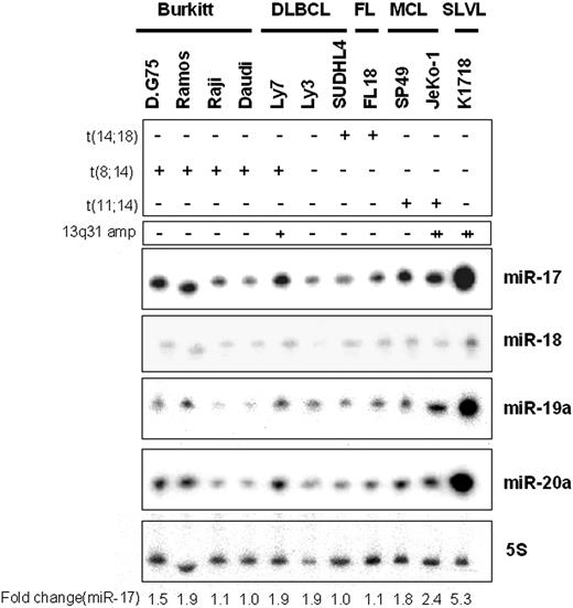
To detect essential target(s) of the miR-17-92 polycistron, we initially carried out Northern blot analyses of miR-17-92 expression in 11 B-cell lymphoma cell lines, including3 DLBCLs (OCI-Ly3, OCI-Ly7, and SUDHL4), 1 follicular lymphoma (FL-18), 4 Burkitt lymphomas (D.G.-75, Ramos, Raji, and Daudi), 2 mantle cell lymphomas (SP-49 and Jeko-1), and 1 splenic lymphoma with circulating villous lymphocytes (Karpas 1718) cell lines (Figure 1). These cell lines generally exhibited high levels of miR-17, miR-19a, miR-19b-1, miR-20a, and miR-92 expression. By contrast, only traces of miR-18 expression were detected. The highest levels of miR-17, miR-19, miR-20a, and miR-92 were found in Karpas 1718 cells. Jeko-1 cells also showed overexpression of the polycistron, as was described previously.9 SP-49, FL-18, OCI-Ly7, Ramos, and D.G.-75 cells all showed greater expression of miR-17-92 than did SUDHL4, Raji, Daudi, or OCI-Ly3 cells.
Expression of miR-17-92 polycistron in various B-cell lymphoma cell lines. Northern blot analysis for 11 B-cell lymphoma cell lines, including DLBCL (3 samples), follicular lymphoma (FL; 1 sample), Burkitt lymphoma (4 samples), mantle cell lymphoma (MCL; 2 samples), and SLVL (1 sample). Fold changes in miR-17 expression are shown below the gels and adjusted to the level in SUDHL4 cells, which was assigned a value of 1.00. The presence or absence of translocations of BCL2, c-MYC, and/or CCND1/CyclinD1 is indicated in the top panel: + indicates positive; −, negative. Copy number counts for 13q31 amplification (13q31 amp) are indicated immediately above the gels: − indicates less than 2; +, 3 or more to less than 10; and ++, 10 or more.
Expression of miR-17-92 polycistron in various B-cell lymphoma cell lines. Northern blot analysis for 11 B-cell lymphoma cell lines, including DLBCL (3 samples), follicular lymphoma (FL; 1 sample), Burkitt lymphoma (4 samples), mantle cell lymphoma (MCL; 2 samples), and SLVL (1 sample). Fold changes in miR-17 expression are shown below the gels and adjusted to the level in SUDHL4 cells, which was assigned a value of 1.00. The presence or absence of translocations of BCL2, c-MYC, and/or CCND1/CyclinD1 is indicated in the top panel: + indicates positive; −, negative. Copy number counts for 13q31 amplification (13q31 amp) are indicated immediately above the gels: − indicates less than 2; +, 3 or more to less than 10; and ++, 10 or more.
We next carried out FISH analyses with each of the cell lines using CCND1/CyclinD1, BCL2, and c-MYC probes. The 11 samples included 4 that were t(8;14) positive (Ramos, D.G.-75, Raji, and Daudi cells), 2 that were t(14;18) positive (SUDHL4 and FL-18 cells), and 2 that were t(11;14)(q13;q32) positive (SP-49 and Jeko-1 cells). Genomic amplification was detected in OCI-Ly7, Jeko-1, and Karpas 1718 cells using a BAC, RP11-121J7 probe, which included the C13orf25/miR-17-92 gene. c-MYC and/or BCL2 rearrangements in SUDHL4, OCI-Ly3, and OCI-Ly7 cells have been described previously,25 as has overexpression of CyclinD1, Bcl2, and c-Myc proteins in Jeko-1 cells.26
Transfection of B-cell lymphoma cell lines with miR-17-19b-1
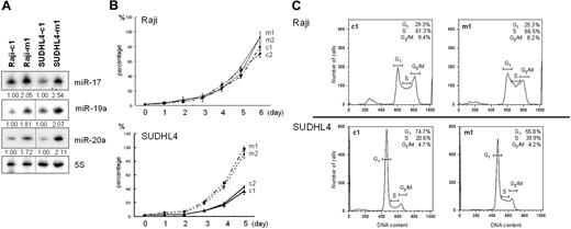
Earlier studies showed that primary B-cell lymphomas possess at least several genetic alterations induced by genomic translocation, loss, and/or amplification.8-13 Additional genomic/genetic alterations (C13orf25/miR-17-92 cluster) on first hit genetic alteration such as c-MYC and BCL2 rearrangements could collaboratively contribute to aggressive progression of B-cell lymphoma. To detect genes targeted by miR-17-92 during B-cell lymphomagenesis, we attempted to use a lentiviral transfection system to transfect miR-17-19b-1 (a miR-17-92 variant) gene into 2 B-cell lymphoma cell lines (Raji and SUDHL4) selected because their endogenous miR-17-92 expression was lower than in the other cell lines tested. We successfully transfected miR-17-19b-1 into both Raji and SUDHL4 cells, after which the transfectants stably overexpressed miR-17-19b-1 (Figure 2A). Transfection of miR-17-19b-1 led to up-regulated expression of miR-17, miR-19a, miR-19b-1, and miR-20a (Figure 2A), but only traces of miR-18 were detected (data not shown).
Transfection of miR-17-19b-1 into B-cell lymphoma cell lines. (A) MiR-17, -19a, and -20a expression in Raji and SUDHL4 cells transfected with miR-17-19b-1 (Raji-m1 and SUDHL4-m1) or empty vector (Raji-c1 and SUDHL4-c1). Fold changes were determined by densitometry and are shown relative to the density obtained with cells transfected with empty vector, which was assigned a value of 1.00. (B) Cell proliferation assays carried out with 2 Raji and 2 SUDHL4 clones transfected with miR-17-19b-1 (Raji-m1 and Raji-m2/SUDHL4-m1 and SUDHL4-m2) or empty vector (Raji-c1 and Raji-c2/SUDHL4-c1 and SUDHL4-c2). Broken lines indicate Raji-c1 and Raji-c2. Cells were transfected using a lentiviral vector and plated at a density 104 cells per well 5 to 7 days after EGFP selection. Cell proliferation was standardized to the fastest proliferating cells and was set to 100%. Symbols and bars indicate means and SDs of 2 independent experiments. (C) Cell-cycle analysis of Raji and SUDHL4 cells transfected with miR-17-19b-1 (Raji-m1 and SUDHL4-m1) or empty vector (Raji-c1 and SUDHL4-c1). Average (n = 3) percentage of empty vector–transfected Raji cells (c1) in G1 phase: 29.3%, S phase: 61.3, and G2 phase: 9.4%; and of miR-17-19b-1–transfected Raji cells (m1) in G1 phase: 25.3%, S phase: 66.5%, and G2 phase: 8.2%. Average (n = 3) percentage of empty vector–transfected SUDHL4 cells (c1) in G1 phase: 74.7%, S phase: 20.6%, and G2 phase: 4.7%; and of miR-17-19b-1–transfected SUDHL4 cells (m1) in G1 phase: 55.8%, S phase: 39.9%, and G2 phase: 4.3%. X-axis represents DNA content; y-axis, number of cells. Horizontal bars represent cell-cycle phases (G1, S, G2/M).
Transfection of miR-17-19b-1 into B-cell lymphoma cell lines. (A) MiR-17, -19a, and -20a expression in Raji and SUDHL4 cells transfected with miR-17-19b-1 (Raji-m1 and SUDHL4-m1) or empty vector (Raji-c1 and SUDHL4-c1). Fold changes were determined by densitometry and are shown relative to the density obtained with cells transfected with empty vector, which was assigned a value of 1.00. (B) Cell proliferation assays carried out with 2 Raji and 2 SUDHL4 clones transfected with miR-17-19b-1 (Raji-m1 and Raji-m2/SUDHL4-m1 and SUDHL4-m2) or empty vector (Raji-c1 and Raji-c2/SUDHL4-c1 and SUDHL4-c2). Broken lines indicate Raji-c1 and Raji-c2. Cells were transfected using a lentiviral vector and plated at a density 104 cells per well 5 to 7 days after EGFP selection. Cell proliferation was standardized to the fastest proliferating cells and was set to 100%. Symbols and bars indicate means and SDs of 2 independent experiments. (C) Cell-cycle analysis of Raji and SUDHL4 cells transfected with miR-17-19b-1 (Raji-m1 and SUDHL4-m1) or empty vector (Raji-c1 and SUDHL4-c1). Average (n = 3) percentage of empty vector–transfected Raji cells (c1) in G1 phase: 29.3%, S phase: 61.3, and G2 phase: 9.4%; and of miR-17-19b-1–transfected Raji cells (m1) in G1 phase: 25.3%, S phase: 66.5%, and G2 phase: 8.2%. Average (n = 3) percentage of empty vector–transfected SUDHL4 cells (c1) in G1 phase: 74.7%, S phase: 20.6%, and G2 phase: 4.7%; and of miR-17-19b-1–transfected SUDHL4 cells (m1) in G1 phase: 55.8%, S phase: 39.9%, and G2 phase: 4.3%. X-axis represents DNA content; y-axis, number of cells. Horizontal bars represent cell-cycle phases (G1, S, G2/M).
We then carried out cell growth assays using 2 individually established clones from each cell line. As shown in Figure 2B, SUDHL4 cells transfected with miR-17-19b-1 grew more aggressively than cells transfected with empty vector. Consistent with that finding, cell cycle analysis of SUDHL4 cells transfected with miR-17-19b-1 showed G1 to S progression (Figure 2C). Raji cells transfected with miR-17-19b-1 also showed slightly greater cell growth than cells transfected with empty vector. MiR-17-19b-1 thus appears to target cell cycle–related gene(s)/protein(s) in B-cell lymphoma cell lines, especially in SUDHL4 cells.
Detection of candidate target genes through analysis of gene expression and RQ-PCR
Because Bim was recently shown to be a possible target of miR-17 and miR-20a in lymphomagenesis,16-18 we next analyzed Bim expression in cells transfected with miR17-19b-1 or empty vector. As shown in Figure 3A, we detected significantly less Bim expression in Raji cells transfected with miR-17-19b-1 than in those transfected with empty vector. Conversely, Bcl2 expression was slightly increased (1.43-fold) in Raji cells transfected with miR-17-19b-1, suggesting miR-17-19b-1 exerts antiapoptotic effects. By contrast, Bim expression in SUDHL4 cells was unaffected by miR-17-19b-1 transfection. Interestingly, however, Bcl2 expression in SUDHL4 cells transfected with miR-17-19b-1 was 1.25-fold higher than in cells transfected with empty vector. That SUDHL4 cells transfected with miR-17-19b-1 showed cell proliferation in the absence of Bim suppression suggests some other target of miR-17-92 is associated with B-cell lymphomagenesis in those cells.
Gene expression and RQ-PCR analyzes. (A) Western blot analysis of Bim and Bcl2 from Raji and SUDHL4 cells transfected with miR-17-19b-1 (Raji-m1 and Raji-m2/SUDHL4-m1 and SUDHL4-m2) or empty vector (Raji-c1 and Raji-c2/SUDHL4-c1 and SUDHL4-c2). Fold changes are shown below the gel normalized to tubulin, which was assigned a value of 1.00. Bim expression in Raji-m1 cells is 0.56 times lower than in Raji-c1 cells. Bcl2 expression in Raji-m1 is 1.43 times higher than in Raji-c1. (B) RQ-PCR analysis of CDKN1A and TP53INP1 expression in SUDHL4 and Raji cells transfected with miR-17-19b-1 (■) or empty vector (□). G6PDH served as an internal control, and the level of each gene's mRNA in each of the samples was normalized to the corresponding G6PDH mRNA level. The significance of the difference between SUDHL4 cells with (SUDHL4-m1) and without (SUDHL4-c1) miR-17-19b-1 transfection is shown above the bars. Values of P were calculated using Student t test. Error bars represent the mean (± SD) of 3 independent experiments. (C) Western blot analysis of p53, p21, CDK4, and CDK6 expression in Raji and SUDHL4 cells transfected with miR-17-19b-1 (Raji-m1 and S4-m1, respectively) or empty vector (Raji-c1 and SUDHL4-c1, respectively). Fold changes are shown below the gels. (D) Luciferase reporter assays of CDKN1A expression in Rat-1 cells transfected with miR-17-19b-1. MiR-17 expression in Rat-1 cells transfected with miR-17-19b-1 or empty vector (control) is shown on the right. The significance of differences between pGL3-CDKN1A 3′UTR wild-type and pGL3-CDKN1A was evaluated using Student t test. Error bars represent the mean (± SD) of 3 independent experiments.
Gene expression and RQ-PCR analyzes. (A) Western blot analysis of Bim and Bcl2 from Raji and SUDHL4 cells transfected with miR-17-19b-1 (Raji-m1 and Raji-m2/SUDHL4-m1 and SUDHL4-m2) or empty vector (Raji-c1 and Raji-c2/SUDHL4-c1 and SUDHL4-c2). Fold changes are shown below the gel normalized to tubulin, which was assigned a value of 1.00. Bim expression in Raji-m1 cells is 0.56 times lower than in Raji-c1 cells. Bcl2 expression in Raji-m1 is 1.43 times higher than in Raji-c1. (B) RQ-PCR analysis of CDKN1A and TP53INP1 expression in SUDHL4 and Raji cells transfected with miR-17-19b-1 (■) or empty vector (□). G6PDH served as an internal control, and the level of each gene's mRNA in each of the samples was normalized to the corresponding G6PDH mRNA level. The significance of the difference between SUDHL4 cells with (SUDHL4-m1) and without (SUDHL4-c1) miR-17-19b-1 transfection is shown above the bars. Values of P were calculated using Student t test. Error bars represent the mean (± SD) of 3 independent experiments. (C) Western blot analysis of p53, p21, CDK4, and CDK6 expression in Raji and SUDHL4 cells transfected with miR-17-19b-1 (Raji-m1 and S4-m1, respectively) or empty vector (Raji-c1 and SUDHL4-c1, respectively). Fold changes are shown below the gels. (D) Luciferase reporter assays of CDKN1A expression in Rat-1 cells transfected with miR-17-19b-1. MiR-17 expression in Rat-1 cells transfected with miR-17-19b-1 or empty vector (control) is shown on the right. The significance of differences between pGL3-CDKN1A 3′UTR wild-type and pGL3-CDKN1A was evaluated using Student t test. Error bars represent the mean (± SD) of 3 independent experiments.
MicroRNAs exert their inhibitory effects on expression not only by inhibiting translation of target proteins but also by reducing the levels of target mRNAs.27 This enabled Voorthoeve et al to use gene expression analysis to determine that the target of miR-373 in a testicular germ cell tumor is LATS2.6 Therefore, to detect target genes of the miR-17-19b-1 cluster, we used a cDNA array platform to analyze gene expression in Raji and SUDHL4 cells. When we analyzed the gene expression in Raji and SUDHL4 cells, with and without miR-17-19b-1 up-regulation, we found that in SUDHL4 cells 17 targets predicted by the TargetScan program28,29 (Whitehead Institute for Biomedical Research, Cambridge, MA) were down-regulated. These included LACE1, CDKN1A, FAM46C, VEGFA, TP53INP1, RPAGD, TESK2, CCNG2, BTG2, FNBP1, AKAP13, WDR26, RYBP, BPTFYPEL2, JAK1, AKAP13, and TGFBRII. Of those, CDKN1A and TP53INP1 may act as tumor suppressors via mechanisms affecting cell progression and apoptosis, respectively. Therefore, to assess the relative expression levels of CDKN1A and TP53INP1, we carried out RQ-PCR analyses in cells transfected with miR-17-19b-1 or empty vector. We found that expression of both CDKN1A and TP53INP1 is down-regulated in SUDHL4 cells transfected with miR17-19b-1 (referred to as SUDHL4-m1 and SUDHL4-m2), compared with cells transfected with empty vector (referred to as SUDHL4-c1 and SUDHL4-c2) (Figure 3B). The CDKN1A product, p21, is known to induce G1 to S arrest via p53-dependent and -independent pathways, whereas the TP53INP1 product, TP53INP1, induces apoptosis.30 In that context, our finding that SUDHL4 cells transfected with miR-17-19b-1 showed aggressive growth facilitated by dysregulation of the cell cycle at the G1 to S phase transition suggests CDKN1A is the most likely target of the expressed miRNA, though TP53INP1 remains a candidate target of miR-17-92 in lymphomagenesis.
By contrast, Raji cell transfectants showed down-regulation of 5 other genes, including RNH1, BAMBI, SYT11, FOXP1, and CASZ1, and transfection of miR-17-19b-1 had no effect on CDKN1A or TP53INP1 expression in either of the Raji cell clones tested.
Expression of both CDKN1A mRNA and p21waf1/Cip1 protein (p21) was reduced and expression of CDK4 was reversibly increased in SUDHL4 cells transfected with miR-17-19b-1, whereas there was almost no change in the level of p53 expression (Figure 3B-C). Expression of p21 was also reduced and CDK4 was reversibly increased in Raji cells transfected with miR-17-19b-1, although there was almost no change in the levels of CDKN1A mRNA (Figure 3B-C). To determine whether CDKN1A is directly regulated by the miR-17-92 polycistron, we carried out luciferase reporter assays using Rat-1 fibroblasts stably expressing miR-17-19b-1. We found that when the cells were transfected with a reporter construct harboring wild-type CDKN1A, luciferase activity was significantly repressed, compared with cells transfected with a construct harboring a CDKN1A mutant (Figure 3D). This suggests that CDKN1A is the direct downstream target of the miR-17-19b-1 polycistron.
Jeko-1 cells exhibit both overexpression of miR-17-92 and homozygous deletion of Bim
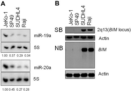
Recent studies have shown that Bim, which may induce apoptosis by negatively regulating Bcl2, is a target of miR-17-92 during lymphomagenesis.16-18 However, Bim appears to be an unlikely target of miR-17-92 in some B-cell lymphoma cell lines. Collectively, the results of our Northern (Figure 4A) and Southern (Figure 4B) blot analyses show the relationship between miR-17-92 expression and Bim deletion in 4 B-cell lymphoma cell lines (Jeko-1, SP-49, SUDHL4, and Raji). As indicated in Figure 4B, Jeko-1 cells exhibit both a loss of BIM expression and overexpression of miR-17-92. The former is caused by homozygous loss of 2q13, whereas the latter is cause by amplification of 13q31. This suggests that Bim is not a target of miR-17-92 in Jeko-1 cells, whereas miR-17-92 must target other gene(s) or protein(s).9,10
Jeko-1 cells show both miR-17-92 overexpression and homozygous deletion of BIM. (A) Northern blot analysis of miR-19 and miR-20a expression in 4 B-cell lymphoma cell lines: lane 1 represents Jeko-1; lane 2, SP49; lane 3, SUDHL4; and lane 4, Raji. Fold changes in miR-17 expression are shown below the gels and normalized to the level in SUDHL4 cells, which was assigned a value of 1.00. (B) Top panels: Southern blot analysis (SB) of the BIM locus carried out using genomic DNA, including BIM, from the indicated 4 cell lines. Bottom panels: Northern blot analysis (NB) of BIM from the 4 indicated cell lines. A cDNA fragment of β-actin was used a control.
Jeko-1 cells show both miR-17-92 overexpression and homozygous deletion of BIM. (A) Northern blot analysis of miR-19 and miR-20a expression in 4 B-cell lymphoma cell lines: lane 1 represents Jeko-1; lane 2, SP49; lane 3, SUDHL4; and lane 4, Raji. Fold changes in miR-17 expression are shown below the gels and normalized to the level in SUDHL4 cells, which was assigned a value of 1.00. (B) Top panels: Southern blot analysis (SB) of the BIM locus carried out using genomic DNA, including BIM, from the indicated 4 cell lines. Bottom panels: Northern blot analysis (NB) of BIM from the 4 indicated cell lines. A cDNA fragment of β-actin was used a control.
Effects of ASOs in Jeko-1 cells reveal that p21 is an essential target of the miR-17-92 polycistron during lymphomagenesis
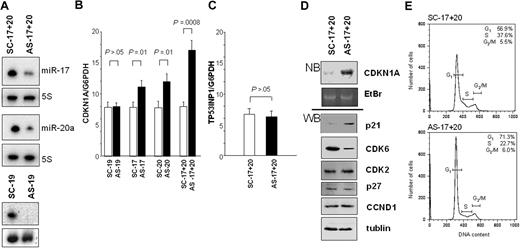
The results summarized so far suggest the most likely target of miR-17-92 in Jeko-1 cells is CDKN1A/p21. To test that idea further, we examined the effects of transfecting the cells with ASOs against miR-19a, miR-17, and miR-20a. As shown in Figure 5A, levels of miR-17 and miR-20a were significantly reduced in cells transfected with ASOs. RQ-PCR analysis of CDKN1A and TP53INP1 expression in Jeko-1 cells treated with ASOs or SCOs revealed that although there was no significant difference between expression levels in cells treated with AS-19 or SC-19, cells treated with AS-17, with or without AS-20, showed greater expression than cells treated with the respective SCOs (Figure 5B). We also carried out RQ-PCR analysis of TP53INP1 expression in SCO- and ASO-treated Jeko-1 cells (Figure 5C), but no up-regulation was detected, indicating that TP53INP1 mRNA is not a target in this cell line. Furthermore, the effects of AS-17 and AS-20 appeared to be additive, as the increase in CDKN1A expression was more pronounced in cells treated with AS-17 plus AS-20 than in those treated with AS-17 or AS-20 alone. Thus CDKN1A expression was unaffected by inhibition of miR-19a but was up-regulated by inhibition of miR-17 and/or miR-20a, which is consistent with the notion that CDKN1A expression is directly regulated by the miR-17 family of miRNAs (miR-17 and miR-20a).
Effects of treating Jeko-1 cells with antisense oligonucleotides (ASOs) and scrambled oligonucleotides (SCOs). (A) Expression of MiR-17 and miR-20a of Jeko-1 cells after targeting by ASOs or SCOs (AS-17+20 and SC-17+20, respectively). Expression of miR-19a of Jeko-1 cells treated with antisense miR-19a (AS-19) and scrambled miR-19a (SC-19) is also shown in this figure. (B) RQ-PCR analysis of CDKN1A expression in Jeko-1 cells treated with SC/AS-19, SC/AS-20, SC/AS-17, or SC/AS-17+20. CDKN1A mRNA levels in each sample were normalized to the corresponding level of G6PDH mRNA. The significance of differences between SC and AS was evaluated using Student t test. Error bars represent the mean (± SD) of 3 independent experiments. (C) RQ-PCR analysis of TP53INP1 expression in Jeko-1 cells treated with SC-17+20 or AS-17+20a. Error bars represent the mean (± SD) of 3 independent experiments. (D) Northern blot analysis (NB) of CDKN1A expression and Western blot analysis (WB) of p21, CDK6, CDK2, p27, Cyclin D1 (CCND1), and tubulin expression in Jeko-1 cells treated with AS-17+20 or SC-17+20. (E) Cell-cycle analysis of Jeko-1 cells treated with SC-17+20 or AS-17+20. Average (n = 3) percentage of SC-treated cells in G1 phase: 56.9%, S phase: 37.6, and G2 phase: 5.5%; and of AS-treated cells in G1 phase: 71.3%, S phase: 22.7%, and G2 phase: 6.0%. X-axis represents DNA content; y-axis, number of cells. Horizontal bars represent cell-cycle phases.
Effects of treating Jeko-1 cells with antisense oligonucleotides (ASOs) and scrambled oligonucleotides (SCOs). (A) Expression of MiR-17 and miR-20a of Jeko-1 cells after targeting by ASOs or SCOs (AS-17+20 and SC-17+20, respectively). Expression of miR-19a of Jeko-1 cells treated with antisense miR-19a (AS-19) and scrambled miR-19a (SC-19) is also shown in this figure. (B) RQ-PCR analysis of CDKN1A expression in Jeko-1 cells treated with SC/AS-19, SC/AS-20, SC/AS-17, or SC/AS-17+20. CDKN1A mRNA levels in each sample were normalized to the corresponding level of G6PDH mRNA. The significance of differences between SC and AS was evaluated using Student t test. Error bars represent the mean (± SD) of 3 independent experiments. (C) RQ-PCR analysis of TP53INP1 expression in Jeko-1 cells treated with SC-17+20 or AS-17+20a. Error bars represent the mean (± SD) of 3 independent experiments. (D) Northern blot analysis (NB) of CDKN1A expression and Western blot analysis (WB) of p21, CDK6, CDK2, p27, Cyclin D1 (CCND1), and tubulin expression in Jeko-1 cells treated with AS-17+20 or SC-17+20. (E) Cell-cycle analysis of Jeko-1 cells treated with SC-17+20 or AS-17+20. Average (n = 3) percentage of SC-treated cells in G1 phase: 56.9%, S phase: 37.6, and G2 phase: 5.5%; and of AS-treated cells in G1 phase: 71.3%, S phase: 22.7%, and G2 phase: 6.0%. X-axis represents DNA content; y-axis, number of cells. Horizontal bars represent cell-cycle phases.
We also carried out Northern and Western blot analyses that, respectively, showed the presence of higher levels of CDKN1A mRNA and p21 protein in cells treated with AS-17 and AS-20. Moreover, expression of CDK6 was suppressed in Jeko-1 cells treated with ASOs, though CDK2 was not (Figure 5D), suggesting that, in this case, p21 induces G1 to S arrest (Figure 5E) by inhibiting the p21-CDK6-CyclinD1 pathway, but not the p27-CDK2-CyclinE pathway. No CDK4 expression was detected in either ASO- or SCO-treated Jeko-1 cells. Thus, overexpression or inhibition of miR-17 family miRNAs is apparently sufficient to, respectively, promote or delay a cell's entry into S phase via targeting p21.
Discussion
MiRNAs encoded by the miR-17-92 polycistron and its paralogs are known to act as oncogenes. Their expression promotes cell proliferation, suppresses cancer cell apoptosis, and induces tumor angiogenesis. Of the more that 1500 miR-17-92 targets predicted by the TargetScan program, 10 (E2F1, E2F3, Pten, AML1, Bim, AIB1, TGFBRII, Tsp1, CTGF, and p130) have been identified.11,31-36 These targets are key players in hematopoiesis, apoptosis, cell-cycle progression, and angiogenesis, and some have been associated with tumorigenesis. Given that each miRNA likely regulates hundreds of mRNAs, the diverse functions of the miR-17-92 cluster in different physiological contexts during tumorigenesis likely involve different subsets of targets.37 In human cancer, for instance, miR-17-92 down-regulates E2F3 in Wilms tumor and AIB1 in breast cancer.35,36 It is unclear whether these targets are associated with lymphomagenesis, however. Inhibition of miR-17 and miR-20a using ASOs selectively induces apoptosis in lung cancer cell lines overexpressing miR-17-92.38 On the other hand, antisense inhibition of miR-17 and miR-20a did not induce apoptosis in Jeko-1 cells; instead, inhibition of these miRNAs appeared to induce G1 arrest, suggesting there are significant differences between targets of the miR-17-92 cluster in terms of their roles in cancer cell growth.
The targets of miR-17-92 might differ not only in cancers of different origin, but also in genetically distinct B-cell lymphoma subtypes. MiR-17-92 is known to be overexpressed in c-MYC rearranged, Bcl2 rearranged, and Cyclin D1 rearranged B-cell lymphomas.8-13 Our experiments demonstrated that upon transfection of miR-17-19b-1 (a miR-17-92 variant), expression of p21, but not Bim, was suppressed in SUDHL4 cells, which were derived from diffuse large B-cell lymphoma with aberrant Bcl2 overexpression. On the other hand, suppression of both of Bim and p21 was detected in transfected Raji cells, which were derived from Burkitt lymphoma with aberrant c-MYC overexpression. It is known that approximately 24% of B-cell lymphomas with c-MYC rearrangement show 13q31 amplification with overexpression of miR-17-92.11 In mice, forcibly expressed miR-17-92 cooperates with the c-myc oncogene to induce B-cell lymphomas by reducing the incidence of apoptosis among lymphoma cells,5 which suggests overexpression of miR-17-92 and down-regulation of Bim might be associated with tumorigenesis via antiapoptotic activity. On the other hand, overexpression of miR-17-92 in B-cell lymphoma with t(14;18) could induce cell proliferation via down-regulation of p21, as aberrantly overexpressed Bcl2 exerts antiapoptotic effects, even in the absence of Bim repression.
It was recently reported that the miR-106b-25 polycistron negatively regulates CDKN1A(p21) and Bim in gastric cancer. Moreover, the targeting of CDKN1A (p21) was recently shown to be an additional mechanism by which miR-106b and miR-93 influence cell-cycle progression.39,40 Given that miR-17-92 is a member of the miR-106b family, we suggest its overexpression through 13q31 genomic amplification also likely leads to down-regulated expression of CDKN1A mRNA and p21 protein during B-cell lymphomagenesis. That said, it remains uncertain whether miR-17-92 does indeed down-regulate CDKN1A in “B-cell lymphomagenesis.” Our present work therefore provides a novel finding that associates B-cell lymphomagenesis with the tumorigenic activity of the miR-17-92 polycistron.
Finally, Pten is also a known target of miR-17-92, and its dysregulation also might contribute to lymphomagenesis.16 However, the fact that we did not detect alteration of Pten expression in our cell lines suggests Pten is not targeted by the miR-17-92 cluster in lymphomagenesis.
In summary, we have shown that CDKN1A/p21 is an essential target of miR-17-92 during B-cell lymphomagenesis, and that miR-17-92 can regulate different subtypes of B-cell lymphomas through down-regulation of different target genes (eg, Bim or/and p21). More detailed analysis will be needed to fully understand the various functions of miR-17-92 in different B-cell lymphoma subtypes.
The publication costs of this article were defrayed in part by page charge payment. Therefore, and solely to indicate this fact, this article is hereby marked “advertisement” in accordance with 18 USC section 1734.
Acknowledgments
We express our appreciation to Mss Keiko Iwamoto, Etuko Kobayashi, Hiromi Kataho, and Mikiko Kikuchi for their outstanding technical assistance. Mr Junichi Yamashita contributed to the RI experiment. pMX vector was kindly provided by Dr Toshio Kitamura (Tokyo University, Tokyo, Japan). Jeko-1 and SP-49 cells were obtained from Hayashibara Research Institute with permission from Drs Tadashi Yoshino (Okayama University) and Masanori Daibata (Kochi University). Karpas 1718 cells were kindly provided from Dr Ablaham Karpas (Cambridge University). SIEW and S-17-19b-IEW were kindly provided from Drs Matthias Eder and Michaela Scherr (Hannover Medical School, Hannover, Germany).
This work was supported by the Grant Global Center of Excellence Program (G-COE) of the Ministry of Education, Culture, Sports, Science and Technology of Japan (Tokyo, Japan), and by the Sagawa Foundation for Promotion of Cancer Research (Kyoto, Japan; H.T.).
Authorship
Contribution: M.I. performed experiments and analyzed data; H.T. designed and performed experiments, analyzed data, and wrote the paper; Y.-M.G. performed experiments; Y.K. and N.T. analyzed data; and K.S. analyzed the data and organized the experiments.
Conflict-of-interest disclosure: The authors declare no competing financial interests.
Correspondence: Hiroyuki Tagawa, Department of Internal Medicine III, Akita University School of Medicine, 1-1-1 Hondo, Akita, Akita 0108543, Japan; e-mail: htagawa0279jp@yahoo.co.jp.


This feature is available to Subscribers Only
Sign In or Create an Account Close Modal