Down-regulation of the Kpm/Lats2 tumor suppressor is observed in various malignancies and associated with poor prognosis in acute lymphoblastic leukemia. We documented that Kpm/Lats2 was markedly decreased in several leukemias that were highly resistant to conventional chemotherapy. Silencing of Kpm/Lats2 expression in leukemic cells did not change the rate of cell growth but rendered the cells more resistant to DNA damage–inducing agents. Expression of p21 and PUMA was strongly induced by these agents in control cells, despite defective p53, but was only slightly induced in Kpm/Lats2-knockdown cells. DNA damage–induced nuclear accumulation of p73 was clearly observed in control cells but hardly detected in Kpm/Lats2-knockdown cells. Chromatin immunoprecipitation (ChIP) assay showed that p73 was recruited to the PUMA gene promoter in control cells but not in Kpm/Lats2-knockdown cells after DNA damage. The analyses with transient coexpression of Kpm/Lats2, YAP2, and p73 showed that Kpm/Lats2 contributed the stability of YAP2 and p73, which was dependent on the kinase function of Kpm/Lats2 and YAP2 phosphorylation at serine 127. Our results suggest that Kpm/Lats2 is involved in the fate of p73 through the phosphorylation of YAP2 by Kpm/Lats2 and the induction of p73 target genes that underlie chemosensitivity of leukemic cells.
Introduction
The Warts (Wts) tumor suppressor gene (also termed Lats after large tumor suppressor) was first identified by mitotic recombination of somatic cells and screening for homozygous mutants with overproliferation phenotype in Drosophila melanogaster.1,2 This discovery initiated a series of genetic studies in Drosophila that led to the delineation of a new signaling network named the Hippo pathway, which is now known to regulate cell growth, cell survival, and organ size in developing animals.3,–5 This pathway consists of a kinase cascade in its core where Hippo (Hpo) phosphorylates and activates Wts/Lats,6 which then in turn phosphorylates and inactivates Yorkie (Yki), a transcription coactivator. Inactivation of Yki results in control of cell survival and cell growth through down-regulation of Drosophila inhibitor of apoptosis 1 (Diap1) and Cyclin E.7 Salvador (Sav),8,9 a scaffold protein for Hpo, and Mats (mob as tumor suppressor),10,–12 a partner and potentiater of Wts, are also essential components of this pathway. Furthermore, recent evidence has placed Expanded (EX), Merlin (Mer),13 both 4.1 family proteins, and Fat (FT),14,,–17 the atypical cadherin, in the upstream of the Hippo pathway although their connection to the kinase cascade is largely based on genetic epitasis. The Hippo pathway is believed to be conserved throughout species because some of the mammalian homologues have been shown to compensate the corresponding defects in the Drosophila Hippo pathway. At present, however, only a small part of the mammalian Hippo pathway has been experimentally substantiated
Kpm (alternatively named Lats2) is one of the 2 human homologues of Drosophila Wts.18,19 In parallel to Drosophila Wts, we and others have shown the critical involvement of Kpm/Lats2 in regulation of cell growth and survival. Kpm/Lats2 overexpression results in the cell cycle arrest in G2/M phase via inhibition of Cdc2–Cyclin B kinase activity leading eventually to apoptosis,20 inhibition of G1/S transition via down-regulation of Cyclin E/Cdk2 kinase activity,21 or apoptosis via down-regulation of Bcl-2 and Bcl-xL.22 Kpm/Lats2 binds to Mdm2 and inhibits its E3 ubiquitin ligase activity, resulting in the stabilization of p53 and leading to the p53-dependent G1/S arrest in nocodazole-treated cells.23 Moreover, Kpm/Lats2 is a target gene of p53 both in mammalian as well as in Drosophila cells,24,25 suggesting that Kpm/Lats2 may be a positive-feedback-loop regulator of p53. Kpm/Lats2 knockout mice are embryonically lethal and fibroblasts isolated from these mice appear to be defective in contact inhibition and display genomic instability through multipolar mitotic spindles.26,27 Mst-2, one of the mammalian orthologues of Drosophila Hippo, has been reported to phosphorylate Kpm/Lats2 as well as its related kinase, Lats1.28 However, the downstream function of Kpm/Lats2 has not been elucidated in the Hippo pathway in mammals, because the interaction between Kpm/Lats2 and Yes-associated protein (YAP), the mammalian orthologue of Yki, has not been shown.
Yes-kinase associated protein (YAP) was initially isolated by virtue of its binding to the Src family member, a nonreceptor tyrosine kinase Yes.29,30 The cloning of YAP revealed a new modular protein domain, known today as the WW domain, which recognizes a specific set of proline-rich ligands. The YAP gene encodes at least 2 isoforms: YAP1 and YAP2, which are generated by differential splicing and differ in the number of WW domains they contain. YAP1 has one WW domain and YAP2 has 2 WW domains.30,31 In human epithelial cells, YAP plays a potentially oncogenic role through several signaling interactions with potent signaling proteins.32,,,–36 Furthermore, a region of human chromosome 11 at position q22 has been reported to be frequently amplified in various cancers, and this amplicon contains YAP and cIAP2 loci.37,38 Curiously, YAP has also been shown to regulate apoptosis. For example, YAP forms a signaling complex with p53-binding protein-2, a known regulator of the apoptotic activity of p53.39,40 YAP interacts with and coactivates p73 to induce transcription of its target genes, leading to apoptosis and cell cycle arrest.41 In sum, YAP has a capacity to function either as an oncogene or as a proapoptotic factor.
Recent clinical studies have indicated that the expression level of Kpm/Lats2 correlates with clinical course of some malignancies. Down-regulation of Kpm/Lats2 was associated with larger tumor size and high number of metastatic lymph nodes in breast cancers,42 and it was significantly associated with poor prognosis in acute lymphoblastic leukemia (ALL).43 The relative level of Kpm/Lats2 expression was shown with high confidence as the most important prognostic factor in predicting disease-free survival in ALL, even compared with the BCR-ABL gene fusion, which is so far the most significant factor for poor prognosis. However, the molecular and cellular mechanisms underlying the poor clinical course in leukemias that have a relatively low level of Kpm/Lats2 expression have not been investigated in detail.
In the present study, we documented that Kpm/Lats2 was down-regulated also in adult T-cell leukemia (ATL) and natural killer (NK) leukemia/lymphoma, both of which are known to be highly resistant to conventional chemotherapy. Then we addressed the molecular mechanism underlying the chemoresistance associated with low Kpm/Lats2 expression. We herewith report that down-regulation of Kpm/Lats2 leads to chemoresistance through insufficient nuclear accumulation of p73, resulting in poor induction of its target genes p21 and p53 up-regulated modulator of apoptosis (PUMA).
Methods
Cells and cell culture
Leukemic cell lines including KG-1a44 were cultured in 10% heat-inactivated fetal bovine serum (FBS; Invitrogen, Paisley, United Kingdom) containing Iscove modified Dulbecco medium (IMDM; Invitrogen) with 2 mM l-glutamine and antibiotics (Invitrogen), and only ED-40515+45 was cultured in the same medium with 100 IU/mL recombinant human interleukin-2 (rhIL-2; Shionogi, Osaka, Japan). Adherent cell lines including 293T, GP2-293, and HeLa were cultured using Dulbecco modified Eagle medium (DMEM; Invitrogen) instead of IMDM. All cells were maintained at 37°C in a 5% CO2 humidified incubator. Clinical samples from patients with leukemia were cryopreserved in our laboratory as described previously.46,–48 Normal peripheral mononuclear cells (PBMCs) were purified from healthy donor with informed consent; normal CD4+ T cells and normal CD56+ cells were purified using magnetic-activated cell sorting (MACS) CD4+ T-cell isolation kit (Miltenyi Biotec, Bergisch Gladbach, Germany) and MACS CD56+ isolation kit (Miltenyi Biotec), respectively. All the clinical samples were taken with informed consent and used only for in vitro study following the guideline of the institutional review board of Kyoto University. This study was conducted in accordance with the Declaration of Helsinki.
Isolation of total RNA and quantitative real-time PCR
The isolation of total RNA was performed using RNeasy Mini kit (Qiagen, Valencia, CA). The cDNA was synthesized from 1 μg total RNA by ImProm-II Reverse Transcription system (Promega, Madison, WI). Quantitative real-time polymerase chain reaction (PCR) was analyzed using SYBR Green (Invitrogen) on an ABI Prism 7900HT instrument (Applied Biosystems, Foster City, CA). More than one internal control was always used in each analysis, and only when relative amounts of internal controls were constant for each sample, were data considered valid. The list of gene-specific primers is provided in Table 1.
Primer sequence for real-time PCR or semiquantitative RT-PCR
| Gene . | Primer sequence . |
|---|---|
| Kpm (for RT-PCR) | |
| Forward | GCTGACTTTGGCCATTGAGAGTGTC |
| Reverse | CATCTACGGGGTCGAAATTCGAGGT |
| Kpm (for real-time) | |
| Forward | GTTCAGGTGGACTCACAATTCC |
| Reverse | CGACAGTTAGACACATCATCCCA |
| Lats1 | |
| Forward | TGGTCATATTAAATTGACTGAC |
| Reverse | CCACATCGACAGCTTGAGGG |
| YAP | |
| Forward | TGGGAGATGGCAAAGACATCTTCTG |
| Reverse | ACACTGGATTTTGAGTCCCACCATC |
| cIAP1 | |
| Forward | CAGCCTGAGCAGCTTGCAA |
| Reverse | CAAGCCACCATCACAACAAAA |
| cIAP2 | |
| Forward | TCCGTCAAGTTCAAGCCAGTT |
| Reverse | TCTCCTGGGCTGTCTGATGTG |
| survivin | |
| Forward | TGCCTGGCAGCCCTTTC |
| Reverse | CCTCCAAGAAGGGCCAGTTC |
| XIAP | |
| Forward | AGTGGTAGTCCTGTTTCAGCATCA |
| Reverse | CCGCACGGTATCTCCTTCA |
| bcl-2 | |
| Forward | AGGAAGTGAACATTTCGGTGAC |
| Reverse | GCTCAGTTCCAGGACCAGG |
| p21 | |
| Forward | CTGTCACTGTCTTGTACCCT |
| Reverse | GGTAGAAATCTGTCATGCTGC |
| PUMA | |
| Forward | GACCTCAACGCACAGTA |
| Reverse | CTAATTGGGCTCCATCT |
| TAp73 | |
| Forward | GCACCACGTTTGAGCACCTCT |
| Reverse | GCAGATTGAACTGGGCCATGA |
| β-actin | |
| Forward | TCCTGTGGCATCCACGAAACT |
| Reverse | GAAGCATTTGCGGTGGACGAT |
| hprt | |
| Forward | TGACACTGGCAAAACAATGCA |
| Reverse | GGTCCTTTTCACCAGCAAGCT |
| gapdh | |
| Forward | GAAGGTGAAGGTCGGAGTC |
| Reverse | GAAGATGGTGATGGGATTTC |
Plasmids
The plasmids for expression of HA-tagged Kpm wild-type (wt) form and Kpm-kinase dead (kd) form (mutant form K697 to A) were described before.18 FLAG-tagged YAP1, YAP1-S127A (mutant form S127 to A; S127 is Akt-phosphorylation site), YAP1-WW* (mutant form of WW domain), YAP2, YAP2-S127A, YAP2-1WW* (mutant form of first WW domain), and YAP2-2WW* (mutant form of second WW domain) inserted into pFLAG-CMV2 vector were described previously 33. TAp73α inserted into pcDNA3 vector, described elsewhere,49 was a gift of Dr Yoshihide Ueda (Kyoto University). The plasmid expressing Kpm/Lats2 shRNA was generated by insertion of target sequence (loop sequence, CTGTGAAGCCACAGATGGG) and target antisense sequence into the retrovirus (RV) vector, pSINsi-hU6 (Takara, Kusatsu, Japan). Kpm/Lats2 target sequence is TTCACCTTCCGAAGGTTCT; control sequence is TCGTACTCTCGTCTTCGAT. Control sequence was constructed by shuffling Kpm/Lats2 target sequence, and it was confirmed using BLASTN that control sequence did not target any other genes.50 p73 target sequence is GGATTCCAGCATGGACGTCTT, as described elsewhere.51 pVSV-G was used as the envelope plasmid.
Plasmid transfection and retrovirus vector transduction
Plasmids were transfected into 293T cells using CalPhos mammalian transfection kit (Clontech, Mountain View, CA) or FuGENE-HD (Roche, Basel, Switzerland) for coimmunoprecipitation assays. Retroviruses were generated by cotransfection of shRNA containing retrovirus vector and pVSV-G into GP2-293 packaging cells with CalPhos kit and collected by ultracentrifugation. Retronectin (Takara) was used to transduce leukemic cells with retroviruses. After transduction, pools of cells in bulk selected with 0.5 mg/mL G-418 (Nacalai Tesque, Kyoto, Japan) were used in the following assays.
MTT assay
To evaluate cell viability, an appropriate number of cells were seeded with doxorubicin (DXR; Pharmacia, Milan, Italy) or etoposide (ETP; Bristol-Myers, New York, NY) in appropriate concentrations. Next MTT assays were performed using WST-8 (Nacalai Tesque) according to the manufacturer's protocol and analyzed with microplate reader Benchmark (Bio-Rad, Hercules, CA) as described previously.20
Antibodies, immunoprecipitation, and Western blotting
The following antibodies were purchased or prepared in our laboratory: anti-FLAG mouse monoclonal antibody (Sigma-Aldrich, St Louis, MO), anti-HA mouse monoclonal antibody (Roche), anti-YAP rabbit polyclonal antibody, anti–phospho-YAP(Ser127) rabbit polyclonal antibody, anti-PUMA rabbit polyclonal antibody, anti-p21 mouse monoclonal antibody (Cell Signaling, Beverly, MA), anti-p73 mouse monoclonal antibody (Ab-4; Lab Vision, Fremont, CA), antiactin goat polyclonal antibody (Santa Cruz Biotechnology, Santa Cruz, CA), HRP conjugated anti–mouse IgG and anti–rabbit IgG (GE Healthcare, Uppsala, Sweden), and anti–goat IgG (Santa Cruz Biotechnology). Anti-Kpm rabbit polyclonal antibody was generated in our laboratory as described previously.18 For immunoprecipitation assay, cells were lysed on ice for 30 minutes with Triton X–based lysis buffer (50 mM Tris-HCl at pH 8.0, 150 mM NaCl, 1% Triton X, 1 mM PMSF, 1 mM EDTA, and protease inhibitor cocktail; Nacalai Tesque), with the optional addition of phosphatase inhibitor cocktail 1 and 2 (Sigma-Aldrich) to detect phosphorylation status of YAP. The lysate, after centrifugation and precleaning, was incubated with 1 μg indicated antibodies overnight at 4°C and precipitated with protein G–sepharose beads (GE Healthcare) at 4°C for 3 hours. After washing 5 times with lysis buffer, the precipitate was boiled in 2 × sample buffer. Phosphatase treatment of the immunoprecipitates was done by incubating beads with 0.2 U/μL calf intestine phosphatase (CIP; Sigma-Aldrich) in 10 μL of 100 mM Tris-HCl (pH 8.0) at 37°C for 1 hour. Western blotting was performed according to the manufacturer's protocol for each antibody, and the protein bands were detected using the enhanced chemiluminescence (ECL) detection system (GE Healthcare).
Immunofluorescence microscopy
KG-1a cells were treated with 0.1 μg/mL ETP for 2.5 days. Then cells were cytospun on glass slides and fixed in ice-cold acetone for 3 minutes. After blocking, cells were incubated with 4 μg/mL anti-p73 mouse monoclonal antibody (Lab Vision) or mouse control IgG (Santa Cruz Biotechnology) in 1% bovine serum albumin containing PBS for 1 hour at room temperature. After extensive wash, cells were incubated with Alexa Fluor-488–conjugated anti–mouse IgG (Invitrogen), and 4′,6-diamino-2-phenylindole (DAPI; Sigma-Aldrich) staining was finally performed. Analysis was performed with fluorescence microscopy BIOZERO B8-8100 (Keyence, Osaka, Japan) that was equipped with a camera as an all-in-one type and with objective lenses (4×/0.2 NA and 20×/0.75 NA). B2-Viewer versus 1.0 (Keyence) and B2-Analyzer BZ-HIA versus 3.5 (Keyence) were used for image acquisition and image processing, respectively.
Chromatin immunoprecipitation assay
Chromatin immunoprecipitation (ChIP) assay of p73 binding to sites in the PUMA promoter region was done using ChIP-IT enzymatic kits (Active Motif, Carlsbad, CA) according to the manufacturer's instructions. Briefly, cells were fixed with 1% formaldehyde for 10 minutes, lysed at 4°C for 30 minutes, homogenized by passing through 27-gauge needles, and centrifuged. Pelleted nuclei were then subjected to enzymatic sharing for the optimized time. One-tenth volume was stored as the input and the remaining was diluted and incubated with 2 μg anti-p73 antibody (Ab-4; Lab Vision) or control IgG (Santa Cruz Biotechnology) at 4°C overnight. Immune complexes were precipitated with protein G–sepharose beads. After intensive washes, beads were treated with elution buffer. The supernatants and the stored input solutions were reverse cross-linked and treated with RNase and proteinase K, and chromatin DNA was purified using the kit-included DNA minicolumns. The following PCR primers were used to amplify the PUMA gene promoter: 5′-tgactgggacccacagatcca-3′ (forward) and 5′-tccaggggaccctgttagtgag-3′ (reverse).
Results
Kpm/Lats2 is down-regulated in various leukemias
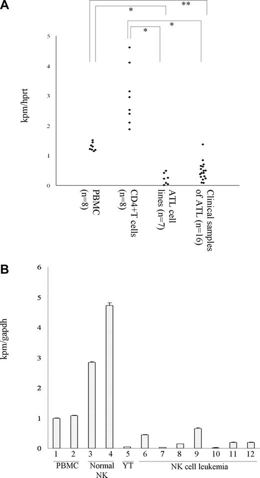
The down-regulation of Kpm/Lats2 has been linked to poor prognosis of ALL. To evaluate the significance of Kpm/Lats2 expression in other types of leukemia, we measured the expression level of Kpm/Lats2 by real-time PCR in leukemic cells that were available in our laboratory. The expression of Kpm/Lats2 was markedly decreased in all adult T-cell leukemia (ATL)–derived cell lines and all clinical samples from ATL patients in comparison with the normal counterpart CD4+ T cells (Figure 1A). Similarly, down-regulation of Kpm/Lats2 was observed in one NK cell line and most of the clinical samples from patients with NK cell leukemia/lymphoma, compared with the normal counterpart CD3− CD56+ NK cells (Figure 1B). Both types of leukemias are known to be clinically aggressive and resistant to conventional chemotherapy. In other leukemic cell lines derived from acute myeloid leukemia (AML) except for KG-1a, B-ALL (Burkitt leukemia), T-ALL, and T-chronic lymphocytic leukemia (T-CLL), the expression level of Kpm/Lats2 was very low or hardly detectable, compared with normal peripheral blood mononuclear cells (PBMCs; Figure S1, available on the Blood website; see the Supplemental Materials link at the top of the online article). These results suggest that the down-regulation of Kpm/Lats2 is rather common in hematologic malignancies and the degree of decrease may be associated with poor prognosis.
Quantitative analysis of Kpm/Lats2 mRNA in clinical samples and cell lines. (A) The amount of Kpm/Lats2 mRNA was measured in ATL cell lines, ATL clinical samples, normal PBMCs, and normal CD4+ T cells. Data normalized to hprt are shown representatively in scale that the value for normal PBMC is 1. Normalization to gapdh gave similar results. The highest one among the ATL cell lines represents ED-40515+. Analyses were performed in duplicate independently 3 times and representative data are shown (Welch t test: *P < .001; **P < .005). (B) The amount of Kpm/Lats2 mRNA was measured in NK cell line (YT), NK cell leukemia clinical samples, normal PBMCs (lanes 1-2), and normal CD3−56+ NK cells (lanes 3-4), NK cell line (YT; lane 5), and NK cell leukemia clinical samples (lane 6-12). Data normalized to gapdh are shown as mean plus or minus SD in scale that the value for normal PBMCs (lane1) is 1. Normalization to hprt gave similar results. Analyses were performed in triplicate independently twice, and representative data are shown.
Quantitative analysis of Kpm/Lats2 mRNA in clinical samples and cell lines. (A) The amount of Kpm/Lats2 mRNA was measured in ATL cell lines, ATL clinical samples, normal PBMCs, and normal CD4+ T cells. Data normalized to hprt are shown representatively in scale that the value for normal PBMC is 1. Normalization to gapdh gave similar results. The highest one among the ATL cell lines represents ED-40515+. Analyses were performed in duplicate independently 3 times and representative data are shown (Welch t test: *P < .001; **P < .005). (B) The amount of Kpm/Lats2 mRNA was measured in NK cell line (YT), NK cell leukemia clinical samples, normal PBMCs (lanes 1-2), and normal CD3−56+ NK cells (lanes 3-4), NK cell line (YT; lane 5), and NK cell leukemia clinical samples (lane 6-12). Data normalized to gapdh are shown as mean plus or minus SD in scale that the value for normal PBMCs (lane1) is 1. Normalization to hprt gave similar results. Analyses were performed in triplicate independently twice, and representative data are shown.
Down-regulation of Kpm/Lats2 by shRNA does not affect the growth rate in 2 different leukemic cell lines
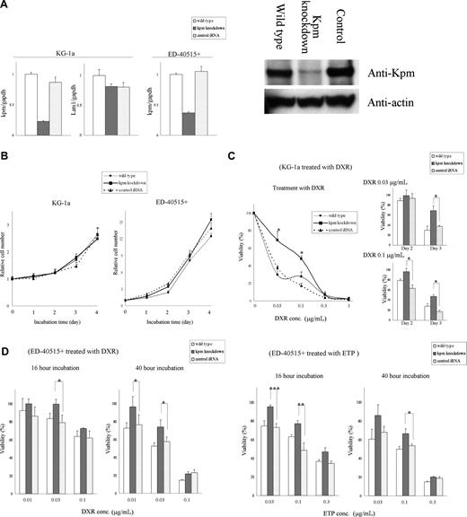
To delineate the cellular changes caused by Kpm/Lats2 down-regulation in leukemic cells, we made pools of Kpm/Lats2-knockdown KG-1a cells, a myeloid cell line, and ED-40515+ cells, an ATL-derived cell line, using Kpm/Lats2-specific shRNA expression retrovirus vector. The reason why we chose these cell lines was because KG-1a and ED-40515+ expressed relatively high levels of Kpm/Lats2 among myeloid and lymphoid cell lines, respectively (Figure 1A; Figure S1). Real-time PCR analyses of Kpm/Lats2 mRNA revealed that the expression levels of Kpm/Lats2 in Kpm/Lats2-knockdown KG-1a cells and ED-40515+ cells was approximately 20% and approximately 30% of basal levels, respectively. There was no difference in the expression level of Lats1, the other human homologue of Drosophila Wts/Lats, between Kpm/Lats2-knockdown cells and wild-type or control cells (Figure 2A). We confirmed that Kpm/Lats2 expression was decreased also at the protein level in Kpm/Lats2-knockdown cells with Western blotting (Figure 2B).
Down-regulation of Kpm/Lats2 renders cells resistant to DNA damage–inducing agents. (A) Kpm/Lats2-knockdown cells were established in KG-1a or ED-40515+ cells using retrovirus (RV) vector containing Kpm/Lats2-specific shRNA. Wild type represents non–RV-transduced cells and control iRNA represents control shRNA-containing RV–transduced cells. Efficiency of Kpm/Lats2-specific shRNA was measured by real-time PCR analyses. Data normalized to gapdh are shown as mean plus or minus SD in scale that the value for wild type is 1. Analyses were performed in triplicate independently twice and representative data are shown. Western blot analysis of Kpm/Lats2 in wild-type, knockdown, or control cells in KG-1a. Analyses were performed independently twice and representative data are shown. (B) Simple growth curve without agents was measured by MTT assay. The assays were performed in quadruplicate independently 3 times and representative data are shown as mean plus or minus SD. (C) Cell viability after treatment with doxorubicin (DXR) or etoposide (ETP) in each KG-1a line was measured by MTT assay. The assays were performed in quadruplicate independently 3 times and representative data are shown as mean plus or minus SD (Welch t test: *P < .01; **P < .05). (D) Cell viability after treatment with DXR or ETP in each ED-40515+ line was measured by MTT assay (Welch t test: *P < .05; **P < .01; ***P < .005). The assays were performed independently in quadruplicate 3 times, and representative data are shown as mean plus or minus SD.
Down-regulation of Kpm/Lats2 renders cells resistant to DNA damage–inducing agents. (A) Kpm/Lats2-knockdown cells were established in KG-1a or ED-40515+ cells using retrovirus (RV) vector containing Kpm/Lats2-specific shRNA. Wild type represents non–RV-transduced cells and control iRNA represents control shRNA-containing RV–transduced cells. Efficiency of Kpm/Lats2-specific shRNA was measured by real-time PCR analyses. Data normalized to gapdh are shown as mean plus or minus SD in scale that the value for wild type is 1. Analyses were performed in triplicate independently twice and representative data are shown. Western blot analysis of Kpm/Lats2 in wild-type, knockdown, or control cells in KG-1a. Analyses were performed independently twice and representative data are shown. (B) Simple growth curve without agents was measured by MTT assay. The assays were performed in quadruplicate independently 3 times and representative data are shown as mean plus or minus SD. (C) Cell viability after treatment with doxorubicin (DXR) or etoposide (ETP) in each KG-1a line was measured by MTT assay. The assays were performed in quadruplicate independently 3 times and representative data are shown as mean plus or minus SD (Welch t test: *P < .01; **P < .05). (D) Cell viability after treatment with DXR or ETP in each ED-40515+ line was measured by MTT assay (Welch t test: *P < .05; **P < .01; ***P < .005). The assays were performed independently in quadruplicate 3 times, and representative data are shown as mean plus or minus SD.
Next we analyzed the growth rates of wild-type, Kpm/Lats2-knockdown, and control KG-1a cells as well as ED-40515+ cells to determine whether the expression level of Kpm/Lats2 affected the duration of cell cycle. It has recently been reported that cell number of mouse embryonic fibroblasts (MEFs) from Kpm/Lats2 knockout mouse (Kpm/Lats2−/−) was approximately 1.25-fold more than those from wild-type mouse (Kpm/Lats2+/+) at day 4.27 Contrary to this, there was no difference between growth rate of Kpm/Lats2-knockdown cells and that of wild-type or control cells during 96 hours as measured by MTT assay (Figure 2C) and cell counting assay with trypan blue dye exclusion (data not shown).
Down-regulation of Kpm/Lats2 by shRNA in 2 different leukemic cell lines renders them resistant to DNA damage–inducing agents
To address whether down-regulation of Kpm/Lats2 renders leukemic cells resistant to DNA damage–inducing agents, we measured the cell viability of wild-type, Kpm/Lats2-knockdown, and control cells by MTT assay after the treatment with anticancer drugs, doxorubicin (DXR) or etoposide (ETP), which are used in standard chemotherapy for leukemia. The viability of these cells decreased after treatment of DXR or ETP in a dose-dependent and time-dependent manner but that of Kpm/Lats2-knockdown KG-1a cells was approximately 20% to 30% higher than that of wild-type or control KG-1a cells (Figure 2D). This tendency was also observed in ED-40515+ cells, although the proportion of dead cells increased faster and the difference in viability was slightly smaller than in KG-1a cells (Figure 2E). In addition, flow cytometric analysis with annexin V–PI dual staining revealed that treatment with ETP induced fewer apoptotic or dead cells in Kpm/Lats2-knockdown KG-1a cells than in control cells (Figure S2). Similar results were obtained with KG-1a cells transduced with microRNA-373,52 which is known to down-regulate Kpm/Lats2 or those transfected with Kpm/Lats2-specific siRNA, although these reagents down-regulated Kpm/Lats expression less efficiently than the shRNA-expressing retrovirus vector (Figure S3A,B). These results clearly indicate that down-regulation of Kpm/Lats2 renders leukemic cells resistant to DNA damage.
Down-regulation of Kpm/Lats2 inhibits transcriptional induction of p21 and PUMA without affecting the IAP family members
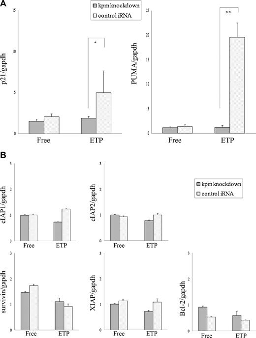
We next examined the expression of several key molecules involved in cell survival. We searched for those genes whose expression level in Kpm/Lats2-knockdown KG-1a cells was significantly different from that in control cells after DNA damage stress. Two genes, p21 and PUMA, were identified by real-time PCR analysis–based screening. The expressions of p21 and PUMA were clearly induced by DNA damage–inducing agents in control KG-1a cells, although KG-1a line was p53 null,53 whereas their inductions were strongly inhibited in Kpm/Lats2-knockdown KG-1a cells (Figure 3A). This finding was also confirmed at the protein level (Figure S4). Since p21 induces cell-cycle arrest54 and PUMA can make Bax localize onto mitochondrial membrane to trigger apoptosis,55,56 these results seemed to be in agreement with what we observed in cell viability assay. On the other hand, none of the members of the IAP family, the major inhibitors of apoptosis,57 was up-regulated by silencing of Kpm/Lats2 despite treatment with ETP (Figure 3B). Of note is that in Drosophila the Hippo signals end up with the inhibition of Yki leading to apoptosis via down-regulation of Diap1.7 In addition, although Bcl-2 was reportedly decreased by ectopic expression of Kpm/Lats2,22 there was no significant difference in its expression level between Kpm/Lats2-knockdown and control cells (Figure 3B).
Induction of p21WAF1 and PUMA but not the IAP family members or bcl-2 is repressed in Kpm/Lats2-knockdown KG-1a cells after treatment with DNA damage–inducing agents. Total mRNA was isolated from Kpm/Lats2-knockdown KG-1a or control cells treated with or without 0.03 μg/mL ETP at day 0 and cultured for 3 days. mRNA expression levels of p21 and PUMA (A), and the IAP family members and bcl-2 (B), were measured by real-time PCR analysis using the specific primers described in Table 1. Data normalized to gapdh are shown as mean plus or minus SD in scale that the value for KG-1a wild-type cells without treatments is 1. Normalization to hprt gave similar results. The analyses were performed in triplicate independently twice, and representative data are shown (Student t test: *P < .05; **P < .005).
Induction of p21WAF1 and PUMA but not the IAP family members or bcl-2 is repressed in Kpm/Lats2-knockdown KG-1a cells after treatment with DNA damage–inducing agents. Total mRNA was isolated from Kpm/Lats2-knockdown KG-1a or control cells treated with or without 0.03 μg/mL ETP at day 0 and cultured for 3 days. mRNA expression levels of p21 and PUMA (A), and the IAP family members and bcl-2 (B), were measured by real-time PCR analysis using the specific primers described in Table 1. Data normalized to gapdh are shown as mean plus or minus SD in scale that the value for KG-1a wild-type cells without treatments is 1. Normalization to hprt gave similar results. The analyses were performed in triplicate independently twice, and representative data are shown (Student t test: *P < .05; **P < .005).
Nuclear accumulation of p73 was suppressed in Kpm/Lats2-knockdown cells
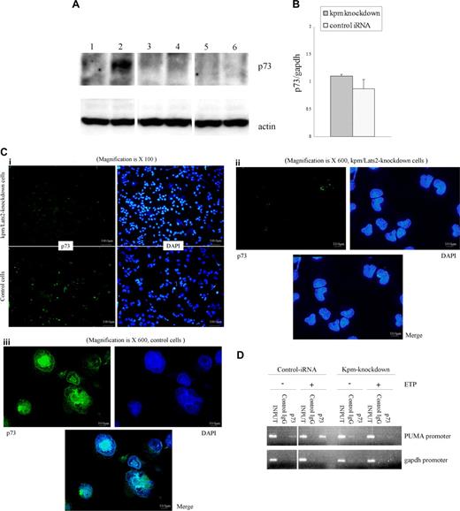
We considered p73 as a molecule that replaced p53 in p53-null cells such as KG-1a and ED-40515+ because p73 is a homologue of p53 and a transcriptional factor for p21 and PUMA expression. Therefore, we measured the amount of p73 at the protein level by Western blot analysis of whole-cell lysates. p73 was hardly detectable under normal conditions but became visible in control cells after treatment with ETP. In contrast, such increase in p73 protein was not observed in Kpm/Lats2-knockdown cells after the same treatment (Figure 4A). There was little difference in the expression level of p73 mRNA after treatment with ETP between Kpm/Lats2-knockdown cells and control cells (Figure 4B). Similar results were obtained with microRNA-373–transduced KG-1a cells (Figure S5). Next, we evaluated the distribution of p73 protein by immunofluorescence microscopy because transcriptional activity of p73 depends on the accumulation of p73 in nucleus due to posttranscriptional modification.58 p73 was scarcely detected in KG-1a cells with or without silencing of Kpm/Lats2 under normal conditions (data not shown) but was significantly induced in control KG-1a cells after treatment with ETP (Figure 4C), In contrast, p73 remained at very low levels in Kpm/Lats2-knockdown KG-1a cells (Figure 4C). Under higher magnification, we visualized more clearly that p73, which should accumulate in the nucleus, was hardly detected or only faintly represented in Kpm/Lats2-knockdown cells (Figure 4C). Moreover, we investigated the recruitment of p73 on the PUMA gene promoter after DNA-damage stress. ChIP assay clearly showed that p73 was recruited to the PUMA gene promoter in control cells but not in Kpm/Lats2-knockdown cells after DNA-damage stress (Figure 4D). In summary, our data suggest that Kpm/Lats2 contributes to the stability of p73 protein, which in turn leads to transcriptional induction of p21 and PUMA to finally trigger cell death after treatment with DNA damage–inducing reagents.
Stabilization of p73 after treatment with ETP is insufficient in Kpm/Lats2-knockdown cells. (A) Western blotting analysis with anti-p73 in whole-cell lysate of control KG-1a cells without treatment of ETP (lane 1) or with treatment of 0.03 μg/mL ETP for 3 days (lane 2), Kpm/Lats2-knockdown KG-1a cells without treatment of ETP (lane 3), or with treatment of 0.03 μg/mL ETP for 3 days (lane 4), and p73-knockdown KG-1a cells without treatment of ETP (lane 5) or with treatment of 0.03 μg/mL ETP for 3 days (lane 6). Twice, independent experiments were performed and representative data are shown. (B) Expression of p73 mRNA in Kpm/Lats2-knockdown KG-1a or control cells treated with 0.03 μg/mL ETP for 3 days was measured by real-time PCR analysis. Data normalized to gapdh are shown as mean plus or minus SD in scale that the value for KG-1a wild-type cells with the same treatment is 1. The analyses were performed in triplicate independently twice and representative data are shown as mean plus or minus SD. (C) Immunofluorescence staining for p73 and DAPI. Kpm/Lats2-knockdown KG-1a cells and control cells treated with 0.1 μg/mL ETP were stained with anti-p73 Ab or DAPI and observed with a fluorescence microscope at the magnification of ×100 or ×600. Four independent experiments were performed and representative data are shown. (D) ChIP assay. ChIP assay was performed as described in “Chomatin immunoprecipatation assay.” p73 was recruited to PUMA gene promoter in control cells but not in Kpm/Lats2-knockdown cells after the treatment with 0.03 μg/mL ETP for 60 hours. Two independent experiments were performed and representative data are shown.
Stabilization of p73 after treatment with ETP is insufficient in Kpm/Lats2-knockdown cells. (A) Western blotting analysis with anti-p73 in whole-cell lysate of control KG-1a cells without treatment of ETP (lane 1) or with treatment of 0.03 μg/mL ETP for 3 days (lane 2), Kpm/Lats2-knockdown KG-1a cells without treatment of ETP (lane 3), or with treatment of 0.03 μg/mL ETP for 3 days (lane 4), and p73-knockdown KG-1a cells without treatment of ETP (lane 5) or with treatment of 0.03 μg/mL ETP for 3 days (lane 6). Twice, independent experiments were performed and representative data are shown. (B) Expression of p73 mRNA in Kpm/Lats2-knockdown KG-1a or control cells treated with 0.03 μg/mL ETP for 3 days was measured by real-time PCR analysis. Data normalized to gapdh are shown as mean plus or minus SD in scale that the value for KG-1a wild-type cells with the same treatment is 1. The analyses were performed in triplicate independently twice and representative data are shown as mean plus or minus SD. (C) Immunofluorescence staining for p73 and DAPI. Kpm/Lats2-knockdown KG-1a cells and control cells treated with 0.1 μg/mL ETP were stained with anti-p73 Ab or DAPI and observed with a fluorescence microscope at the magnification of ×100 or ×600. Four independent experiments were performed and representative data are shown. (D) ChIP assay. ChIP assay was performed as described in “Chomatin immunoprecipatation assay.” p73 was recruited to PUMA gene promoter in control cells but not in Kpm/Lats2-knockdown cells after the treatment with 0.03 μg/mL ETP for 60 hours. Two independent experiments were performed and representative data are shown.
Kpm/Lats2 can associate with YAP2 via the first WW domain and induce its phosphorylation
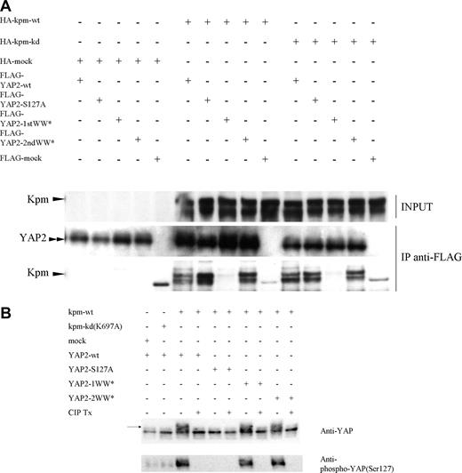
We investigated whether Kpm/Lats2 could interact with and phosphorylate YAP as demonstrated in Drosophila. YAP has 2 major functional isoforms, YAP1 and YAP2, and we found that YAP2 was a dominant form expressed in hematopoietic lineage cells (Figure S2). Coimmunoprecipitation analyses indicated that Kpm/Lats2 interacted with YAP2 via its first WW domain (Figure 5A) and YAP1 via its only WW domain (Figure S5). In addition, coexpression of Kpm/Lats2 and YAP2 resulted in a mobility shift of YAP2. This mobility shift was abrogated by phosphatase treatment and was not caused by coexpression of the Kpm-kinase–dead form and YAP2. Therefore, this shift is indeed due to the phosphorylation of YAP protein (Figure 5B). It was unexpected, however, that this mobility shift occurred with the first WW domain mutant but not with the S127A mutant form of YAP2. Phosphorylation of YAP2 at serine 127 was further confirmed by Western blot using anti–phospho-YAP, which could recognize phosphorylated serine 127 of YAP2 specifically. In accordance with mobility shift assay, the phosphorylated YAP2 was clearly detected in coexpression of wild-type Kpm/Lats2 but barely detectable in coexpression of kinase-dead form of Kpm/Lats2 or mock. It was also detected in coexpression of wild-type of Kpm/Lats2 with first WW domain mutant or second WW domain mutant of YAP2. These data suggest that Kpm/Lats2 phosphorylates YAP2 at serine 127 regardless of interaction via the WW domains.
Kpm/Lats2 can associate with YAP2 via its first WW domain and phosphorylate YAP2, but it requires not this association but kinase function of Kpm/Lats2 to stabilize p73. (A) Any one of HA-tagged Kpm-wild type (wt), Kpm-kinase dead (kd), or mock was cotransfected with any one of FLAG-tagged YAP2-wild type (wt), YAP2-S127A (mutant form S127 to A; S127 is Akt-phosphorylation site), YAP2-1WW* (mutant form of first WW domain), YAP2-2WW* (mutant form of second WW domain), or mock into 293T cells by the calcium phosphate method. Upper lanes represent Western blotting with anti-HA in cell lysates. Middle lanes and lower lanes represent Western blotting with anti-FLAG and anti-HA, respectively, in the immunoprecipitates by anti-FLAG. Three independent experiments were performed and representative data are shown. (B) The coimmunoprecipitate fraction by anti-FLAG from each lysate was treated with or without CIP and then analyzed by Western blotting with anti-YAP polyclonal antibody and anti–phospho-YAP (Ser127) polyclonal antibody. Arrow indicates mobility shift band of YAP2. Three independent experiments were performed and representative data are shown.
Kpm/Lats2 can associate with YAP2 via its first WW domain and phosphorylate YAP2, but it requires not this association but kinase function of Kpm/Lats2 to stabilize p73. (A) Any one of HA-tagged Kpm-wild type (wt), Kpm-kinase dead (kd), or mock was cotransfected with any one of FLAG-tagged YAP2-wild type (wt), YAP2-S127A (mutant form S127 to A; S127 is Akt-phosphorylation site), YAP2-1WW* (mutant form of first WW domain), YAP2-2WW* (mutant form of second WW domain), or mock into 293T cells by the calcium phosphate method. Upper lanes represent Western blotting with anti-HA in cell lysates. Middle lanes and lower lanes represent Western blotting with anti-FLAG and anti-HA, respectively, in the immunoprecipitates by anti-FLAG. Three independent experiments were performed and representative data are shown. (B) The coimmunoprecipitate fraction by anti-FLAG from each lysate was treated with or without CIP and then analyzed by Western blotting with anti-YAP polyclonal antibody and anti–phospho-YAP (Ser127) polyclonal antibody. Arrow indicates mobility shift band of YAP2. Three independent experiments were performed and representative data are shown.
Stabilization of p73 requires YAP2 and kinase activity of Kpm
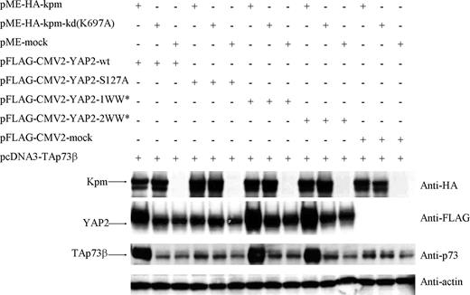
Finally, we addressed how the interaction between Kpm/Lats2 and YAP2 contributes to stabilization of p73. p73 was cotransfected with wild type as well as mutant clones of Kpm/Lats2 and YAP2 into 293T cells, and the protein levels of these molecules were estimated by Western blot. These transient coexpression experiments showed that the protein level of p73 was high in coexpression of wild-type YAP2 and wild-type Kpm/Lats2 but not the kinase-dead form or mock. High expression of p73 was observed not only in coexpression of the second WW domain mutant of YAP2 that was able to interact with Kpm/Lats2 but also in that of the first WW domain mutant of YAP2 that was unable to do that. On the other hand, the expression level of p73 was not increased in coexpression of the S127A mutant of YAP2 compared with mock. Furthermore, coexpression of wild-type Kpm/Lats2 but not the kinase-dead form augmented the protein level of YAP2 regardless of the presence of the first WW domain, and even wild-type Kpm/Lats2 did not affect the protein level of the S127A mutant of YAP2 (Figure 6). Similar results were obtained in the experiments of coexpression of Kpm/Lats2 and YAP1 (Figure S6). Considering that YAP is an important cofactor that associates with and stabilizes p73, the increase in p73 expression may be due to its stabilization by facilitated interaction with YAP2 mediated by its phosphorylation by Kpm/Lats2. Overall, these findings suggest that Kpm/Lats2 is involved in the stabilization of p73 by increasing the protein level of YAP2, which is dependent on the serine 127 of YAP2 and the kinase function of Kpm/Lats2.
The protein level of p73 is increased by coexpression of YAP2 and wild-type Kpm/Lats2. Any one of HA-tagged Kpm-wild type (wt), Kpm-kinase dead (kd), or mock was cotransfected with TAp73α and any one of FLAG-tagged YAP2-wt, YAP2-S127A, YAP2-1WW*, YAP2-2WW*, or mock into 293T cells by FuGENE-HD reagent. Four rows of lanes from the top in this order represent Western blotting with anti-HA, anti-FLAG, anti-p73, and antiactin in cell lysates. Two independent experiments were performed and representative data are shown.
The protein level of p73 is increased by coexpression of YAP2 and wild-type Kpm/Lats2. Any one of HA-tagged Kpm-wild type (wt), Kpm-kinase dead (kd), or mock was cotransfected with TAp73α and any one of FLAG-tagged YAP2-wt, YAP2-S127A, YAP2-1WW*, YAP2-2WW*, or mock into 293T cells by FuGENE-HD reagent. Four rows of lanes from the top in this order represent Western blotting with anti-HA, anti-FLAG, anti-p73, and antiactin in cell lysates. Two independent experiments were performed and representative data are shown.
Discussion
Drosophila Warts/Lats is considered to be a tumor suppressor since its loss of function mutation results in overproliferation phenotype in mosaic animals.1,2 The equivalent experiment has not been reproduced with Kpm/Lats2 in mammals because Kpm/Lats2 knockout mice are embryonically lethal.26,27 Nevertheless, down-regulation of Kpm/Lats2 has been described in a variety of human cancers.42,43,59 Notably, low level of Kpm/Lats2 expression correlated with poor prognosis in ALL.43 Therefore, we focused on the cellular changes caused by down-regulation of Kpm/Lats2 expression. The most intriguing finding was that silencing of Kpm/Lats2 in 2 types of leukemic cells resulted in increased resistance to DNA damage–inducing agents. The viability of Kpm/Lats2-knockdown KG-1a cells after treatment with ETP or DXR was consistently higher than that of wild-type or control cells. This tendency was also observed in Kpm/Lats2-knockdown ED-40515+ cells. Based on the findings of Hippo pathway in Drosophila, we expected that a direct executor may be one of the IAP family members because IAPs are major inhibitor of caspases, but the mRNA expression levels of IAP family members were not altered in Kpm/Lats2-knockdown cells. To find out what molecules could directly cause such resistance, we used real-time PCR analysis to search for altered expression of several candidate genes related to cell-cycle arrest or apoptosis. Among the candidates we screened, induction of p21 and PUMA by treatment with ETP was markedly suppressed in Kpm/Lats2-knockdown cells compared with control shRNA-transduced cells that responded with high levels of these genes. p21 is a well-known CDK inhibitor that induces cell cycle arrest,60 and PUMA is a final executor that localizes BAX onto mitochondrial membrane to trigger apoptosis.56 Therefore, the suppressed induction of these genes in Kpm/Lats2-knockdown KG-1a cells seems to be compatible with their resistance to DNA damage–inducing agents.
Expression of both p21 and PUMA is induced by p53 and its family members. Since KG-1a cells53 and ED-40515+ cells are p53 null (Dr M. Matsuoka, Kyoto University, e-mail communication, April 16, 2007), we thought that another p53 family member, presumably transcriptionally active p73 (TAp73), should function in the induction of p21 and PUMA in these cells. p73 is able to bind promoters of several p53 responsible genes involved in cell cycle arrest and apoptosis including p21 and PUMA.61 Unlike p53, it is rarely mutated or deleted in human malignant cells.62,63 In addition, it has been reported that knockdown of p73 leads to enhanced resistance to DNA damage–inducing agents including DXR, ETP, and cisplatinum.64 Protein levels of p73 are kept low due to rapid degradation through the ubiquitin pathway under physiological conditions,65 whereas TAp73 accumulates in response to stress caused by DNA damage.66 Therefore, we regarded p73 as a p53-substituting molecule that mediated induction of p21 and PUMA, and examined the amount and the distribution of p73 in Kpm/Lats2-knockdown KG-1a cells and in control cells after treatment with ETP. As expected, after treatment of ETP, p73 protein level was kept very low in Kpm/Lats2-knockdown KG-1a cells compared with control cells, despite no difference in the mRNA level of p73. Immunofluorescence microscopic analysis revealed that nuclear accumulation of p73 was clearly detected in control cells but not, or only faintly, in Kpm/Lats2-knockdown cells. These results suggest that Kpm/Lats2 is involved in the stability and the nuclear accumulation of p73.
It has been shown that several steps are required for DNA damage–induced activation and stabilization of p73. DNA damage induces p73 gene expression via Chk1,67 Chk2,68 and E2F1,69,70 and in parallel activates c-Abl and p38 MAP kinase (MAPK). Activated c-Abl and p38 MAPK phosphorylates p73 at Tyr99 and at Ser/Thr-Pro, respectively, to stabilize it in the nucleus.71,,–74 In addition to phosphorylation, binding of phosphorylated p73 to promyelocytic leukemia protein (PML),65 acetylation of p73 mediated by p300,75 and prolyl isomerization of p73 mediated by peptidyl-prolyl cis/trans isomerase Pin176 are essential for functional activation of p73. YAP is essential for coactivation of p73 with PML and for recruitment of p300 and contributes to the DNA damage–induced nuclear accumulation of p73. Trapping of YAP in the cytosol due to phosphorylation by Akt (protein kinase B) and binding to 14-3-3 represents a physiological mechanism for the inhibition of inappropriately accumulated p73.77,78 Among these factors, we concentrated our efforts on YAP because it should mediate the downstream signals of Kpm/Lats2 based on the analogy of Drosophila. We confirmed that Kpm/Lats2 could interact with YAP2 through its first WW domain and induce phosphorylation of YAP2, which indicates that the Hpo-Wts-Yki axis is evolutionally conserved. However, the downstream signals of YAP (Yki) in mammals may be different from that in Drosophila, because, as mentioned in “Introduction,” YAP has dual and opposite functions, growth-promoting or proapoptotic. In addition, as shown in this study, no alterations of the IAP family members were detected in Kpm/Lats-knockdown cells, although Diap1 is considered to be one of the final target molecules in the Drosophila Hippo pathway. There is no report describing YAP-dependent regulation of the IAP family members in mammalian cells.
With regard to the relationship among Kpm/Lats2, YAP2, and p73, the transient expression experiments suggested that coexpression of Kpm/Lats2 positively regulates the protein amounts of both YAP2 and p73 in whole-cell lysates regardless of interaction with YAP2 via the WW domains and this effect requires phosphorylation of YAP2 at serine 127 by Kpm/Lats2. After submission of our original paper, Matallanas et al have reported that RASSF1A activates Lats1 through enhanced phosphorylation by MST2 and activated Lats1 phosphorylates and activates YAP1, allowing YAP1 to translocate to the nucleus and associate with p73, resulting in transcription of the proapoptotic target gene PUMA.79 According to the authors, phosphorylation of YAP1 by Lats1 is needed to enable the formation of a YAP1-p73 complex, whereas, in unstimulated cells, Lats1 can negatively regulate YAP1 by sequestering YAP1 in the cytosol. Thus, it is suggested that Lats1 has dual functions as an anchoring protein that retains YAP1 in the cytosol and as a kinase that phosphorylates YAP1 to facilitate its interaction with p73. The former function has been emphasized by 2 recent papers insisting that inactivation of YAP by the Hippo pathway is involved in organ size control and cell contact inhibition.80,81 These papers describe that Lats1 or Kpm/Lats2 but not AKT directly phosphorylates YAP at serine 127, increasing the interaction between YAP2 and 14-3-3. Our results are compatible with those with RASSF1A activation in that Lats1 or Kpm/Lats2 is crucial for the induction of proapoptotic gene PUMA that is mediated by the YAP-p73 complex. It should be noted that such function of Lats1 or Kpm/Lats2 becomes dominant only in the presence of proapoptotic stimulations such as RASSF1A activation, anti-Fas, and DNA damage–inducing agents. Evidence has indicated that DNA damage stress induces activation of c-Jun N-terminal kinase (JNK) and activated JNK then phosphorylates 14-3-3, resulting in release of several 14-3-3–binding proteins including c-Abl, which is known to activate and stabilize p73 in the nucleus.82,83 Thus, it seems possible that phosphorylation of 14-3-3 by activated JNK is involved in Kpm/Lats2-dependent nuclear translocation of YAP and accumulation of the YAP-p73 complex after DNA damage. Further studies are needed to clarify how DNA damage signals intersect the Hippo pathway.
In conclusion, we demonstrate that Kpm/Lats2 is required for the stabilization of p73 and subsequent induction of p21 and PUMA in response to DNA damage–inducing agents, suggesting that the down-regulation of Kpm/Lats2 contributes to the instability of p73 through the insufficient phosphorylation of YAP2 at serine 127, resulting in chemoresistance and poor prognosis for some leukemias.
The online version of this article contains a data supplement.
The publication costs of this article were defrayed in part by page charge payment. Therefore, and solely to indicate this fact, this article is hereby marked “advertisement” in accordance with 18 USC section 1734.
Acknowledgments
We thank Dr Masao Matsuoka and Dr Michiyuki Maeda (Laboratory of Virus Immunology, Research Center for AIDS, Institute for Virus Research, Kyoto University) for ATL-derived cell lines and the personal communication concerning ED-40515+; Dr Akira Nakagawara and Dr Toshinori Ozaki (Biochemistry, Chiba Cancer Center Research Institute, Chiba, Japan) for their experimental advice on p73; Dr Syohei Yamaoka for his help in preparation of retrovirus vectors; Dr Yoshihide Ueda (Department of Gastroenterology and Hepatology, Kyoto University) for TAp73α; Virginia Mazack for expert technical assistance; and Dr Akifumi Takaori-Kondo (Department of Hematology and Oncology, Kyoto University) for the immunofluorescence microscope and his continuous help.
This work was supported in part by grants-in-aid from the Ministry of Education, Culture, Sports, Science, and Technology of Japan (T.H. and T.U.), and by a Pennsylvania Department of Health–sponsored Breast Cancer Research Grant (Philadelphia, PA; SAP no. 41-000-37378, M.S.).
Authorship
Contribution: M.K. performed most experiments and cowrote the paper; T.H. designed the research and cowrote the paper; K.C. performed some experiments; T.O. and M.S. contributed vital plasmids; and T.U. supervised the research.
Conflict-of-interest disclosure: The authors declare no competing financial interests.
Correspondence: Toshiyuki Hori, Department of Hematology and Oncology, Graduate School of Medicine, Kyoto University, 54 Shogoin-Kawara-cho, Sakyo-ku, Kyoto 606-8507, Japan; e-mail: thori@kuhp.kyoto-u.ac.jp.

