Abstract
B-cell receptor (BCR) signaling activates a number of intracellular signaling molecules including phospholipase C–γ2 (PLC-γ2), which generates membrane diacylglycerol (DAG). DAG recruits both protein kinase C (PKC) and RasGRP family members to the membrane and contributes to their activation. We have hypothesized that membrane colocalization facilitates activation of RasGRP3 by PKC. Here we demonstrate that PKCθ phosphorylates RasGRP3 on Thr133 in vitro, as determined by mass spectrometry. RasGRP3 with a Thr133Ala substitution is a poor PKC substrate in vitro and a poor Ras activator in vivo. Antiphosphopeptide antibodies recognize Thr133-phosphorylated RasGRP3 in B cells after BCR stimulation or DAG analog treatment, but much less so in resting cells. PKC inhibitors block RasGRP3 Thr133 phosphorylation and Ras–extracellular signal-related kinase (Erk) signaling with a similar pattern. After stimulation of T-cell receptor (TCR) or DAG analog treatment of T cells, PKC-catalyzed phosphorylation of RasGRP1 occurs on the homologous residue, Thr184. These studies shed light on the proposed “PKC-Ras pathway” and support the hypothesis that RasGRP phosphorylation by PKC is a mechanism that integrates DAG signaling systems in T and B cells. PKC-mediated regulation of RasGRPs in lymphocytes may generate cooperative signaling in response to increases in DAG. The mast- and myeloid-selective family member RasGRP4 is regulated by different means.
Introduction
The small membrane-bound GTPase Ras acts as a guanyl nucleotide-dependent switch, cycling between guanosine diphosphate (GDP)– and guanosine triphosphate (GTP)–bound forms. Ras guanyl nucleotide exchange factors (Ras GEFs) catalyze release of GDP, allowing Ras to bind the relatively prevalent cellular GTP. Conversely, Ras GTPase activating proteins (Ras GAPs), such as P120GAP and neurofibromin, enhance the hydrolysis of GTP by Ras, returning the GTPase to its inactive state.1 Between phases of activation and inactivation, Ras-GTP can bind downstream effectors. For example, recruitment of Raf to the plasma membrane by Ras-GTP activates the Raf-Mek–extracellular signal-regulated kinase (Erk) kinase cascade. This and other effector pathways bring about alterations in gene transcription and other biochemical changes that underlie the Ras-regulated cellular processes. Ras signaling plays an essential role in immune-receptor signaling. Antigen interactions with both B-cell receptor (BCR) and T-cell receptor (TCR) initiate complex intracellular signaling events, including Ras activation, and these play important roles in both the development and activation of lymphocytes.2-5
In principle, Ras can be regulated by controlling either the rate of GTP hydrolysis or the rate of nucleotide exchange. The seminal Ras regulation experiments involved the analysis of metabolically labeled guanyl nucleotides associated with immune-precipitated Ras from human peripheral blood lymphocytes and the Jurkat T-cell line.6 These studies showed that Ras-GTP levels rise rapidly after stimulation of T cells with anti-TCR antibodies. Furthermore, strong and prompt Ras activation was observed after treatment with the diacylglycerol (DAG) analog phorbol 12-myristate 13-acetate (PMA). This latter result is in line with the idea that TCR stimulation is linked to phosphatidylinositol hydrolysis and DAG second messenger generation. Using a permeable cell assay, Ras guanyl nucleotide exchange rates were constitutively high; the major effect of prior cell stimulation was to decrease the GTPase rate. In line with this hypothesis, cellular extracts prepared from PMA-treated cells had lower cellular Ras GAP activity. It was surmised that PKC down-regulated a Ras GAP, although the mechanistic details were never uncovered. A second report from the same group indicated that, indeed, a sizable fraction of the Ras activation in lymphocytes was eliminated by PKC inhibitors, although evidence for a PKC-independent component was also apparent.7
Subsequent work in many labs, including important work by some of the authors cited above,6 provided a rather different view of Ras regulation in nonlymphoid cells. In most cells, there does not appear to be a link between DAG or PKC and Ras activation. Furthermore, in most cells Ras is regulated by the recruitment of the Ras GEF Sos (son of sevenless) to the plasma membrane by means of adaptor proteins and tyrosine-phosphorylated docking sites.1
The discovery of RasGRP1 and the demonstration that this Ras GEF protein is expressed in T cells, but not expressed in many other cell types, provided a convenient explanation for the original and unique observations on DAG activation of Ras in lymphocytes.8-10 RasGRP1 has a catalytic domain similar to Sos. In addition, RasGRP1 possesses a pair of atypical EF (helix E and F prototypic calcium binding protein) hands that bind calcium in vitro and a C1 domain that binds DAG and DAG analogs with high affinity.11 The primary structure of RasGRP1 suggests that RasGRP1 can respond to second messengers derived from phosphatidylinositol hydrolysis. Although the functional significance of the EF hands is still unclear, it has been confirmed that the C1 domain facilitates RasGRP1 membrane recruitment by DAG and interaction with substrate Ras.8-10
The study of RasGRP1 in Jurkat T cells and the analysis of Rasgrp1 null mutant mice have provided substantial evidence that this Ras GEF is the link between TCR, phospholipase C–γ1 (PLC-γ1), and DAG on the one hand, and Ras signaling on the other.9,12,13 Nonetheless, this new model for Ras activation in T cells did not account for the original evidence from inhibitor studies that PKC was somehow involved.
A harmonious hypothesis emerged from our analysis of the Ras signaling in the Ramos B-cell line.14 B cells express RasGRP3, which has a similar domain structure to RasGRP1. By analogy with our T-cell work, we hypothesized that this Ras GEF would be linked to a BCR–PLC-γ2–DAG pathway. However, an unexpected observation provided a possible resolution to the long-standing puzzle of the “PKC-Ras pathway”; stimulation of BCR in Ramos B cells resulted in high stoichiometry, multisite phosphorylation of RasGRP3 contemporaneously with Ras activation. Furthermore, with PMA or BCR stimulation, in the presence or absence of various PKC inhibitors, we found a striking concordance between RasGRP3 phosphorylation and Ras activation. Extrapolation from these B-cell studies to T cells provides a plausible explanation for the original TCR signaling studies: membrane accumulation of DAG leads to recruitment of both RasGRP1 and one or more forms of PKC, with regulatory phosphorylation of the former by the latter.14
Here we have identified Thr133 in RasGRP3 as a BCR-regulated, PKC-catalyzed phosphorylation site and have investigated the functional significance of this modification.
Materials and methods
Reagents and generic antibodies
The PKC inhibitors Go6976 and Ro318220 were purchased from Calbiochem (San Diego, CA). Okadaic acid was either purchased from Calbiochem or was a gift from Dr Charles Holmes (University of Alberta). Phorbol 12-myristate 13-acetate (PMA) was purchased from Sigma-Aldrich (St Louis, MO).
For immunoblotting, we used the pan-Ras antibody (clone RAS10) from Upstate Biotechnology (Lake Placid, NY). Anti–PKC-θ (no. P15120) and anti–PKC-δ (no. NP-997704) were purchased from Transduction Laboratories (Mississauga, ON, Canada). The phospho p44/p42 ERK antibody (no. 9101) was from Cell Signaling Technology (Beverly, MA). The Rap1 (121) and ERK1 antibodies (no. K-23) were from Santa Cruz Biotechnology (Santa Cruz, CA).
Stimulatory goat antihuman immunoglobulin M (IgM) F(ab′)2 fragment was purchased from Jackson ImmunoResearch Laboratories (West Grove, PA). Anti–mouse IgM (μ-chain specific) was purchased from Southern Biotechnology Associates (Birmingham, AL). Mouse- and human-reactive anti-CD3 antibodies 145-2C11 and OKT3 were purified from hybridomas.
Concanavalin A was purchased from Pharmacia (Kirkland, QC) and lipopolysaccharide (LPS) from Sigma-Aldrich.
RasGRP-specific antibodies
Polyclonal anti-RasGRP1 (H176) and anti-RasGRP3 (L247-9) antibodies have been described,9,14 as has m199, a rodent-specific anti-RasGRP1 monoclonal antibody.15 The m133 monoclonal antibody was derived from mice that had been immunized with rat RasGRP1, and it reacts with human as well as rodent protein. However, the epitope maps to the highly conserved EF hand region and the antibody also immune-precipitates RasGRP3. Despite this cross-reaction, note that RasGRP1 (90 kDa) and RasGRP3 (78 kDa) can be readily distinguished by their electrophoretic mobility. The m404 anti-RasGRP3 monoclonal antibody was derived from mice immunized with bacterially expressed full-length human RasGRP3. RasGRP1 and RasGRP were immune-precipitated with one or more monoclonal antibodies before immunoblotting with polyclonal antibodies, except in the experiment described in Figure 2B, where RasGRP3 was immune-precipitated with polyclonal phospho-specific antibodies.
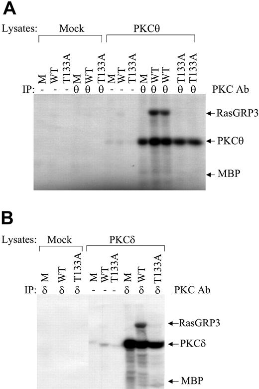
RasGRP3 Thr133Ala substitution blocks in vitro phosphorylation by nPKC forms PKCθ and PKCδ. (A) The effect of a RasGRP3 Thr133Ala substitution on phosphorylation by PKCθ is shown. Parental MBP (M), MBP-RasGRP3 (WT), or MBP-RasGRP3 Thr133Ala (T133A) were used as substrates in an immune complex kinase assay, as described in “Materials and methods.” Arrows show, from top to bottom, phosphorylation of MBP-RasGRP3, autophosphorylation of PKCθ, and the position of parental MBP protein. Some lanes are in duplicate. IP indicates immunoprecipitation; Ab, antibody. (B) Similar experiments as in panel A were performed with PKCδ. Results are representative of 3 independent experiments.
RasGRP3 Thr133Ala substitution blocks in vitro phosphorylation by nPKC forms PKCθ and PKCδ. (A) The effect of a RasGRP3 Thr133Ala substitution on phosphorylation by PKCθ is shown. Parental MBP (M), MBP-RasGRP3 (WT), or MBP-RasGRP3 Thr133Ala (T133A) were used as substrates in an immune complex kinase assay, as described in “Materials and methods.” Arrows show, from top to bottom, phosphorylation of MBP-RasGRP3, autophosphorylation of PKCθ, and the position of parental MBP protein. Some lanes are in duplicate. IP indicates immunoprecipitation; Ab, antibody. (B) Similar experiments as in panel A were performed with PKCδ. Results are representative of 3 independent experiments.
To generate antiserum that selectively recognizes Thr133-phosphorylated RasGRP3, rabbits were immunized with an 11-residue phosphopeptide (WMRRVpTQRKKV) coupled to KLH (keyhole limpet hemocyanin; Alberta Peptide Institute, Edmonton, AB, Canada). The resulting serum, CV4, was diluted and used directly without purification. Similarly, an 11-residue phosphopeptide (WSRKLpTQRIKS) was used to generate 3B7, an antiserum that recognizes RasGRP1 phosphorylated on Thr184. In this case, it was necessary to affinity purify specific antibodies using the phosphopeptide coupled to bovine serum albumin (BSA). For clarity in the text and figures, we have termed these antibodies “anti-3/pThr133” (reacts with RasGRP3 phosphorylated on Thr133) and “anti-1/pThr184” (reacts with RasGRP1 phosphorylated on Thr184).
Plasmids
Expression of full-length human RasGRP3 cDNA in rat2 cells using the pBabePuro expression vector has been described,16 as has the use of the pMAL system for expression of maltose-binding protein (MBP)–RasGRP3 fusion protein in Escherichia coli.14 RasGRP3 cDNA containing a threonine 133 to alanine (Thr133Ala) point mutation was generated using a polymerase chain reaction (PCR) method, sequence verified, and incorporated into the plasmids pMAL-c2 and pBabepuro. Full-length RasGRP4 cDNA (GenBank accession no. AY048119) was cloned as an EcoRI/SalI fragment into the retrovirus vector pBabeHygro.17
Sequence alignments
Sequence alignments were performed with Macvector 7.1 (Accelrys, San Diego, CA).
Mass spectrometry analysis
RasGRP3 purified from E coli was phosphorylated with PKCθ using an immune-complex kinase assay and resolved by sodium dodecyl sulfate (SDS)/7.5% polyacrylamide gel electrophoresis (PAGE), followed by analysis as described elsewhere.18 After staining with Biosafe (Biorad, Hercules, CA), the protein band was excised, reduced, and digested with trypsin. After in-gel digestion, eluted phosphorylated peptides were enriched by open tubular immobilized metal ion affinity chromatography (IMAC). Peptides eluted from IMAC were subjected to matrix-assisted laser desorption ionization (MALDI) mass spectrometry (MS) and MS/MS analysis on an Applied Biosystems MDS-Sciex QSTAR Pulsar QqTOF instrument equipped with an orthogonal MALDI source using a 337-nm nitrogen laser (Mississauga, ON, Canada). ProteinProspector Tools MS-Digest and MS-Product (http://prospector.ucsf.edu/) were used for peptide mapping and calculation of the theoretical protonated mass values of peptides (phosphorylated and nonphosphorylated).
Cell culture and gene transfer
Jurkat human T-cell and Ramos human B-cell lines were grown in RPMI supplemented with 10% heat-inactivated fetal bovine serum (FBS), 2 mM l-glutamine, 5 × 10⁻⁵ M 2-mercaptoethanol, and antibiotics at 37°C in a 10% CO2 atmosphere, as described previously.9,14 Rat2 fibroblasts were cultured in Dulbecco modified Eagle medium containing 4.5 g/L glucose with 10% fetal bovine serum, and antibiotics at 37°C in a 10% CO2 atmosphere and transduced with retrovirus vectors as we have described. Splenocytes from B6 × 129/J mixed background mice were allowed to proliferate in media containing 20 μg/mL LPS for 72 hours to generate B-cell cultures. Likewise, to generate primary T-cell cultures, splenocytes were incubated in media containing 5 μg/mL concanavalin A plus 20 ng/mL interleukin-2 (IL-2) for 48 hours, then maintained in media containing 20 ng/mL IL-2 for 4 days.
Immune-precipitations, immune-complex kinase assays, signaling assays, Ras and Rap activation assay, and inhibitor studies
As a source of active PKC, Sf9 cells were infected with either PKC-θ– or PKC-δ–expressing baculovirus, or they were mock infected and cultured for 48 hours. After stimulation with 100 nM PMA, cells were lysed in buffer A (20 mM Tris [tris(hydroxymethyl)aminomethane, pH 8.0], 150 mM NaCl, 10% glycerol, 1% nonidet P-40 [NP-40], 10 mM NaF, 40 mM beta-glycerolphosphate, 1 mM sodium orthovanadate, plus protein inhibitors) and nuclei were removed by centrifugation. Specific forms of PKC were immune-precipitated and used in immune-complex kinase assays with 5 μg MBP-RasGRP3 wild type or Thr133Ala, as we have described.14 To prepare MBP-RasGRP3 for mass spectrometry, the assay used unlabeled adenosine triphosphate (ATP) and reactions were scaled up to include 100 μg substrate.
After stimulation of T and B cells, RasGRP1 and RasGRP3 were immune-precipitated and blotted as we have described.9,14 To prevent dephosphorylation of RasGRP1 and RasGRP3 during immune-precipitation, we added 1 μM okadaic acid to the lysis buffer A. Ras activation assays were performed as we have described.14
Ras activation was assayed by comparing the amount of Ras-GTP and the amount of total Ras in cell lysates, as described previously.12 Rap1 activation was similarly determined by RalGDS pull-down assay as previously described.19 Some immunoblot bands were quantified using NIH ImageJ 1.32j program (W. S. Rasband, ImageJ, National Institutes of Health, Bethesda, MD, http://rsb.info.nih.gov/ij/, 1997-2004).
For cell stimulation, PMA was used at 100 nM. Soluble anti–human IgM and anti–mouse IgM were used to stimulate Ramos and mouse B cells, respectively. Likewise, antihuman CD3 (OKT3) and antimouse CD3 (clone 145-2C11) were used to stimulate Jurkat and mouse T cells, respectively. CD3 is a component of TCR. Stimulatory antibodies were used at 10 μg/mL. Pan-PKC inhibitor Ro318220 was used at 10 μM and conventional PKC inhibitor Go6976 was used at 20 μM. Cells were pretreated with inhibitors for 15 minutes before stimulation.
Proliferation assays
Rat2 cells expressing vector, wild-type RasGRP3, or mutant RasGRP3 (Thr133Ala) were seeded at 2 × 10⁴ cells per well in 24-well tissue-culture plates in medium containing 10% FBS followed by incubation at 37°C. Cells were then harvested later the same day and counted with a Coulter counter (Coulter Electronics, Hialeah, FL) or grown for 3 days in media containing 0.5% FBS plus 100 nM PMA or solvent, then counted.
Results
RasGRP3 is phosphorylated by PKCθ on threonine 133 in vitro
To identify PKC phosphorylation sites in RasGRP3, recombinant protein was phosphorylated in vitro by PKC using an immune complex kinase assay and product was analyzed by mass spectrometry. Lymphocytes express several conventional PKCs (cPKCs), which are DAG- and calcium-regulated, and novel forms (nPKC), which are DAG-but not calcium-regulated. The nPKC family member PKCθ, which is expressed in Ramos B cells, can phosphorylate and activate RasGRP3.14 Parallel work with RasGRP3 using an ectopic expression system in HEK293 cells has provided evidence that the novel form of PKCδ regulates RasGRP3.20 We focused on these 2 nPKCs using MBP-RasGRP3 fusion proteins expressed in E coli as substrate.
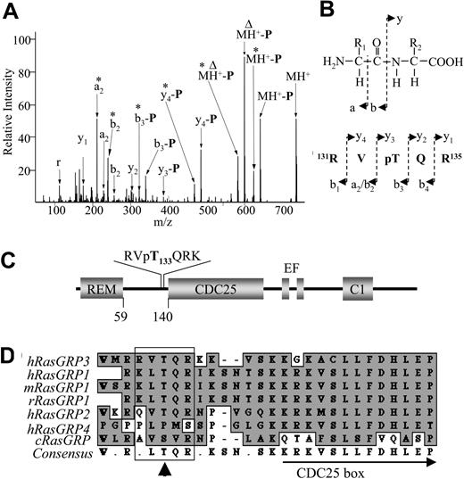
PKCθ-phosphorylated RasGRP3 was resolved by SDS-PAGE and subjected to in-gel digestion with trypsin. Peptides were eluted and phosphorylated peptides were enriched by open tubular immobilized metal ion affinity chromatography before analysis by mass spectrometry. We observed 2 related tryptic peptides represented by singly charged ions at m/z 739.36 (131-RVpTQR-135) and 867.45 (131-RVpTQRK-136) in phosphorylated RasGRP3. These 2 phosphopeptides were selected for tandem mass spectroscopy (MS/MS) analysis for detailed structural elucidation and sequence confirmation. In this method, particular tryptic peptides selected during mass spectropic analysis are broken down and these peptide fragment ions are further analyzed by a second round of mass spectrometry. The MS/MS spectrum along with peak assignments for m/z 739.36 is shown in Figure 1A. A fragment ion at m/z 641.42 resulting from loss of the phosphomoiety (H3PO4, molecular weight = 98) from the parental ion (phosphopeptide at m/z 739.36) was observed, verifying single phosphorylation of this peptide. The observed m/z values of other reduced fragment ions were also listed in Table 1, and were compared with their predicted theoretic values. The results further confirmed that RasGRP3 is phosphorylated on Thr133 in vitro. Similar data were collected for the other related ion (data not shown).
Threonine 133 in RasGRP3 is phosphorylated in vitro by PKCθ. (A) MS/MS spectrum of phosphopeptide m/z 739.36 (131-RVpTQR-135) derived from analysis of RasGRP3 that was subjected to phosphorylation by recombinant PKCθ. The fragment ions are labeled with peak assignments (a, b, or y ions) conforming to the common notation used for peptide fragment ions. The peak labeled MH+ corresponds to the protonated ion of the phosphorylated parental tryptic peptide. Peaks resulting from neutral loss of a phosphate group from an ion are marked “-P.” * denotes the loss of an “-NH3”; Δ, the loss of a “-C(NH)2”; and r, immonium ion of arginine. (B) Generic (top) and specific (bottom) structural representations of a, b, and y fragment ions are shown. (C) A schematic diagram of RasGRP3 with the position of the deduced phosphorylation and the sequence of the diagnostic peptide shown. REM indicates Ras exchange motif; CDC25, Ras activator domain; EF, calcium-binding domain; and C1, DAG-binding domain. (D) The region surrounding the PKC phosphorylation site in human RasGRP3 is shown aligned with other RasGRP family members including mouse and rat RasGRP1. cRasGRP indicates C elegans RasGRP. Consensus sequence is summarized at the bottom (consensus). The position of the partially conserved Thr is shown with a vertical arrow, while the box highlights flanking basic residues. The amino terminal region of the CDC25 box is also indicated. Conserved sequences are indicated by shading.
Threonine 133 in RasGRP3 is phosphorylated in vitro by PKCθ. (A) MS/MS spectrum of phosphopeptide m/z 739.36 (131-RVpTQR-135) derived from analysis of RasGRP3 that was subjected to phosphorylation by recombinant PKCθ. The fragment ions are labeled with peak assignments (a, b, or y ions) conforming to the common notation used for peptide fragment ions. The peak labeled MH+ corresponds to the protonated ion of the phosphorylated parental tryptic peptide. Peaks resulting from neutral loss of a phosphate group from an ion are marked “-P.” * denotes the loss of an “-NH3”; Δ, the loss of a “-C(NH)2”; and r, immonium ion of arginine. (B) Generic (top) and specific (bottom) structural representations of a, b, and y fragment ions are shown. (C) A schematic diagram of RasGRP3 with the position of the deduced phosphorylation and the sequence of the diagnostic peptide shown. REM indicates Ras exchange motif; CDC25, Ras activator domain; EF, calcium-binding domain; and C1, DAG-binding domain. (D) The region surrounding the PKC phosphorylation site in human RasGRP3 is shown aligned with other RasGRP family members including mouse and rat RasGRP1. cRasGRP indicates C elegans RasGRP. Consensus sequence is summarized at the bottom (consensus). The position of the partially conserved Thr is shown with a vertical arrow, while the box highlights flanking basic residues. The amino terminal region of the CDC25 box is also indicated. Conserved sequences are indicated by shading.
Fragment ions detected by ms/ms analysis of RVpTQR
Ions . | Sequences . | T. m/z . | O. m/z . |
|---|---|---|---|
| MH+ | RVpTQR | 739.36 | 739.44 |
| MH+-P | RVTQR | 641.38 | 641.42 |
| MH+-P* | RVTQR* | 624.36 | 624.41 |
| MH+-PΔ | RVTQRΔ | 599.36 | 599.42 |
| MH+-P*Δ | RVTQR*Δ | 582.34 | 582.38 |
| y4-P | VTQR | 485.28 | 485.34 |
| y4-P* | VTQR* | 468.26 | 468.28 |
| y3-P | TQR | 386.22 | 386.25 |
| b3-P | RVT | 339.21 | 339.25 |
| b3-P* | RVT* | 322.19 | 322.23 |
| y2 | QR | 303.18 | 303.19 |
| b2 | RV | 256.18 | 256.22 |
| b2* | RV* | 239.15 | 239.20 |
| a2 | RV | 228.18 | 228.21 |
| a2* | RV* | 211.18 | 211.18 |
| y1 | R | 175.12 | 175.14 |
The corresponding peptide sequences (Sequences), the theoretical m/z value (T. m/z), and the observed m/z value (O. m/z) of each fragment ion in Figure 1A are shown. The information derived from these fragment ions confirms that Thr133 in this peptide is phosphorylated.
-P indicates peaks resulting from neutral loss of a phosphate group from an ion; *, the loss of an “-NH3”; Δ, the loss of a “-C(NH)2”; and r, immonium ion of arginine.
The phosphorylation site is near the amino terminal region of the CDC25 box, named for the prototypic Ras activator domain from Saccharomyces cerevisiae, and corresponds to a consensus PKC phosphorylation sequence RXXS/T (Figure 1C-D). Mammalian forms of RasGRP1 and RasGRP2 have threonine at the corresponding site, while the Caenorhabditis elegans orthologue has serine. RasGRP4, which is expressed in mast cells and cell lines derived from the myeloid lineage,21,22 lacks this sequence and has several prolines in the region (Figure 1D).
To confirm that Thr133 was a major site of phosphorylation by PKCθ in vitro, we generated a mutant cDNA containing a substitution encoding alanine at position 133. Parental MBP, MBP-RasGRP3, and MBP-RasGRP3-Thr133Ala (T133A) were each individually incorporated into immune complex kinase reactions with baculovirus-expressed PKCθ serving as the modifying enzyme. In these experiments, we used [32Pγ]-ATP as cofactor to radiolabel product, and the reaction was monitored by autoradiography after resolution of products by SDS/7.5% PAGE. Sf9 cells that were mock infected and immune-precipitations that lacked the anti-PKCθ antibody served as negative controls. As described previously,14 PKCθ-dependent phosphorylation of MBP-RasGRP3 was readily detected with this assay (Figure 2A). In contrast, labeling of parental MBP and mutant RasGRP3 was undetectable. Similar results were obtained with PKCδ (Figure 2B). Collectively, these results confirm that Thr133 in RasGRP3 is a major phosphorylation site for nPKC forms in vitro.
RasGRP3 is phosphorylated on threonine 133 in vivo
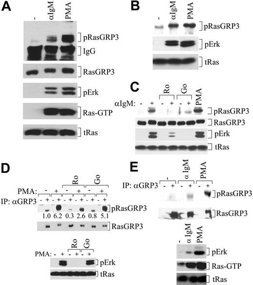
In order to confirm that Thr133 serves as a RasGRP3 phosphorylation site in vivo, we generated polyclonal anti–phospho-peptide antibodies, anti-3/pThr133. Ramos B cells were left unstimulated, treated with anti-IgM antibodies to stimulate the BCR, or treated with PMA to activate DAG signaling proteins directly. To ensure the specificity of our assay, we immune-precipitated RasGRP3 from lysates with monoclonal antibodies and then resolved recovered material by SDS/10% PAGE before blotting with anti-3/pThr133 antibodies.
The weak reactivity observed with RasGRP3 from untreated Ramos B cells could reflect either a low level of basal phosphorylation or weak reactivity of the antibodies with the nonphosphorylated RasGRP3 (Figure 3A). Much stronger reactivity was observed 5 minutes after treatment with anti-IgM antibodies or 10 minutes after treatment with PMA. Both of these treatments resulted in strong Ras and Erk activation, as expected. To confirm the results, we also showed that anti-3/pThr133 antibodies selectively immune-precipitate RasGRP3 after cell stimulation (Figure 3B).
PKC-dependent RasGRP3 phosphorylation on Thr133 occurs in B cells after stimulation and is correlated with Ras-Erk signaling. (A) Ramos B cells were left untreated or stimulated with PMA for 10 minutes or anti-IgM for 5 minutes, and the levels of Thr133-phosphorylated RasGRP3 (pRasGRP3), immunoglobulin G (IgG), RasGRP3, phosphorylated Erk, Ras-GTP, and total Ras (tRas) were determined. (B) Ramos B cells were treated as in panel A. Phosphorylated RasGRP3 was immune-precipitated with anti-3/pThr133 antibodies and precipitated RasGRP3 was then detected by immunoblotting. Levels of phospho-Erk and total Ras in lysates were also determined. (C) Ramos B cells were pretreated with inhibitors followed by stimulation with anti-IgM antibodies, as indicated, and lysates were probed for Thr133-phosphorylated RasGRP3, total RasGRP3, and phosphorylated Erk. PMA-treated cells served as a positive control. Ro indicates Ro318220; Go, Go6976. (D) Ramos B cells were pretreated as in panel C, and then stimulated with PMA for 10 minutes followed by analysis of RasGRP3 (top) and other signaling molecules (bottom). Numbers are quantification of band intensity. (E) Mouse B cells were stimulated with anti-IgM antibodies for 5 minutes or PMA for 10 minutes. RasGRP3 phosphorylation and RasGRP3 recovery (top) were determined as in panel A, as were the levels of phospho-Erk, Ras-GTP, and total Ras in lysates (bottom). Results are representative of at least 2 independent experiments.
PKC-dependent RasGRP3 phosphorylation on Thr133 occurs in B cells after stimulation and is correlated with Ras-Erk signaling. (A) Ramos B cells were left untreated or stimulated with PMA for 10 minutes or anti-IgM for 5 minutes, and the levels of Thr133-phosphorylated RasGRP3 (pRasGRP3), immunoglobulin G (IgG), RasGRP3, phosphorylated Erk, Ras-GTP, and total Ras (tRas) were determined. (B) Ramos B cells were treated as in panel A. Phosphorylated RasGRP3 was immune-precipitated with anti-3/pThr133 antibodies and precipitated RasGRP3 was then detected by immunoblotting. Levels of phospho-Erk and total Ras in lysates were also determined. (C) Ramos B cells were pretreated with inhibitors followed by stimulation with anti-IgM antibodies, as indicated, and lysates were probed for Thr133-phosphorylated RasGRP3, total RasGRP3, and phosphorylated Erk. PMA-treated cells served as a positive control. Ro indicates Ro318220; Go, Go6976. (D) Ramos B cells were pretreated as in panel C, and then stimulated with PMA for 10 minutes followed by analysis of RasGRP3 (top) and other signaling molecules (bottom). Numbers are quantification of band intensity. (E) Mouse B cells were stimulated with anti-IgM antibodies for 5 minutes or PMA for 10 minutes. RasGRP3 phosphorylation and RasGRP3 recovery (top) were determined as in panel A, as were the levels of phospho-Erk, Ras-GTP, and total Ras in lysates (bottom). Results are representative of at least 2 independent experiments.
Both the pan-PKC inhibitor Ro318220 and the cPKC-selective inhibitor Go6976 largely blocked both anti-3/pThr133 reactivity and Erk activation in BCR-stimulated cells (Figure 3C). For comparative purposes, we also looked at the effect of inhibitors on PMA-induced RasGRP3 phosphorylation (Figure 3D). In this case, we observed substantial inhibition of anti–3/p-Thr133 reactivity with Ro318220 but negligible inhibition with Go6976. Despite this difference, the inhibition of PMA-induced Thr133 phosphorylation matched the effect on PMA-induced Erk activation.
The phosphorylation of RasGRP3 coincident with Ras-Erk signaling after BCR and PMA stimulation was confirmed in primary mouse B cells (Figure 3E).
RasGRP3 Thr133Ala is functionally impaired in an ectopic expression system
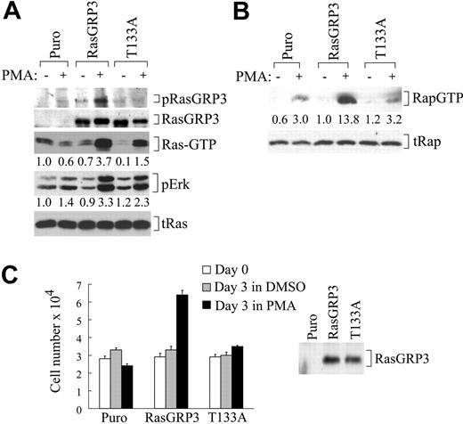
To study the functional properties of the RasGRP3 Thr133Ala substitution in vivo, we expressed wild-type and mutant RasGRP3 using a retrovirus vector system in rat2 cells. Rat2 cells do not express endogenous RasGRP3. Expression of wild-type RasGRP3 endows rat2 cells with the ability to couple DAG analog treatment to activation of Ras and Erk (Figure 4A) as has been reported previously.16 When we tested rat2 cells that express RasGRP3 Thr133Ala, we found that Ras and Erk activation were impaired relative to wild type, although signaling was higher than seen in empty vector cells. As expected, the mutant protein exhibited no reactivity with the anti-3/pThr133 antibody.
RasGRP3 Thr133Ala expressed in rat2 cells is defective at PMA-induced Ras-Erk signaling and PMA-induced growth. (A) Rat2 cells expressing empty vector (Puro), RasGRP3, or RasGRP3Thr133Ala (T133A) were either left unstimulated or stimulated with PMA for 10 minutes. Lysates were assayed for Thr133-phosphorylated RasGRP3, RasGRP3, phospho-Erk, Ras-GTP, and total Ras by immunoblotting. Numbers are quantification of band intensity. (B) Rat2 cells similar to those in panel A were assayed for Rap-GTP and total Rap (tRap). Numbers are quantification of band intensity. (C, left) Rat2 cells similar to those in panel A were plated in complete medium. One set of plates was harvested later the same day (□), while 2 sets were incubated for 3 days in medium containing 0.5% FBS alone (▦) or supplemented with 100 nM PMA (▪) before performing cell counts. Values represent the means ± standard deviations of triplicates within a single experiment (P < .01). (Right) RasGRP3 expression levels in these cells were estimated using anti-RasGRP3 antibodies. Results are representative of at least 2 independent experiments.
RasGRP3 Thr133Ala expressed in rat2 cells is defective at PMA-induced Ras-Erk signaling and PMA-induced growth. (A) Rat2 cells expressing empty vector (Puro), RasGRP3, or RasGRP3Thr133Ala (T133A) were either left unstimulated or stimulated with PMA for 10 minutes. Lysates were assayed for Thr133-phosphorylated RasGRP3, RasGRP3, phospho-Erk, Ras-GTP, and total Ras by immunoblotting. Numbers are quantification of band intensity. (B) Rat2 cells similar to those in panel A were assayed for Rap-GTP and total Rap (tRap). Numbers are quantification of band intensity. (C, left) Rat2 cells similar to those in panel A were plated in complete medium. One set of plates was harvested later the same day (□), while 2 sets were incubated for 3 days in medium containing 0.5% FBS alone (▦) or supplemented with 100 nM PMA (▪) before performing cell counts. Values represent the means ± standard deviations of triplicates within a single experiment (P < .01). (Right) RasGRP3 expression levels in these cells were estimated using anti-RasGRP3 antibodies. Results are representative of at least 2 independent experiments.
RasGRP3 has been reported to catalyze activation of the Ras-related GTPase Rap1A.23 Although the significance of this function of endogenous RasGRP3 is uncertain, we have found that the Thr133Ala mutant was defective relative to wild type in PMA-induced Rap1 activation, at least when ectopically expressed in rat2 cells (Figure 4B).
Unlike RasGRP1 and RasGRP4, RasGRP3 does not induce any of the signs of morphologic transformation associated with activated Ras in rat2 cells. Nonetheless, RasGRP3 expression does influence growth in these cells. Proliferation of parental rat2 cells is strictly dependent on serum growth factors. In medium containing 10% serum, rat2 cells undergo mitosis daily, but in medium containing 0.5% serum, very little net increase in cell number is observed over 3 days. PMA does not substitute for serum in rat2 cells and in fact may be slightly detrimental to cell recovery. In rat2 cells expressing wild-type RasGRP3, however, PMA can induce substantial growth (Figure 4C). This effect is largely abrogated by the Thr133Ala mutation in RasGRP3. These results support the idea that phosphorylation of Thr133 plays a role in RasGRP3 activation in response to DAG signals.
Implications of RasGRP3 Thr133 phosphorylation for regulation of RasGRP1 and RasGRP4
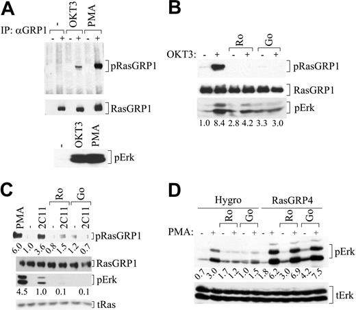
RasGRP3 amino acid Thr133 corresponds to RasGRP1 Thr184 (Table 1), so we generated a second polyclonal antibody, anti-1/pThr184, that recognizes phosphorylated RasGRP1. Extracts were prepared from unstimulated Jurkat T cells, from cells treated with PMA, and from cells stimulated with a stimulatory antibody that recognizes CD3, a component of TCR. As we predicted, anti-1/pThr184 antibodies recognized RasGRP1, but only after stimulation (Figure 5A). Ro318220 and Go6976 individually inhibited phosphorylation of RasGRP1 in Jurkat T cells after immune receptor stimulation (Figure 5B). This result was confirmed in primary mouse T cells (Figure 5C). Again, there was a good correspondence between inhibition of phosphorylation and inhibition of Erk activation. However, functional analysis of the RasGRP1 Thr184Ala mutation has so far not revealed a signaling defect, and ongoing work indicates that regulation of RasGRP1 by phosphorylation may be complex.
RasGRP1 in T cells is subject to PKC-dependent regulatory phosphorylation on Thr184, while RasGRP4 is regulated by other means. (A) Jurkat T cells were left untreated or were stimulated with anti-TCR antibodies (OKT3) for 5 minutes or PMA for 10 minutes, and lysates were probed for Thr184-phosphorylated RasGRP1 and total RasGRP1 (top). Signaling to Erk was also monitored (bottom). (B) Jurkat T cells were pretreated with PKC inhibitors and stimulated with the anti-TCR antibody OKT3. Lysates were assayed for phospho-RasGRP1 and total RasGRP1, as in panel A. Lysates were also probed for phospho-Erk to monitor signaling. (C) Mouse T cells were pretreated with inhibitors and stimulated with anti-TCR antibodies (2C11) or PMA, and lysates were probed for signaling molecules as in panel B. Numbers are quantification of band intensity. (D) Rat2 cells expressing empty vector pBabeHygro (Hygro) or RasGRP4 in the same vector were pretreated with inhibitors, followed by PMA treatment, as indicated. Total cell lysates were blotted for phospho-Erk to monitor Ras-Erk signaling and total Erk (tErk) as a loading control. Numbers are quantification of band intensity. Results are representative of at least 2 independent experiments.
RasGRP1 in T cells is subject to PKC-dependent regulatory phosphorylation on Thr184, while RasGRP4 is regulated by other means. (A) Jurkat T cells were left untreated or were stimulated with anti-TCR antibodies (OKT3) for 5 minutes or PMA for 10 minutes, and lysates were probed for Thr184-phosphorylated RasGRP1 and total RasGRP1 (top). Signaling to Erk was also monitored (bottom). (B) Jurkat T cells were pretreated with PKC inhibitors and stimulated with the anti-TCR antibody OKT3. Lysates were assayed for phospho-RasGRP1 and total RasGRP1, as in panel A. Lysates were also probed for phospho-Erk to monitor signaling. (C) Mouse T cells were pretreated with inhibitors and stimulated with anti-TCR antibodies (2C11) or PMA, and lysates were probed for signaling molecules as in panel B. Numbers are quantification of band intensity. (D) Rat2 cells expressing empty vector pBabeHygro (Hygro) or RasGRP4 in the same vector were pretreated with inhibitors, followed by PMA treatment, as indicated. Total cell lysates were blotted for phospho-Erk to monitor Ras-Erk signaling and total Erk (tErk) as a loading control. Numbers are quantification of band intensity. Results are representative of at least 2 independent experiments.
RasGRP4 is not highly conserved in the region corresponding to the sites phosphorylated in RasGRP1 and RasGRP3. Rather than a consensus PKC phosphorylation sequence, RasGRP4 has several prolines in the corresponding region (Figure 1D), arguing that this protein cannot be subject to the same sort of regulation. We noted a slight activation of Erk in empty vector cells exposed to PMA (Figure 5D). This response likely reflects a direct activation of Raf by PKC that occurs without Ras activation, although it may depend on basal Ras-GTP levels.24 Erk activation is much more robust in RasGRP4-expressing rat2 cells as a result of Ras activation. Strikingly, PMA-induced Erk activation in rat2 cells expressing RasGRP4 was completely resistant to PKC inhibitors (Figure 5D).
Discussion
The engagement of BCR by antigen is tightly linked to the activation of PLC-γ2 and the generation of DAG. Accumulation of membrane DAG is expected to recruit and activate various PKCs, RasGRPs, and other C1 domain proteins.25 In principle, PKC and RasGRP3 pathways might operate independently. We have hypothesized, however, that colocalization of these proteins on the same membrane surfaces may facilitate regulatory phosphorylation of RasGRP3 by PKC. B cells express RasGRP1 and RasGRP3, although the analysis of mutant chicken DT40 pre-B cells indicates that the latter protein is the major Ras activator responding to BCR signals.26 B cells also express a variety of PKC isozymes and information about the functions of some has been gleaned from the analysis of knock-out mice. For example, PKCβ null mutant mice have impaired humoral response and their B cells exhibit impaired proliferation in vitro.27 PKCδ mutant B cells fail to establish an antigen-tolerant state and can predispose mice to an autoimmune disease.28,29
We previously demonstrated that PKCθ phosphorylates RasGRP3 in vitro and that cotransfection of genetically activated PKCθ and RasGRP3 into HEK293 cells resulted in phosphorylation of the latter protein and cooperative Ras signaling.14 Furthermore, although PKCθ expression is typically associated with T cells, we showed that Ramos B cells express this isozyme. For these reasons, in the present study we first mapped the position of phosphate incorporated into RasGRP3 by PKCθ in vitro using mass spectrometry. These studies provided physical evidence for phosphorylation on Thr133. The result was confirmed by the observation that recombinant RasGRP3 bearing the Thr133Ala substitution was virtually inert as a PKCθ substrate. This RasGRP3 substitution also blocks phosphorylation by PKCδ. The latter observation is intriguing in light of the evidence that this PKC isozyme is an effective partner of RasGRP3 in HEK293 cells20 and the proposal that BCR signaling through PKCδ is required for induction of B-cell antigen tolerance, a process that depends on active BCR signaling and Erk activation.30 Recently it was shown that PKCδ plays a role in BCR-induced pp90rsk activation and subsequent phosphorylation of the cyclic AMP response element binding protein (CREB) transcription factor.31 The involvement of RasGRP3-Erk intermediates between PKCδ and pp90rsk could neatly explain these observations.
The development of antibodies that are specific for phospho-Thr133 in RasGRP3 has allowed us to verify that this modification occurs in vivo promptly after both BCR stimulation and DAG agonist treatment, in both the Ramos B-cell line and primary mouse B cells. In both cell types, phosphorylation was more intense after PMA treatment. Importantly, PKC inhibitors that interfere with RasGRP3 phosphorylation on Thr133 also diminish Ras-Erk signaling. Evidence for the role of an nPKC was evident in our PMA stimulation protocol, as only the pan-PKC inhibitor Ro318220 was effective. The results observed after BCR stimulation are more puzzling. Although the pan-PKC inhibitor was the most effective at blocking RasGRP3 phosphorylation, we have consistently found that this drug was less effective than the cPKC-selective drug Go6976 at blocking BCR-stimulated signaling. We previously demonstrated that this anomaly can be seen at the level of Ras-GTP and can be duplicated with a second pan-PKC inhibitor, Bisindolylmaleimide.14 Possibly, pan-PKC inhibitors have effects on other Ras regulators in Ramos B cells such as Ras GAPs, as originally proposed for T cells.6
Consistent with the idea that Thr133 phosphorylation represents a positive regulatory modification in RasGRP3, we found that the Thr133Ala mutant protein has reduced Ras-Erk signaling in rat2 cells after brief PMA treatment. RasGRP3 Thr133Ala-expressing cells also exhibited defective PMA-induced growth, which provides a longer-term functional assay. However, we cannot exclude the possibility that the Thr133Ala substitution has some effect on RasGRP3 other than preventing phosphorylation. We did find that GFP-tagged wild-type and Thr133Ala mutant forms of RasGRP3 had similar subcellular distribution when expressed in HEK293 cells and showed similar recruitment to cellular membranes in response to PMA (Y.Z., unpublished data, February 2004). In some respects, then, the mutant form behaves normally and may serve as a specific model of nonphosphorylated RasGRP3. Recently, BCR-induced phosphorylation of RasGRP3 on Thr133 was demonstrated in the DT40 chicken pre-B cell line.32 This modification was dependent upon PLC-γ2 and PKC activity. Furthermore, the Thr133Ala mutation impaired Ras activation as shown in the present study.
Our initial analysis of RasGRP1 phosphorylation in T cells indicates that the homologous position, Thr184, is phosphorylated after both TCR stimulation and treatment with DAG agonist. Again, PKC inhibitors block both phosphorylation and Ras-Erk signaling, consistent with the idea that phosphorylation of RasGRP1 requires both membrane recruitment and regulatory phosphorylation by a membrane-recruited PKC for activation. PKCθ plays an important role in TCR signaling and is an obvious candidate for a RasGRP1 regulator. However, the effectiveness of Go6976 implies a role for cPKC, and our initial studies indicate that the Thr184Ala mutation does not significantly impair RasGRP1 phosphorylation by PKCθ in vitro (Y.Z., unpublished data, May 2003).
The position of Thr133 suggests that phosphorylation might affect guanyl nucleotide exchange activity. Our analysis of RasGRP4 offers an interesting comparison. This species has an unusual density of prolines in this region and lacks a homologous PKC phosphorylation site. The insensitivity of RasGRP4 signaling to PKC inhibitors is in line with the idea that this protein is not subject to regulatory phosphorylation. We speculate the RasGRP4 is constitutively in the activated state, as a result of the structural effect of these prolines. Consistent with this idea, when we individually express all 4 family members in the same retrovirus vector in rat2 cells, RasGRP4 is readily the most transforming species.
Phosphorylation may be a common mechanism for Ras GEF regulation. Erk phosphorylates Sos, and this may provide a negative feedback mechanism to uncouple Ras signaling in later stages of growth factor responses.33 RasGRF1, a neuronal Ras GEF is phosphorylated and positively regulated by cyclic adenosine monophosphate (cAMP)–dependent kinase, at a site close to that corresponding to Thr133 in RasGRP3.34 It has been reported that RasGRP3 is phosphorylated by a src-type protein tyrosine kinase in HEK 293 cells as part of an epidermal growth factor (EGF) receptor–pp60src–PLC-γ1–RasGRP3–Rap2B–PLC-ϵ pathway.35 Using our anti-RasGRP3 antibodies, however, we found no expression of RasGRP3 in these cells. Furthermore, we were unable to demonstrate BCR-induced tyrosine phosphorylation of immune-precipitated RasGRP3 in Ramos cells, using an antiphosphotyrosine antibody in an immunoblotting assay. This induced modification was readily demonstrated in our control protein, PLC-γ2 (Stacy Stang, unpublished data, January 2002).
It is unclear what purpose PKC regulation of RasGRP3 serves. In principle, this Ras GEF could be regulated solely by recruitment, as may be the case in RasGRP4. One possibility is that while the level of membrane recruitment of RasGRP3 (or PKC) may be a linear function of DAG membrane concentration, the membrane concentration of active, phosphorylated RasGRP may be nonlinear, as it would depend on the surface concentrations of PKC and substrate RasGRP3, as well as the activities of phosphatases. The ability to respond to DAG in a nonlinear fashion might effectively turn an otherwise analog system into a digital switch, a useful feature for a biochemical pathway that is used to control procession through various stages of lymphocyte differentiation, activation, and death. The present identification of RasGRP phosphorylation sites provides a specific link in the PKC-RasGRP axis, from which further mechanistic and physiologic insights may be gained.
Prepublished online as Blood First Edition Paper, January 18, 2005; DOI 10.1182/blood-2004-10-3916.
Supported by grants from the Canadian Institutes of Health Research to J.C.S. and from the Protein Engineering Network of Centers of Excellence and Alberta Cancer Board to L.L. and J.C.S.
The publication costs of this article were defrayed in part by page charge payment. Therefore, and solely to indicate this fact, this article is hereby marked “advertisement” in accordance with 18 U.S.C. section 1734.
We thank Ms Stacey Stang for her expert advice on the use of anti-RasGRP antibodies.

This feature is available to Subscribers Only
Sign In or Create an Account Close Modal