Abstract
Tissue factor (TF), the cellular receptor for factor VIIa (FVIIa), besides initiating blood coagulation, is believed to play an important role in tissue repair, inflammation, angiogenesis, and tumor metastasis. Like TF, the chemokine interleukin-8 (IL-8) is shown to play a critical role in these processes. To elucidate the potential mechanisms by which TF contributes to tumor invasion and metastasis, we investigated the effect of FVIIa on IL-8 expression and cell migration in a breast carcinoma cell line, MDA-MB-231, a cell line that constitutively expresses abundant TF. Expression of IL-8 mRNA in MDA-MB-231 cells was markedly up-regulated by plasma concentrations of FVII or an equivalent concentration of FVIIa (10 nM). Neither thrombin nor other proteases involved in hemostasis were effective in stimulating IL-8 in these cells. Increased transcriptional activation of the IL-8 gene is responsible for increased expression of IL-8 in FVIIa-treated cells. PAR-2–specific antibodies fully attenuated TF-FVIIa–induced IL-8 expression. Additional in vitro experiments showed that TF-FVIIa promoted tumor cell migration and invasion, active site–inactivated FVIIa, and specific antibodies against TF, PAR-2, and IL-8 inhibited TF-FVIIa–induced cell migration. In summary, the studies described herein provide insight into how TF may contribute to tumor invasion. (Blood. 2004;103:3029-3037)
Introduction
Cells that express tissue factor (TF) are usually not exposed to the blood. However, in normal response to vessel injury, TF exposure is an initial event of a strictly regulated process resulting in fibrin deposition, inflammation, angiogenesis, and tissue repair. Carcinomas exploit a normal physiologic response in a way that allows tumor growth and dissemination. It has long been presumed that tumors may take advantage of the hemostatic system. A relationship between increased clotting and malignancy was recognized more than a century ago.1 Numerous clinical observations suggest that the hemostatic system is frequently activated in cancer patients.2-5 Many tumor types have been shown to express TF.6,7 Further, the level of TF expression in various tumor types has been shown to correlate with their metastatic potential.8-10 Studies carried out with mouse tumor metastasis models establish that TF plays a critical role in tumor metastasis.11,12 TF is the cellular receptor for coagulation factor VIIa (FVIIa). TF-induced metastasis requires participation of the cytoplasmic tail of TF and assembly of an active TF-FVIIa complex,13,14 indicating a dual function for TF in tumor metastasis. The TF cytoplasmic domain, through its specific interaction with ABP-280, has been shown to support cell adhesion and migration.15 At present it is unclear how TF on tumor cells contributes to tumor metastasis and whether the TF-FVIIa complex plays a direct role or whether its sole requirement is for the downstream generation of active coagulation factors, particularly thrombin, which have been implicated in tumor metastasis.16-18
Recent studies show that proteolytic hydrolysis mediated by the TF-FVIIa complex induces cell signaling through G-protein–coupled receptors in a number of cell types (for reviews, see Prydz et al,19 Pendurthi and Rao,20 Ruf et al21 ). TF-FVIIa–induced signaling in various cell types was shown to alter the expression of specific genes that encode transcription factors, growth factors, and proteins related to cellular reorganization.22-27 These studies suggest that TF-FVIIa–induced signaling may play a role in growth-promoting settings, such as wound healing and cancer. However, it has yet to be shown how TF-FVIIa–induced regulation of gene expression actually affects cell phenotype or pathophysiologic processes. Moreover, a considerable overlap in signaling induced by TF-FVIIa and various other proteases, especially a high-magnitude response generated by thrombin, raises a valid question about the potential significance of TF-FVIIa–induced signaling in pathophysiology.
Similar to the correlation between TF expression and metastatic potential, a strong correlation between metastatic potential and ectopic expression of the chemokine interleukin-8 (IL-8) has been found in many tumor cells, including those of breast carcinoma.28-30 IL-8, a member of the CXC chemokine family, initially shown to be a chemoattractant for neutrophils and lymphocytes, can act multifunctionally to induce tumor growth and metastasis.31,32 IL-8 was shown to act as an autocrine growth factor and to stimulate invasion and chemotaxis of many tumor cell types.33-37 Further, IL-8 secreted by tumor cells may also promote vascularization by enhancing endothelial cell proliferation, survival, and matrix metalloproteinase production.38 In this context, it is interesting to note that recent studies showed that TF-FVIIa induced the expression of IL-8 in keratinocytes.26
Induction of IL-8 provides a putative link between blood coagulation and diverse processes such as inflammation, wound healing, angiogenesis, and cancer. To further understand the role of coagulation in tumor cell migration and invasion, we investigated in the present study whether FVIIa, thrombin, and other proteases involved in hemostasis affect the expression of IL-8 in a breast carcinoma cell line that constitutively expresses TF and whether IL-8 production by these proteases leads to increased tumor cell migration and invasion in vitro. Our data revealed that FVIIa, but not factor Xa (FXa) or thrombin, markedly up-regulated IL-8 expression, resulting in increased cell migration and invasion, and that these effects are attenuated when FVIIa binding to TF is prevented.
Materials and methods
Reagents
Dulbecco modified Eagle medium (DMEM), fetal bovine serum (FBS), trypsin-EDTA (ethylenediamine tetraacetic acid), and penicillin-streptomycin were obtained from Gibco-BRL Life Technologies (Grand Island, NY). Matrigel was from BD Biosciences (San Diego, CA). Other chemicals, of reagent grade or better, were from Sigma Chemical (St Louis, MO). IL-8 antibodies and ELISA assay kits were from R&D Systems (Minneapolis, MN). PAR-1–specific monoclonal antibodies WEDE-15 and ATAP-2 were obtained from Beckman Coulter (Fullerton, CA) and Santa Cruz Biotechnology (Santa Cruz, CA), respectively. TF monoclonal antibodies (TF9-10H10) (used in flow cytometry and binding studies) and polyclonal neutralizing antibodies of PAR-2 were kindly provided by Wolfram Ruf (Scripps Research Institute, La Jolla, CA). PAR-2–specific antibodies were raised in rabbits by immunizing a PAR-2 peptide (sequence covering the region of PAR-2 activation site) conjugated to keyhole limpet hemocyanin (KLH). The antibodies inhibited specific responses mediated by PAR-2 (activated with FXa or FVIIa) but not by PAR-1 (activated with thrombin or plasmin). Polyclonal antibodies against TF, used for neutralizing TF, were described earlier.39,40 PAR-1 and PAR-2 agonist peptides TFLLRN-NH2 and SLIGKV-NH2, respectively, were custom synthesized and high-performance liquid chromatography (HPLC) purified (Biosynthesis, Lewisville, TX). Recombinant human FVIIa41 and FVIIa blocked in the active site with phenylalanyl-phenylalanyl-arginyl chloromethyl ketone (FFR-FVIIa)42 were obtained from Novo Nordisk A/S (Maaloev, Denmark). Zymogen FVII, which contains less than 0.1% FVIIa, was purified from human plasma as described earlier.43 Thrombin, FXa, and other proteases were from Enzyme Research Laboratory (South Bend, IN).
Cell line and cell culture
MDA-MB-231 and NIH 3T3 cells were obtained from ATCC (Rockville, MD). Cells were maintained in DMEM with glutamax and high-glucose medium supplemented with 1% glutamine, 1% penicillin/streptomycin, and 10% FBS. The cells were cultured at 37°C and 5% CO2 in a humidified incubator to near confluence and were deprived of serum for 16 hours before they were stimulated with agonists.
Northern blot analysis
Northern blot analysis was performed on total RNA extracted with Trizol reagent (Invitrogen, Carlsbad, CA) according to the manufacturer's instructions. RNA was dissolved in 1× RNA Secure (Ambion, Austin, TX). Ten micrograms of each RNA sample was precipitated, dissolved in RNA sample buffer (20 mM MOPS, pH 7.0, 1 mM EDTA, 5 mM sodium acetate, and 50% formamide), and denatured at 55°C before it was run on a denaturing gel in formaldehyde buffer (20 mM MOPS, pH 7.0, 1 mM EDTA, 5 mM sodium acetate, and 10% formaldehyde). Northern blot analysis was performed essentially as described earlier23 using 32 P-labeled IL-8 cDNA probe. Hybridized membranes were exposed to x-ray film, and hybridization signal intensities were quantified exposing the membranes to phosphor screens and analyzing them using a PhosphorImager (Molecular Imager; Bio-Rad, Richmond, CA).
Nuclear run off
Quiescent monolayers of MDA-MB-231 were treated with a control serum-free medium or the serum-free medium containing FVIIa for 1 hour. Cells were then washed once with ice-cold DMEM, scraped into the medium using a rubber policeman, and spun down at 1800g for 5 minutes at 4°C. Cell pellets were resuspended in cell lysis buffer (10 mM HEPES [N-2-hydroxyethylpiperazine-N′-2-ethanesulfonic acid], pH 8, 10 mM KCl, 0.1 mM EDTA, and 1 mM dithiothreitol [DTT]) and kept on ice for 1 hour. The lysed cell suspension was transferred to the top of 3 mL sucrose cushion (1.3 M sucrose in 10 mM HEPES, pH 8, containing 5 mM MgCl2, 2 mM DTT, and 0.1% Triton X-100) and was centrifuged at 10 000g for 15 minutes at 4°C. The supernatant was removed, and the nuclei were resuspended in 100 μL nuclei storage buffer (10 mM HEPES, pH 8, containing 5 mM MgCl2, 0.1 mM EDTA, 2 mM DTT, and 30% glycerol). Run-off assays were performed with [α-32 P]UTP (3000 Ci/mmol [111 TBq/mmol])–labeled RNA, as described previously.44 Labeled nuclear RNA was hybridized to 10 μg DNA (linearized vectors containing β-actin and IL-8) immobilized on nitrocellulose membranes using a slot-blot apparatus.
IL-8 protein measurement
IL-8 protein levels in conditioned media from cells that were serum starved for 24 hours and exposed to FVIIa and other proteases for another 24 hours were measured using an IL-8 Quantikine enzyme-linked immunosorbent assay (ELISA) from R&D Systems (Oxon, United Kingdom) according to the manufacturer's instructions.
Flow cytometry
Breast carcinoma cells were washed once with serum-free medium and detached from the culture flask by a brief incubation with versene solution (3 minutes; 0.5 mM EDTA). Cells were pelleted and washed once with fluorescence-activated cell sorter (FACS) buffer (phosphate-buffered saline containing 1% bovine serum albumin and 0.05% sodium azide) by centrifugation at 200g for 5 minutes. Cells suspended in FACS buffer (0.5 × 10⁶ cells/100 μL) were incubated with a control mouse immunoglobulin G (IgG) or monoclonal antibodies (5 μg/mL) against PAR-1 (ATAP2) or PAR-2 (SAM11) for 60 minutes at 4°C. Unbound antibodies were removed, and the cells were washed with FACS buffer before they were incubated with fluorescein isothiocyanate (FITC)–conjugated goat antimouse IgG (1:50 dilution; Molecular Probes, Eugene, OR). After 30 minutes' incubation at 4°C in the dark, the secondary antibody was removed and the cells were washed with FACS buffer and fixed in 0.5% paraformaldehyde in FACS buffer for 2 hours at 4°C in the dark. Cells were analyzed for fluorescence using a Coulter Epics flow cytometer (Beckman Coulter).
Ca2+ measurements
Cells were seeded in 96-well plates (ViewPlate-96 Black; PerkinElmer, Shelton, CT). When cells reached 80% confluence, the medium was removed, and 3.6 μM fluo-4/am (Molecular Probes) in 100 μL DMEM containing 10% fetal calf serum (FCS) was added. Cells were incubated for 30 minutes at 37°C, 5% CO2, followed by 1 wash with 200 μL Hanks balanced salt solution (HBSS: 1.3 mM CaCl2, 5.4 mM KCl, 0.4 mM KH2PO4, 0.5 mM MgCl2, 0.4 mM MgSO4, 137 mM NaCl, 4.1 mM NaHCO3, 0.3 mM Na2HPO4, 5.6 mM d-glucose, pH 7.5) without phenol red. Cells were treated with test compounds (in a final volume of 200 μL), and fluorescence was measured as the increase in fluorescence after excitation at 485 nm and emission at 520 nm using a microplate fluorometer with integrated pipetting system (NOVOstar; B&L Systems, Maarssen, The Netherlands). Results are presented as relative change (fold differences) in fluorescence after the addition of test compound compared with basal levels observed (before test compounds were added).
Cell migration
A transwell system (8-μm pore size, polycarbonate filter, 6.5-mm diameter) was used to evaluate cell migration. Both sides of the membrane filter of the upper chamber were coated with collagen type IV (1 mg/mL, 50 μL) for 2 minutes and then were air-dried. Cells (50 000 in 200 μL serum-free medium) were added to the upper well, and 500 μL serum-free medium was added to the lower chamber. Unless otherwise specified, FVIIa and other stimulants were added to the lower chamber. At the end of 24-hour incubation at 37°C/5% CO2, cells on the top of the membrane were removed by swiping with a damp cotton swab. The membrane was rinsed once with distilled water and stained with hematoxylin (Hema3 staining kit; Fisher Scientific, Hampton, NH) according to the instructions provided with the kit. Cells on the underside of the membrane were counted under a microscope, in 4 different viewing fields, at 20× magnification.
Matrigel invasion assay
A transwell system, similar to that used for the cell migration assays, was used for tumor cell invasion assays. Cells were starved overnight in serum-free medium. Matrigel (50 μg, ie, 50 μL of 1 mg/mL) was polymerized in the upper well at 37°C for 4 hours and then rinsed once with serum-free medium. MDA-MB-231 cells (25 000 cells in 200 μL serum-free medium) were added to the upper well. Conditioned medium (500 μL) from NIH3T3 fibroblasts were added to the lower well with or without an agonist (conditioned media were obtained by culturing NIH3T3 cells overnight in serum-free medium). Cells were incubated for 48 hours at 37°C/5% CO2 in a humidified culture incubator. Cells in the top well were removed by peeling of the Matrigel and swiping the top of the membrane with cotton swabs. Cells on the underside of the membrane were stained with hematoxylin and were counted as described in “Cell Migration.”
Results
TF expression by MDA-MB-231 breast carcinoma cells
MDA-MB-231 cells were shown to constitutively express high levels of TF mRNA.45 Consistent with this, flow cytometry and the binding of (125I)–labeled TF monoclonal antibodies (TF9-10H10) indicated abundant TF exposure on the surfaces of these cells. This was further supported by 125I-FVIIa binding studies and by FVIIa-mediated FX activation assays (data not shown). Binding studies with 125I-FVIIa revealed that MDA-MB-231 cells contained approximately 1.2 × 10⁶ binding sites/cell.
FVIIa induces IL-8 expression in MDA-MB-231 cells
Recent studies indicate that IL-8 can act multifunctionally in the invasiveness of various cancers.46 To address the possibility that the role of TF-FVIIa in tumor metastasis is mediated by IL-8, we investigated the effect of TF-FVIIa on IL-8 expression in a cancer cell line. Quiescent monolayers of MDA-MB-231 cells were exposed to varying concentrations of FVIIa for 75 minutes, and IL-8 mRNA steady-state levels were determined by Northern blot analysis. As shown in Figure 1A, FVIIa induced the expression of IL-8 mRNA in MDA-MB-231 cells in a dose-dependent manner. The increased expression of IL-8 mRNA was clearly evident in cells treated with 5 nM FVIIa and reached maximum levels with 25 to 50 nM FVIIa. In cells treated with 10 nM FVIIa, a concentration equivalent to a plasma concentration of FVII, IL-8 mRNA steady-state levels were 3- to 5-fold higher than IL-8 mRNA levels observed in control cells. Time course studies indicate that FVIIa-induced IL-8 mRNA expression peaked between 75 to 120 minutes and thereafter declined slowly (data not shown). We observed a similar increase in IL-8 mRNA expression in cells treated with a plasma concentration of zymogen FVII (10 nM), except that induction was delayed by 1 hour, presumably the time required for autoactivation of FVII (Figure 1D). Additional studies show that FVIIa treatment dose dependently increased IL-8 antigen levels in overlying conditioned media (Figure 1C). The increase in IL-8 antigen levels in the conditioned medium was time dependent; it was detected 2 hours after the addition of FVIIa and increased linearly up to 16 hours (data not shown).
FVIIa-induced IL-8 expression in breast carcinoma cells. Quiescent monolayers of MDA-MB-231 cells were treated with varying concentrations of FVIIa (A-C), a plasma concentration (10 nM) of zymogen FVII or FVIIa (D), or varying concentrations of active site-inactivated FVIIa (ASIS/FFR-FVIIa), followed by 10 nM FVIIa (E). (F) Cells were pretreated with anti-TF IgG or control IgG (100 μg/mL) for 45 minutes before FVIIa (10 nM) was added to the cells. Unless otherwise specified, cells were treated with FVIIa for 75 minutes at 37°C, and total RNA was isolated and subjected to Northern blot analysis. (A, D-F) Representative Northern blot analysis of IL-8 mRNA; (B) quantitative data from such experiments. (C) IL-8 antigen levels in overlying conditioned media of MDA-MB-231 cells treated with varying concentrations of FVIIa. Error bars indicate SEM from 2 to 3 experiments.
FVIIa-induced IL-8 expression in breast carcinoma cells. Quiescent monolayers of MDA-MB-231 cells were treated with varying concentrations of FVIIa (A-C), a plasma concentration (10 nM) of zymogen FVII or FVIIa (D), or varying concentrations of active site-inactivated FVIIa (ASIS/FFR-FVIIa), followed by 10 nM FVIIa (E). (F) Cells were pretreated with anti-TF IgG or control IgG (100 μg/mL) for 45 minutes before FVIIa (10 nM) was added to the cells. Unless otherwise specified, cells were treated with FVIIa for 75 minutes at 37°C, and total RNA was isolated and subjected to Northern blot analysis. (A, D-F) Representative Northern blot analysis of IL-8 mRNA; (B) quantitative data from such experiments. (C) IL-8 antigen levels in overlying conditioned media of MDA-MB-231 cells treated with varying concentrations of FVIIa. Error bars indicate SEM from 2 to 3 experiments.
Blockage of TF, but not inhibition of FXa or thrombin, prevents IL-8 expression
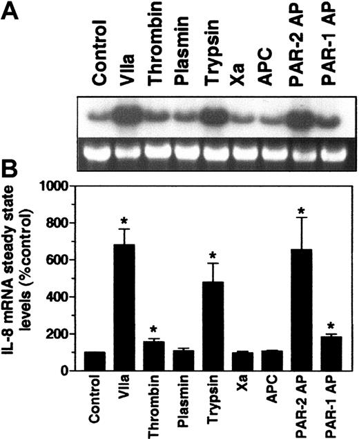
Active site-inactivated FVIIa (FFR-FVIIa) competes with FVIIa for TF.47 Blockage of FVIIa binding to TF with FFR-FVIIa inhibited the FVIIa-induced expression of IL-8 mRNA (Figure 1E). Similarly, FVIIa-induced IL-8 expression was also prevented when the MDA-MB-231 cells were pretreated with an antibody against TF (Figure 1F). In contrast, pretreatment of cells with hirudin or recombinant tick anticoagulant protein (TAP), specific inhibitors of thrombin and FXa, respectively, had no effect on FVIIa-induced IL-8 mRNA expression or antigen production (data not shown). To further examine the specificity of protease-induced IL-8 expression, we treated MDA-MB-231 cells with thrombin, FXa, activated protein C, or plasmin. They all failed to induce or only minimally induced IL-8 mRNA (Figure 2) or antigen (data not shown).
Effect of FVIIa and other agonists on the induction of IL-8 mRNA. Quiescent monolayers of MDA-MB-231 cells were treated with FVIIa (50 nM), thrombin (10 nM), plasmin (50 nM), trypsin (50 nM), FXa (50 nM), APC (50 nM), PAR-2–specific peptide agonist SLIGKV (25 μM) and PAR-1–specific peptide agonist TFLLRN (25 μM) for 75 minutes. Total RNA was analyzed for IL-8 expression by Northern blot analysis. (A) Representative autoradiograph. (B) Quantitative data (mean ± SEM, n = 4 to 7). *Value significantly higher (P < .05) than the control value.
Effect of FVIIa and other agonists on the induction of IL-8 mRNA. Quiescent monolayers of MDA-MB-231 cells were treated with FVIIa (50 nM), thrombin (10 nM), plasmin (50 nM), trypsin (50 nM), FXa (50 nM), APC (50 nM), PAR-2–specific peptide agonist SLIGKV (25 μM) and PAR-1–specific peptide agonist TFLLRN (25 μM) for 75 minutes. Total RNA was analyzed for IL-8 expression by Northern blot analysis. (A) Representative autoradiograph. (B) Quantitative data (mean ± SEM, n = 4 to 7). *Value significantly higher (P < .05) than the control value.
Involvement of PAR-2 in FVIIa-induced IL-8 expression
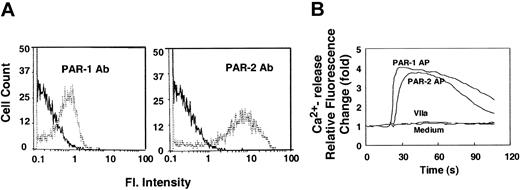
As with FVIIa and trypsin, treating cells with PAR-2–specific peptide agonist (SLIGKV) markedly enhanced IL-8 mRNA expression and antigen production. In contrast, PAR-1–specific peptide agonist (TFLLRN) treatment had only a minimal effect on IL-8 mRNA expression (Figure 2) and antigen production (data not shown). These observations suggest that FVIIa-induced IL-8 expression is mediated by PAR-2. At present, data conflict about whether TF-FVIIa–induced cell signaling involves the activation of PAR-1, PAR-2, or a putative PAR.23,24,48-51 To investigate the role of PAR-1 and PAR-2 in FVIIa-induced IL-8 expression in MDA-MB-231 cells, we first examined the expression of PAR-1 and PAR-2 in these cells by flow cytometry and by Ca2+ signaling (increase in intracellular Ca2+ in response to PAR-1– and PAR-2–specific peptide agonists). Data from these experiments revealed that MDA-MB-231 cells expressed PAR-1 and PAR-2 on their surfaces and that these receptors were functionally active (Figure 3). Consistent with our earlier observations with BHK-TF50 and fibroblasts,52 we did not detect any increase in intracellular Ca2+ in MDA-MB-231 cells in response to FVIIa (Figure 3). In additional experiments, we investigated the effect of thrombin (10 nM), trypsin (10 nM), and FXa (50 nM) on the release of intracellular Ca2+ in MDA-MB-231 cells. Both thrombin and trypsin increased intracellular Ca2+ with kinetics similar to that of PAR-1–and PAR-2 peptide agonists, whereas no detectable increase in intracellular Ca2+ was observed in cells treated with FXa (data not shown).
Expression and functional activity of PAR-1 and PAR-2 in MDA-MB-231 cells. (A) MDA-MB-231 cells were probed with anti–PAR-1 (ATAP2) or anti–PAR-2 (SAM11) monoclonal antibodies, followed by FITC-labeled secondary antibody. FITC-labeled cells were analyzed by flow cytometry. Solid lines represent background fluorescence (control IgG), whereas dotted lines represent fluorescence shift attributable to PAR expression. (B) Intracellular calcium fluxes in response to PAR-1– and PAR-2–specific peptide agonists, or FVIIa. Fluo-4–loaded cells were exposed to a control medium, PAR-1–, or PAR-2–specific peptide agonists (50 μM) or FVIIa (100 nM). The resultant change in fluorescence at 520 nm after excitation at 485 nm is presented as relative fluorescence change compared with basal level fluorescence measured before the addition of compounds.
Expression and functional activity of PAR-1 and PAR-2 in MDA-MB-231 cells. (A) MDA-MB-231 cells were probed with anti–PAR-1 (ATAP2) or anti–PAR-2 (SAM11) monoclonal antibodies, followed by FITC-labeled secondary antibody. FITC-labeled cells were analyzed by flow cytometry. Solid lines represent background fluorescence (control IgG), whereas dotted lines represent fluorescence shift attributable to PAR expression. (B) Intracellular calcium fluxes in response to PAR-1– and PAR-2–specific peptide agonists, or FVIIa. Fluo-4–loaded cells were exposed to a control medium, PAR-1–, or PAR-2–specific peptide agonists (50 μM) or FVIIa (100 nM). The resultant change in fluorescence at 520 nm after excitation at 485 nm is presented as relative fluorescence change compared with basal level fluorescence measured before the addition of compounds.
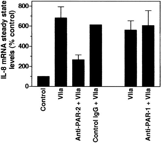
Next, we investigated the effect of PAR-1– and PAR-2–neutralizing antibodies on FVIIa-induced expression of IL-8. As shown in Figure 4, PAR-2–neutralizing antibodies markedly attenuated FVIIa-induced IL-8 expression. In contrast, PAR-1–neutralizing antibodies had no effect on FVIIa-induced IL-8 mRNA accumulation. It should be noted that the same concentration of PAR-1–neutralizing antibodies completely blocked thrombin-induced increase in intracellular Ca2+ in MDA-MB-231 cells (data not shown), strongly indicating that FVIIa-induced IL-8 expression in MDA-MB-231 cells is mediated through the activation of PAR-2.
PAR-2 antibody inhibits FVIIa-induced IL-8 mRNA induction. MDA-MB-231 cells were pretreated with control rabbit IgG (500 μg/mL), rabbit anti PAR-2 IgG (500 μg/mL), or monoclonal antibodies against PAR-1 (10 μg/mL ATAP2 plus 25 μg/mL WEDE15) for 1 hour before they were stimulated with FVIIa (50 nM) for 75 minutes. Total RNA was analyzed by Northern blot analysis for IL-8 mRNA, and the hybridization signals were quantitated. Data shown in the figure represent mean ± SEM from 3 to 6 experiments.
PAR-2 antibody inhibits FVIIa-induced IL-8 mRNA induction. MDA-MB-231 cells were pretreated with control rabbit IgG (500 μg/mL), rabbit anti PAR-2 IgG (500 μg/mL), or monoclonal antibodies against PAR-1 (10 μg/mL ATAP2 plus 25 μg/mL WEDE15) for 1 hour before they were stimulated with FVIIa (50 nM) for 75 minutes. Total RNA was analyzed by Northern blot analysis for IL-8 mRNA, and the hybridization signals were quantitated. Data shown in the figure represent mean ± SEM from 3 to 6 experiments.
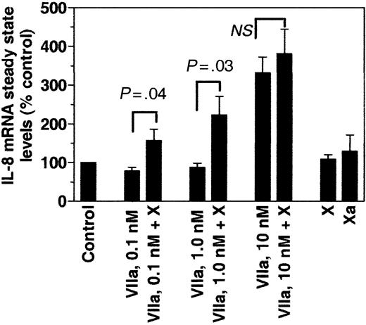
Recent studies suggest that the ternary complex, TF-FVIIa-FXa, is a more potent inducer of PAR-1/2–mediated signaling than the binary TF-FVIIa complex.53 To test this possibility, we compared the induction of IL-8 mRNA in cells treated with FVIIa, FXa, or FXa complexed with TF-FVIIa. To generate TF-FVIIa-FXa complexes transiently, FVIIa and FX were added to the cells. As shown in Figure 5, we found no significant increase in IL-8 mRNA levels in cells treated with FVIIa and FX, compared with FVIIa alone, when these reagents were used at their plasma concentration equivalents. However, the effect of FX was clearly evident at low concentrations of FVIIa. FVIIa at 0.1 or 1.0 nM failed to induce IL-8 expression when used alone, whereas in the presence of FX, we detected a significant induction of the IL-8 gene (Figure 5). These data suggest that including FXa as a transient partner in TF-FVIIa complex formation may potentiate FVIIa-induced IL-8 production in MDA-MB-231 cells when the FVIIa concentration is limited but not at saturating concentrations.
Effect of FXa on FVIIa-induced IL-8 expression. MDA-MB-231 cells were stimulated for 75 minutes with varying concentrations of FVIIa in the presence and absence of FX (175 nM). As controls, cells were stimulated with FX (175 nM) or FXa (175 nM). Induction of IL-8 mRNA was analyzed by Northern blot analysis and quantitated using a PhosphorImager (mean ± SEM, n = 4). NS indicates not statistically significant (P = .3).
Effect of FXa on FVIIa-induced IL-8 expression. MDA-MB-231 cells were stimulated for 75 minutes with varying concentrations of FVIIa in the presence and absence of FX (175 nM). As controls, cells were stimulated with FX (175 nM) or FXa (175 nM). Induction of IL-8 mRNA was analyzed by Northern blot analysis and quantitated using a PhosphorImager (mean ± SEM, n = 4). NS indicates not statistically significant (P = .3).
FVIIa-induced IL-8 mRNA accumulation is the result of de novo transcription of the IL-8 gene
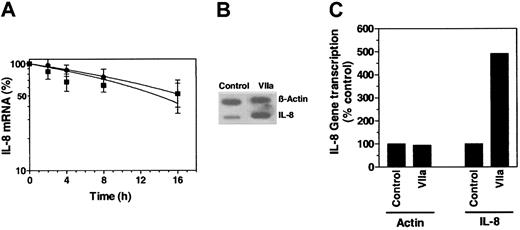
To investigate whether increased IL-8 mRNA steady-state levels observed in cells treated with FVIIa resulted from increased stabilization of IL-8 mRNA or from increased de novo transcription of the IL-8 gene, we evaluated IL-8 mRNA stability and IL-8 gene transcription in control cells and in cells treated with FVIIa. IL-8 mRNA stability was analyzed by arresting ongoing transcription of the IL-8 gene in control cells and in cells stimulated with FVIIa (for 75 minutes) with actinomycin D. Subsequently, IL-8 mRNA levels were determined at various time periods. The half-life of IL-8 mRNA in control cells (t½, approximately 14 hours) and in cells stimulated with FVIIa (t½, approximately 16.5 hours) appeared to be essentially similar (Figure 6A). Measuring ongoing IL-8 gene transcription by nuclear runoff analysis showed a 5-fold increase in IL-8 gene transcription in FVIIa-stimulated cells compared with nontreated cells (Figure 6B-C). Therefore, our results strongly indicated that the increased accumulation of IL-8 transcripts in FVIIa-treated cells was caused by de novo mRNA synthesis and not by stabilization of existing mRNAs.
Effect of FVIIa on IL-8 mRNA stability and gene transcription. (A) MDA-MB-231 cells were first treated with a control vehicle or FVIIa (50 nM) for 75 minutes. Then 10 μg/mL actinomycin D was added to inhibit RNA synthesis. Total RNA was harvested at indicated times after the addition of actinomycin D and was subjected to Northern blot analysis using IL-8 probe. IL-8 mRNA levels measured 10 minutes after the addition of actinomycin D were taken as 100% (mean ± SEM, n = 3). (B) Nuclei were isolated from unstimulated MDA-MB-231 cells or cells stimulated with FVIIa (50 nM) for 1 hour. Two identical blots containing β-actin and IL-8 DNAs were hybridized with equal amounts of labeled transcripts of nuclear RNA. (C) Quantitative representation of the data shown in panel B (mean values of 2 experiments).
Effect of FVIIa on IL-8 mRNA stability and gene transcription. (A) MDA-MB-231 cells were first treated with a control vehicle or FVIIa (50 nM) for 75 minutes. Then 10 μg/mL actinomycin D was added to inhibit RNA synthesis. Total RNA was harvested at indicated times after the addition of actinomycin D and was subjected to Northern blot analysis using IL-8 probe. IL-8 mRNA levels measured 10 minutes after the addition of actinomycin D were taken as 100% (mean ± SEM, n = 3). (B) Nuclei were isolated from unstimulated MDA-MB-231 cells or cells stimulated with FVIIa (50 nM) for 1 hour. Two identical blots containing β-actin and IL-8 DNAs were hybridized with equal amounts of labeled transcripts of nuclear RNA. (C) Quantitative representation of the data shown in panel B (mean values of 2 experiments).
FVIIa promotes tumor cell migration and invasion
To test whether FVIIa-induced IL-8 expression could play a role in tumor cell migration, we first evaluated the effect of FVIIa on MDA-MB-231 cell migration using a modified Boyden chamber. Adding FVIIa, at 10 and 50 nM concentrations, to the bottom well increased the number of cells that migrated across the membrane by 3- to 6-fold (Figure 7). Checkerboard analysis revealed that FVIIa must be added to the bottom well for migration to occur, indicating that FVIIa acts as a chemotactic for MDA-MB-231 cells and does not stimulate chemokinesis (data not shown). Including a 10-fold molar excess of FFR-FVIIa, which inhibited FVIIa binding to TF, markedly inhibited FVIIa-induced MDA-MB-231 cell chemotaxis (Figure 7A). Similarly, neutralizing antibodies against TF also attenuated FVIIa-induced cell migration (Figure 7A). In addition to FVIIa, trypsin and PAR-2 peptide agonist enhanced tumor cell migration (Figure 7B). However, the fold increase in cell migration observed with PAR-2 peptide and trypsin was consistently lower than that observed with FVIIa. In contrast to FVIIa, thrombin reduced the cell migration (Figure 7B). PAR-1 peptide agonist, FXa, and plasmin had no significant effect on tumor cell migration.
Effect of FVIIa and other agonists on cancer cell migration: involvement of PAR-2 and IL-8 in FVIIa-induced cell migration. MDA-MB-231 cells were placed in the upper well, and various concentrations of FVIIa (A) or other agonists (B) or FVIIa and antibodies against IL-8 or PAR-2 (C) were added to the lower well. In additional experiments, ASIS or anti-TF IgG was included with FVIIa in the lower well (A). The number of cells that migrated to the underside of the membrane in 20 hours at 37°C was determined as described in “Materials and methods” (mean ± SEM, n = 3 to 5). Concentrations of various reagents were: FVIIa, 50 nM (unless specified otherwise); ASIS, 500 nM; anti-TF IgG, 100 μg/mL; thrombin, 10 nM; trypsin, 10 nM; plasmin, 50 nM; PAR-1 AP, 50 μM; PAR-2 AP, 50 μM; rabbit antihuman IL-8 IgG, 50 μg/mL; rabbit anti–PAR-2 IgG, 500 μg/mL; control IgG, 50 μg/mL (1) or 500 μg/mL (2); and IL-8, 100 ng/mL. *Difference in value is statistically significant (P < .05) from the value obtained in control treatment (panel B) or corresponding control IgG (panel C).
Effect of FVIIa and other agonists on cancer cell migration: involvement of PAR-2 and IL-8 in FVIIa-induced cell migration. MDA-MB-231 cells were placed in the upper well, and various concentrations of FVIIa (A) or other agonists (B) or FVIIa and antibodies against IL-8 or PAR-2 (C) were added to the lower well. In additional experiments, ASIS or anti-TF IgG was included with FVIIa in the lower well (A). The number of cells that migrated to the underside of the membrane in 20 hours at 37°C was determined as described in “Materials and methods” (mean ± SEM, n = 3 to 5). Concentrations of various reagents were: FVIIa, 50 nM (unless specified otherwise); ASIS, 500 nM; anti-TF IgG, 100 μg/mL; thrombin, 10 nM; trypsin, 10 nM; plasmin, 50 nM; PAR-1 AP, 50 μM; PAR-2 AP, 50 μM; rabbit antihuman IL-8 IgG, 50 μg/mL; rabbit anti–PAR-2 IgG, 500 μg/mL; control IgG, 50 μg/mL (1) or 500 μg/mL (2); and IL-8, 100 ng/mL. *Difference in value is statistically significant (P < .05) from the value obtained in control treatment (panel B) or corresponding control IgG (panel C).
Next, we determined the role of FVIIa-induced IL-8 production in mediating the enhancing effect of FVIIa on tumor cell migration. Adding saturating concentrations of IL-8 (100 ng/mL) to the lower well increased cell migration by approximately 3- to 4-fold, indicating that IL-8 can promote MDA-MB-231 cell migration. Adding IL-8–with FVIIa to MDA-MB-231 cells did not further enhance FVIIa-induced cell migration (data not shown). More important, adding IL-8–neutralizing antibodies, along with FVIIa, to the lower well markedly inhibited FVIIa-induced cell migration (Figure 7C). These data strongly suggested that FVIIa-induced IL-8 expression leads to increased cell migration of these cancer cells. In contrast to FVIIa-induced cell migration, the basal cell migration observed in these cells appeared to be independent of IL-8 given that the IL-8 antibodies failed to inhibit basal migration (Figure 7C). Because the specific antibodies against PAR-2 were shown to inhibit FVIIa-induced IL-8 expression (Figure 4), we next examined whether PAR-2 antibodies also inhibit FVIIa-induced cell migration. As expected, PAR-2 antibodies fully attenuated FVIIa-induced cell migration. Adding IL-8 reversed the PAR-2 antibody blocking effect of FVIIa-stimulated cell migration.
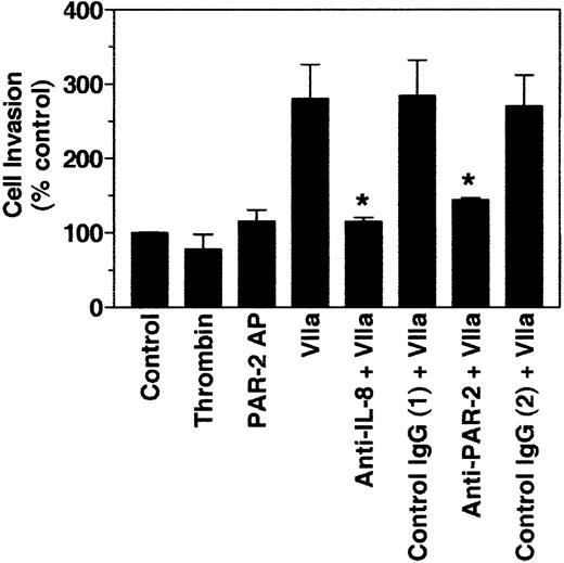
We also determined the effect of FVIIa on the invasion of MDA-MB-231 cells in an in vitro invasion assay in which the upper well of the transwell was layered with Matrigel (1.5-mm thickness) and cells were seeded on top. Including FVIIa in the bottom chamber significantly increased the invasion of MDA-MB-231 cells through the Matrigel. IL-8 and PAR-2 antibodies attenuated the FVIIa-induced invasion of cells (Figure 8). In contrast to FVIIa, thrombin had no significant effect on cell invasion, and PAR-2 peptide agonist minimally increased cell invasion.
Effect of FVIIa on tumor cell invasion. MDA-MB-231 cells were placed on top of a Matrigel barrier. FVIIa and other agonists were added to a lower well that contained NIH3T3 cell–conditioned media. At the end of a 48-hour incubation period at 37°C, the number of cells that migrated across the Matrigel barrier to the underside of the membrane was determined. Concentrations of various reagents used were: FVIIa, 50 nM; thrombin, 10 nM; PAR-2 AP, 50 μM; rabbit antihuman IL-8 IgG, 50 μg/mL; rabbit anti–PAR-2 IgG, 500 μg/mL; control IgG, 50 μg/mL (1) or 500 μg/Ml (2). Mean ± SEM, n = 3 to 5. *Inhibition was statistically significant.
Effect of FVIIa on tumor cell invasion. MDA-MB-231 cells were placed on top of a Matrigel barrier. FVIIa and other agonists were added to a lower well that contained NIH3T3 cell–conditioned media. At the end of a 48-hour incubation period at 37°C, the number of cells that migrated across the Matrigel barrier to the underside of the membrane was determined. Concentrations of various reagents used were: FVIIa, 50 nM; thrombin, 10 nM; PAR-2 AP, 50 μM; rabbit antihuman IL-8 IgG, 50 μg/mL; rabbit anti–PAR-2 IgG, 500 μg/mL; control IgG, 50 μg/mL (1) or 500 μg/Ml (2). Mean ± SEM, n = 3 to 5. *Inhibition was statistically significant.
Discussion
In the present study, we have used a breast carcinoma cell line (MDA-MB-231) with abundant TF expression to show that exposure of these cells to FVIIa leads to markedly increased IL-8 expression and cell migration/invasion. The FVIIa-induced effects are fully attenuated when FVIIa binding to TF is blocked either by FFR-FVIIa or by anti-TF antibodies. Further, FVIIa failed to up-regulate IL-8 expression in the MDA-MB-435 breast carcinoma cell line, which has little or no TF. The data indicate that TF-FVIIa proteolytic activity–dependent signaling is responsible for the increased expression of IL-8 and that TF expression at the cell surface is a primary determining factor for this process. TF expression, and not the plasma FVII/FVIIa concentration, is expected to be decisive for the pathophysiologic response, since previous studies have documented that TF functionality on cells is saturated at low concentrations of FVII/FVIIa54,55 and that FVII is converted to FVIIa when bound to TF on the cell surface.56,57
Earlier studies showed that stimulating keratinocytes with FVIIa26 or trypsin58 induced IL-8 secretion. In other studies, activating PAR-2 by agonist peptides and trypsin was shown to induce IL-8 expression in other cell types.59,60 Although it is generally believed that TF-FVIIa–induced signaling is mediated through activation of a PAR, it is unclear whether a known PAR (PAR-1, PAR-2, PAR-3, and PAR-4) or a putative PAR is responsible for TF-FVIIa–induced signaling. Because of a lack of specific inhibitors or neutralizing antibodies to known PARs, earlier studies relied on heterologous desensitization or transfection studies to address whether TF-FVIIa–induced signaling involves the activation of a known PAR, particularly PAR-1 or PAR-2. Heterologous desensitization studies with PAR peptide agonists, thrombin, and trypsin indicated that activation of a known PAR may not be responsible for TF-FVIIa–induced signaling.23,24,48-50 Consistent with this, FVIIa failed to elicit signaling in Chinese hamster ovary (CHO) cells transfected with PAR-2 and TF.24 In contrast to these data, FVIIa was shown to induce p44/42 mitogen–activated protein kinase (MAPK) phosphorylation in CHO cells transfected with TF and PAR-253 and to trigger Ca2+ release in Xenopus oocytes transfected with TF plus PAR-1 or PAR-2.51 The latter study also showed that transfection of PAR-2 into lung fibroblasts derived from PAR-1 knockout mice resulted in responsiveness to FVIIa as evidenced by intracellular Ca2+ release and phosphoinositol-3 hydrolysis.51 Although these data suggest that TF-FVIIa could activate PAR-2 (and, to a lesser degree, PAR-1), they do not allow for a firm conclusion about whether TF-FVIIa–induced gene expression in cells that constitutively express TF is mediated by PAR-2. The data presented in this manuscript show that neutralizing antibodies against PAR-2, but not against PAR-1, markedly inhibited TF-FVIIa–induced IL-8 expression in MDA-MB-231 cells, indicating that TF-FVIIa–induced IL-8 expression is mediated by PAR-2. We believe this is the first report that documents the involvement of PAR-2 in TF-FVIIa–induced gene expression in cells that constitutively express TF. It remains to be unraveled why the activation of PAR-2 by TF-FVIIa, in contrast to the activation by PAR-2 agonist peptide (and trypsin), fails to give rise to Ca2+ mobilization.
In general, PAR-1 and PAR-2 activation produce similar secondary messages, such as Ca2+ mobilization, phosphoinositide hydrolysis, activation of MAPKs, and transcriptional activation of immediate-early genes.61 Recent large-scale gene expression profiling of endothelial cells with PAR-1 and PAR-2 agonists showed that activating PAR-1 and PAR-2 results in an almost identical alteration in gene expression profile.62 Consistent with this, TF-FVIIa, thrombin, FXa, and plasmin were shown to up-regulate the expression of Cyr61 in fibroblasts.23,63,64 In contrast, none of the coagulation proteases, except FVIIa, were effective in up-regulating IL-8 expression in MDA-MB-231 cells. Lack of PAR-1 expression could not be a reason for the ineffectiveness of other proteases because flow cytometry and functional activity assays confirm that MDA-MB-231 cells express PAR-1 and PAR-2 (Figure 3).65 Because thrombin, FXa, activated protein C, and plasmin transmit their signals primarily through the activation of PAR-1, the data suggest that activation of PAR-1 plays no role in IL-8 expression in these cells. Recent reports show that FXa can also activate PAR-2.66,67 However, in contrast to FVIIa, FXa had no significant effect on IL-8 induction in MDA-MB-231 cells. A probable explanation for this discrepancy could be the absence of a specific cellular receptor for FXa on MDA-MB-231 cells that may be essential for docking FXa to gain access to PAR-2. Effector cell protease receptor-1 (EPR-1) has been shown to play a role in localizing FXa in proximity to PAR-2 in endothelial cells.66 Even when the receptor is present, FXa appears to be a poor activator of PAR-2, because 100 to 1000 nM concentration of FXa is required for robust activation of PAR-2 in endothelial cells.66,67
Work from several laboratories, including our own, shows that the TF-FVIIa complex, independent of other downstream proteases, activates typical G-protein–coupled receptor pathways (for a review, see Pendurthi and Rao20 ). However, recent studies by Riewald and Ruf53 suggest that the ternary complex of TF-FVIIa-FXa, and not the binary complex of TF-FVIIa, efficiently activates PAR-1 and PAR-2. It was shown that the activation of substrate FX by the TF-FVIIa complex produces enhanced cell signaling in comparison with the TF-FVIIa complex alone or with free FXa. The same investigators, in a more recent study with human keratinocytes (HaCaT), showed that even a supraphysiologic concentration of FVIIa (50 nM) failed to induce PAR-2–mediated TR3 gene induction, whereas initiation of coagulation at near-plasma concentrations of FVIIa and FX effectively induced expression of this gene in HaCaTs.21 In contrast to these recent reports, our present data provide convincing evidence that a TF-FVIIa binary complex, even at plasma concentrations of FVII or FVIIa, is sufficient to induce prominent PAR-2–mediated signaling in MDA-MB-231 breast carcinoma cells. Moreover, adding FX at plasma concentrations did not further enhance TF-FVIIa–induced IL-8 expression when FVIIa was used at 10 nM. However, when FVIIa was limited, adding FX promoted IL-8 gene expression, indicating that the TF-FVIIa–FXa ternary complex may be more efficient than TF-FVIIa in inducing IL-8 gene expression. We do not know the reason for the discrepancy between the present findings and those of earlier studies (at saturating concentrations of FVIIa), but differences in cell type and functional readouts used to evaluate PAR activation could be contributing factors. Recent studies with FVIIa, FXa, and plasmin suggest that the capability of these proteases to induce PAR-mediated cell signaling depends not only on the expression of PARs but also on additional expression of cellular receptors that support their binding and proteolytic activity. Because of the restricted mobility of a protease bound to its cellular receptor, it can only activate PARs in spatial proximity to the receptor. Therefore, in addition to expressing PARs and specific protease receptors, their spatial localization on the membrane determines whether a particular protease can activate a PAR. This might explain why FVIIa failed to induce signaling in cells that express TF, PAR-1, and PAR-2.21,24
Similar to our present observation in carcinoma cells, a recent study also showed FVIIa-induced up-regulation of IL-8 expression in a human keratinocyte cell line.26 However, the mechanism by which FVIIa up-regulates IL-8 expression in keratinocytes and breast carcinoma cells appears to differ. In the earlier study,26 TF-FVIIa was shown to up-regulate IL-8 primarily through IL-8 mRNA stabilization. FVIIa treatment prolonged the half-life of IL-8 transcripts from 40 minutes to 140 minutes, whereas transcriptional activity was increased only minimally (15%-80%). In contrast to short-lived IL-8 transcripts in keratinocytes, IL-8 transcripts in carcinoma cells have a relatively longer half-life (t½, 14 hours in unstimulated cells), and FVIIa treatment had no significant effect on IL-8 mRNA stabilization (t½, 16.5 hours). More important, FVIIa increased the transcriptional activation of IL-8 in carcinoma cells by approximately 5-fold, and a similar fold increase of IL-8 mRNA steady-state levels was noted. It is possible that the TF-FVIIa-induced increase in IL-8 expression in keratinocytes could be the result of TF-FVIIa–induced transcriptional activation of early genes that encode mRNA stabilizing proteins or phosphorylation of these proteins. It is unclear which PAR mediated TF-FVIIa–induced IL-8 expression in keratinocytes because no data were given on this aspect.26
Tumor cell proliferation and migration, in addition to angiogenesis, are pivotal steps in the intricate process of tumor growth and metastasis. IL-8 produced locally by tumor cells was shown to stimulate angiogenesis and tumor invasion in human prostate cancer, thereby increasing tumorigenicity and metastasis.68 A number of studies showed that IL-8 can stimulate cell migration and invasion in a variety of tumor cell types, including breast carcinoma cells, in autocrine and paracrine fashions.34-37,68,69 IL-8, similar to TF, is thought to play a role in angiogenesis and tumor metastasis.46 Furthermore, TF expression8-10 and IL-8 production28-30 correlate strongly with metastatic potential in many tumors. Therefore, it is likely that some of the pathophysiologic functions of TF are mediated by TF-FVIIa–induced IL-8 expression. It is, however, of interest to note that other cancer cell lines, including U87 and U373 brain glioblastoma cells, despite functional PAR-1 and PAR-2, respond with increased IL-8 production on stimulation with thrombin and not with FVIIa (results not shown). This suggests that in addition to the receptors expressed on the surface of the cell, different intracellular signaling pathways in different cell types are also important for the final expression levels of many genes, including IL-8. Therefore, it remains to be seen how the different response patterns are orchestrated in various carcinomas.
Similar to our present findings in tumor cells, earlier studies showed that TF-FVIIa can induce or enhance migration of vascular smooth muscle cells (VSMCs)70 and fibroblasts.71 However, it is unclear from these data whether FVIIa-induced cell migration involves FVIIa-induced expression of IL-8 in these cells. In addition, a number of differences exist between the present findings and those of earlier studies. In fibroblasts, FVIIa alone had no effect on cell migration, but it enhanced platelet-derived growth factor (PDGF)–BB–stimulated cell migration.71 No information presented in that study71 indicated whether the FVIIa-induced effect is mediated by PAR-2. Although PAR-2 was shown to be responsible for TF-FVIIa–induced cell migration in VSMCs,70 earlier studies from the same group indicated that TF can induce cell migration in VSMCs independent of its coagulant activity and FVIIa.72 Further, these studies were carried out with exogenously added TF, which raises a valid question regarding whether TF expressed in VSMCs, either constitutively or induced by pathophysiologic stimuli, could induce cell migration.
Although activation of PAR-2 by TF-FVIIa and by PAR-2 peptide agonist induced similar levels of IL-8 mRNA and antigen, we consistently observed that PAR-2 peptide agonist was less effective than FVIIa in promoting cell migration or invasion. These data raise the possibility that TF-FVIIa may induce an additional component that could complement IL-8–mediated cell migration. In contrast to FVIIa, thrombin had no effect on the migration of MDA-MB-231 breast carcinoma cells. In fact, thrombin appeared to inhibit the basal migration of these cells. These data are consistent with a recent report that showed activation of PAR-1 by thrombin inhibited the migration and invasion of MDA-MB-231 cells.65 Recent studies showed that PAR-1 and PAR-2 differ in coupling to Rho and Rac, and this differential regulation of the Rho/Rac pathway is thought to be responsible for differential actions of PAR-1 and PAR-2 in stimulating endothelial cell permeability.73,74 Therefore, it is possible that the differential effect of PAR-1 and PAR-2 on MDA-MB-231 cell migration could result from differential regulation of Rho-GTPases or other distinct cytoskeletal responses induced by the activation of PAR-1 and PAR-2. However, Even-Ram et al75 showed that the activation of PAR-1 promotes the invasiveness of melanoma cells and that PAR-1 mediates these functions through selective cross-talk with αvβ5 integrin to confer a focal adhesion complex. At present it is unknown whether cross-talk exists between PAR-2 and αvβ5 integrin and whether it plays a role in TF-FVIIa–induced tumor cell invasion. Overall, our present findings coupled with earlier observations suggest that selective stimulation of PARs could lead to different migratory outcomes, depending on tumor cell types.
Recent studies show that FVIIa acts as an antiapoptotic agent in BHK cells transfected with TF76 and enhances cell proliferation in smooth muscle cells,77 and a valid question arises regarding whether increased cell migration observed in FVIIa-stimulated cells in the present study could have been the indirect effect of increased cell survival in FVIIa-stimulated cells. However, this is unlikely. In contrast to BHK-TF cells, serum deprivation did not induce apoptosis in MDA-MB-231 cells (analyzed by caspase-3 activation and lactate dehydrogenase [LDH] release assays), and FVIIa had no effect on proliferation of these cells, as measured in 3H-thymidine incorporation (data not shown).
In summary, our present data show that FVIIa binding to TF on breast carcinoma cells up-regulates the expression of IL-8 through a PAR-2–dependent mechanism, and the increased expression of IL-8 leads to increased cell migration and invasion. These cellular events could explain how TF plays a role in tumor metastasis. Further work is needed to determine whether this is a general mechanism or is restricted to specific tumor cell types. The present data raise the possibility that therapeutics that prevent TF-FVIIa complex formation and its protease activity may be useful not only for managing thrombotic complications associated with malignancy but also for preventing tumor growth and dissemination.
Supported by National Institutes of Health grant HL65500 (U.R.P.).
Several of the authors (G.M.H., L.C.P., T.A., B.B.S., P.L.N.) are employed by Novo Nordisk, Denmark, whose product and potential product were studied in the present work.
The publication costs of this article were defrayed in part by page charge payment. Therefore, and solely to indicate this fact, this article is hereby marked “advertisement” in accordance with 18 U.S.C. section 1734.
Prepublished online as Blood First Edition Paper, January 8, 2004; DOI 10.1182/blood-2003-10-3417.
We thank Mylinh Ngyuen, Elke Gottfriedsen, Berit Lassen, and Lone Langhoff for their skilled technical assistance.



This feature is available to Subscribers Only
Sign In or Create an Account Close Modal