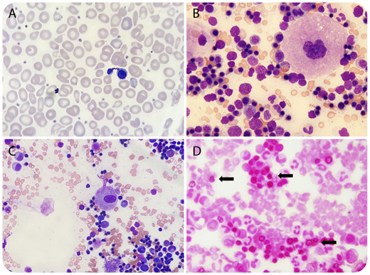
Two unrelated males experiencing microcytic anemia since birth presented for bone marrow examination. Their peripheral blood smears demonstrated anisocytosis and nucleated red blood cells (panel A; original magnification ×1000, Wright-Giemsa stain) with normal platelet counts. Their bone marrow aspirate smears showed abnormal megakaryocytes consisting of both hypolobated (panel B, 10-month-old patient; original magnification ×1000, Wright-Giemsa stain) and monolobated (panel C, 22-month-old patient; original magnification ×400, Wright-Giemsa stain) forms with mature cytoplasm and chromatin. Iron stain demonstrated numerous ring sideroblasts (panel D; original magnification ×1000). Both bone marrow core biopsies were normocellular with morphologically abnormal megakaryocytes present in normal numbers and normal distribution. Cytogenetic studies revealed normal male karyotypes. The patients were found to have congenital sideroblastic anemia, B-cell immunodeficiency, periodic fevers, developmental delay (SIFD), along with biallelic TRNT1 mutations.
TRNT1 encodes a ubiquitously expressed enzyme required for nuclear and mitochondrial transfer RNA function, attesting to its multisystemic roles and the varying clinical phenotypes of SIFD patients. Although platelet counts and functions are not affected to date, an alteration in hematopoiesis beyond erythroid lineage, including megakaryopoiesis, appears to be biologically plausible. Examination of additional cases with longer follow-up will be helpful to further evaluate the prevalence and significance of morphologically abnormal megakaryocytes in SIFD patients.
For additional images, visit the ASH Image Bank, a reference and teaching tool that is continually updated with new atlas and case study images. For more information, visit http://imagebank.hematology.org.


