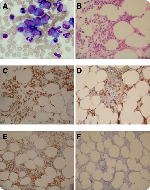
A 64-year-old woman presented to our institution for increasing neck pain without headache, dizziness, or blurred vision. Magnetic resonance imaging revealed an osteolytic lesion at the C6 vertebra with a compression fracture, as well as lymphadenopathy around the upper middle esophagus. She underwent C6 vertebral resection and local reconstruction, and the pathological findings indicated plasmacytoma with expression of CD20, CD79a, VS38C, CD138, and κ light chain. Serum immunofixation electrophoresis was positive for immunoglobulin G (IgG)-κ paraprotein with serum IgG4 surging to an astounding 4.88 g/dL. No anemia or renal insufficiency was found. Bone marrow smear (panel A) and biopsy (panel B) showed accumulation of mature and immature plasma cells up to 28.5%. Immunohistochemical study revealed membrane staining of CD138 (panel C) as well as cytoplasmic staining of IgG4 (panel D) among myeloma cells, which demonstrated κ (panel E) instead of λ (panel F) light chain restriction.
IgG4-related diseases (IgG4-RD) are characterized by chronic fibro-inflammatory lesions affecting multiple organs, which might be misinterpreted as neoplasm. Meanwhile, plasma cell neoplasms with monoclonal IgG4 production have seldom been described. The accumulation and neoplastic transformation of IgG4 secreting plasma cells in this patient seem not sufficient to induce any clinical manifestations mimicking IgG4-RD, which suggests that elevated IgG4 alone is not diagnostic for IgG4-RD.
For additional images, visit the ASH IMAGE BANK, a reference and teaching tool that is continually updated with new atlas and case study images. For more information visit http://imagebank.hematology.org.


