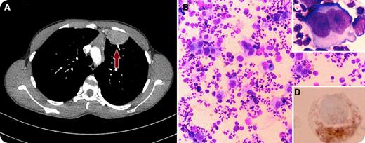
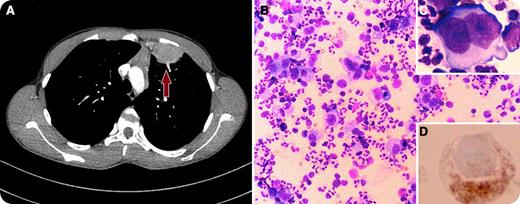
A 15-year-old young man presented to the emergency department with a 3-week history of sharp left-sided chest pain, shortness of breath on exertion, and a low-grade intermittent fever. Chest radiograph revealed increased density in the upper left lobe, thus he was treated with antibiotics for query pneumonia. Although his fever subsided, the patient continued to have chest pain, and subsequently developed night sweats and fatigue. Chest computed tomography revealed complex soft tissue, mostly located in his left lung, and minimally enlarged mediastinal lymph nodes (panel A). The differential diagnosis included sarcoma, congenital cyst, and lymphoma. The suspicious lung mass was resected for pathology. The cytospin results showed numerous large atypical cells in a background of neutrophils, eosinophils, lymphocytes, and histiocytes (panel B). The atypical cells often had abundant cytoplasm, and the nuclei often contained prominent nucleoli. Typical Reed-Sternberg cells are noted with a bi-lobed nucleus, giving the cell a typical “owl’s eyes” appearance (panel C). Cells showed CD30 positivity with immunohistochemistry stains (panel D).
A diagnosis of nodular sclerosis Hodgkin lymphoma stage IIB was made. The patient was started on adriamycin-bleomycin-vinblastine-dacarbazine combination chemotherapy, with favorable response. Primary pulmonary Hodgkin lymphoma is a very rare entity whose prognosis depends on its stage.
For additional images, visit the ASH IMAGE BANK, a reference and teaching tool that is continually updated with new atlas and case study images. For more information visit http://imagebank.hematology.org.


