Abstract
Immune thrombocytopenia induced by quinine and many other drugs is caused by antibodies that bind to platelet membrane glycoproteins (GPs) only when the sensitizing drug is present in soluble form. In this disorder, drug promotes antibody binding to its target without linking covalently to either of the reacting macro-molecules by a mechanism that has not yet been defined. How drug provides the stimulus for production of such antibodies is also unknown. We studied 7 patients who experienced severe thrombocytopenia after ingestion of quinine. As expected, drug-dependent, platelet-reactive antibodies specific for GPIIb/IIIa or GPIb/IX were identified in each case. Unexpectedly, each of 6 patients with GPIIb/IIIa-specific antibodies was found to have a second antibody specific for drug alone that was not platelet reactive. Despite recognizing different targets, the 2 types of antibody were identical in requiring quinine or desmethoxy-quinine (cinchonidine) for reactivity and in failing to react with other structural analogues of quinine. On the basis of these findings and previous observations, a model is proposed to explain drug-dependent binding of antibodies to cellular targets. In addition to having implications for pathogenesis, drug-specific antibodies may provide a surrogate measure of drug sensitivity in patients with drug-induced immune cytopenia.
Introduction
Drug-induced immune thrombocytopenia (DITP) is a relatively common, sometimes life-threatening disorder, characterized by drug-dependent antibodies (DDAbs) that bind to platelets and cause their destruction when the responsible drug is ingested or injected.1,2 Less often, drug-induced antibodies target erythrocytes3 or neutrophils4,5 and cause immune hemolytic anemia or neutropenia. Quinine, usually taken to prevent nocturnal leg cramps, is a common cause of DITP, but many other medications, including antibiotics, nonsteroidal anti-inflammatory drugs, sedatives, and anticonvulsants have been implicated.6,7 DITP induced by quinine and other drugs (with the exception of heparin) is typically caused by an unusual type of antibody that is nonreactive in the absence of drug but binds tightly to specific epitopes on platelet membrane glycoproteins (GPs) IIb/IIIa8,9 or Ib/IX9-11 when the sensitizing drug is present in soluble form.
It is generally thought that low-molecular-weight compounds are immunogenic only when linked covalently to a carrier protein, in which state they can induce the formation of drug (hapten)–specific antibodies.12,13 Accordingly, early studies to define the pathogenesis of DITP considered the possibility that the drug became coupled in vivo to certain membrane proteins that were then recognized as “foreign” by the immune system, leading to the production of drug-specific antibodies. On subsequent administration, the drug could then reassociate with the membrane protein to create a target for antibody, leading to blood cell destruction.14 This mechanism may explain episodes of immune hemolysis in some patients given large doses of penicillin or penicillin derivatives, because antibiotics containing a β-lactam ring can link spontaneously to free amino groups on proteins.15,16 Hapten-specific antibodies have never been convincingly demonstrated in patients with DITP, however.2,7 For some time, a second possible mechanism was considered: that antibodies causing DITP might react directly with soluble drug to form immune complexes that somehow reacted with platelets as “innocent bystanders” to cause their destruction.17,18 However, the putative immune complexes were never demonstrated experimentally and it was later shown that DDAbs bind to platelets via their Fab rather than Fc domains as would be expected of immune complexes.19,20
Failure of these mechanisms to explain binding of DDAbs to their targets requires that other mechanisms be considered to explain this interaction, mechanisms that take into account observations showing that DDAb binding occurs without covalent linkage of drug to the target and that the reaction is not inhibited by excess soluble drug at the highest concentrations that can be achieved in vitro.2,21 One possibility consistent with these facts is that binding of drug to a target GP induces a structural modification elsewhere in the molecule for which the antibody is specific.2,7 A second is that the DDAb recognizes the drug itself in the context of its binding site; that is, the antibody may interact both with a structural element of the drug and with adjacent peptide sequences.21,22 The latter possibility is supported by studies done with quinine- and quinidine-dependent antibodies showing that drug is “trapped” at the antigen-antibody interface when antibody binds23 and that seemingly minor alterations in drug structure can abolish immunologic reactivity.24 Yet a third possibility is that the drug may react first with the antibody itself and modify it in such a way that it acquires specificity for an epitope on a membrane GP. Evidence for and against these and other mechanisms proposed to explain drug-dependent binding of antibodies to cellular targets has been reviewed in detail in several publications.3,21,22,25,26
In this report, we show that 6 of 7 patients with quinine-induced immune thrombocytopenia had a previously undescribed type of antibody (drug-specific) that directly recognizes quinine itself and is distinct from the platelet-binding antibodies thought to be responsible for platelet destruction. Although these “drug-specific” antibodies may not be capable of causing thrombocytopenia, they appear to provide clues to how pathogenic antibodies are induced and how the sensitizing drug promotes binding of antibody to its target. Additionally “drug-specific” antibodies may act as surrogate markers for drug sensitization.
Materials and methods
Source of DDAbs
Antibodies studied were from 7 patients who developed acute, severe (platelet count < 10 × 109/L) thrombocytopenia within 6 to 24 hours of ingesting quinine for prevention or treatment of nocturnal leg cramps. In each case, there had been intermittent exposure to quinine for several months prior to the thrombocytopenic episode. One patient (no. 2) had severe neutropenia (< 0.1 × 10⁹/L) in addition to thrombocytopenia and 2 patients (nos. 5 and 6) had accompanying hemolytic uremic syndrome27 requiring hemodialysis. All 7 patients eventually achieved a stable platelet count in the normal range after drug was discontinued. Approval for these studies was obtained from the BloodCenter of Wisconsin's institutional review board. Informed consent was provided according to the Declaration of Helsinki.
Reagents
Quinine, quinine hydrochloride, succinic anhydride, 6-methoxyquinolone, and other quinine analogues were from Aldrich Chemical (Milwaukee, WI). Human serum albumin (HSA), bovine serum albumin (BSA), monoclonal antibody (mAb) HSA-11, and Tween-20 were from Sigma Chemical (St Louis, MO). Alkaline phosphatase, horseradish peroxidase (HRP), and fluorescein (FITC)–conjugated, affinity-purified F(ab′)2 goat anti–human IgG (H&L) were from Jackson ImmunoResearch Laboratory (West Grove, PA). Antiquinine antiserum (rabbit) was from Biodesign (Kennebunk, ME). The mAbs were AP1 (anti-GPIb) from Dr Robert Montgomery (BloodCenter of Wisconsin, Milwaukee, WI); AP2 (anti-GPIIb/IIIa complex) and AP6 (anti-GPIIIa) from Dr Thomas Kunicki (Scripps Medical Research Institute, La Jolla, CA); AP3 (anti-GPIIIa) from Dr Peter Newman (BloodCenter of Wisconsin); and MBC 142.11 (anti-GPIb) from the Monoclonal Antibody Laboratory of the BloodCenter of Wisconsin.
Detection and characterization of quinine-dependent antibodies
Drug-dependent binding of patient antibodies to intact platelets was detected by flow cytometry as described previously.28,29 For maximum sensitivity, it was essential that quinine (0.4 mM) be present not only in the primary reaction mixture, but also in all subsequent washing steps prior to adding the FITC-labeled F(ab′)2 anti-immunoglobulin reagent used to detect immunoglobulin binding to platelets.11,28 Specificity of antibodies for the GPIIb/IIIa or GPIb/IX complexes was characterized by the modified antigen capture enzyme-linked immunosorbent (MACE) assay.11,30 In brief, platelets were sensitized by antibody in the presence of 0.4 mM quinine and washed in phosphate-buffered saline (PBS) containing drug. The sensitized platelets were then lysed in Triton X-100 detergent and solubilized GPs were captured in microtiter wells by immobilized mAbs AP3 (anti-GPIIIa), AP2 (anti-GPIIbIIIa), AP1 (anti-GPIb), and 142.11 (anti-GPIb). Human immunoglobulin bound to the captured GPs was then detected by enzyme-linked immunosorbent assay (ELISA).
Absorption of serum and preparation of antibody-containing eluates
Washed platelets (3 × 10⁹) were suspended in 1 mL patient serum and 1 mL 0.8 mM quinine. After 1 hour, the platelets were pelleted and the supernatant was removed and saved. The platelets were then washed 3 times with buffer containing 0.4 mM quinine and resuspended in 1 mL isotonic NaCl. The pH was adjusted to 3.0 with HCl. After 5 minutes, the platelets were pelleted and the supernatant containing eluted antibody was neutralized with 0.5 M Tris buffer, pH 8.8. Serum remaining from the first absorption was absorbed once more with 3 × 10⁹ platelets to remove remaining antibody. Both the eluate and the adsorbed serum were dialyzed 3 times against 5 L isotonic PBS, pH 7.2, to remove quinine.
Conjugation of quinine to HSA
Quinine was covalently linked to human serum albumin (Qn-HSA) via quinine hemisuccinate as described previously for linkage of quinine to hemocyanine.31,32 Quinine hemisuccinate was combined with sulfo-NHS and 1-ethyl-3-(3-dimethylaminopropyl)carbodiimide (EDAC) in PBS, pH 5.0, for 2 hours. This material was then added to HSA in PBS, pH 8.0, and allowed to mix overnight. The mixture was dialyzed against PBS with 3 exchanges of buffer and was concentrated. High-performance liquid chromatography (HPLC) analysis for quinine following chemical cleavage of the quinine succinate bond and analysis by a bicinchoninic acid protein analysis (BCA) indicated that the molar ratio of Qn to HSA was about 13:1.
Western blot analysis
Western blot analysis was performed as previously described.33 Briefly, platelets lysed in 1% Triton X-100 or Qn-HSA were subjected to SDS electrophoresis in 8% polyacrylamide and transferred to a polyvinylidene fluoride (PVDF) membrane. The membrane was blocked overnight with buffer containing 3% BSA, 0.05% Tween 20, 150 mM NaCl, and 20 mM Tris, pH 7.6 (blocking buffer). Antibody-containing plasma was diluted 1:280 in blocking buffer with or without quinine (0.4 mM) and incubated with the membrane for 3.5 hours at room temperature. The membranes were then washed 4 times with Tris-buffered saline/Tween 20 (TBS/Tw) with or without quinine. Bound antibody was detected with HRP-labeled goat anti–human IgG (H&L) diluted 1:5000 in TBS/Tw with or without Qn followed by visualization with enhanced chemiluminescence (ECL) reagent (Amersham, Arlington Heights, IL).
Results
Characterization of quinine-dependent antibodies
Each patient had strong IgG antibodies (titer, 1:400-1:16 000) that reacted with normal platelets in the presence but not in the absence of quinine using flow cytometry to detect antibody binding. Although reactions were strongest with quinine concentrations of 0.4 mM or higher, each antibody could be detected using quinine at subpharmacologic concentrations (10 μM). With the MACE assay, it was found that 6 of the 7 patients had antibodies recognizing both GPIIb/IIIa and GPIb/IX (data not shown). The antibody from patient 4 recognized only the GPIb/IX complex. Previous studies have shown that reactions with different GP complexes are mediated by separate antibodies.11
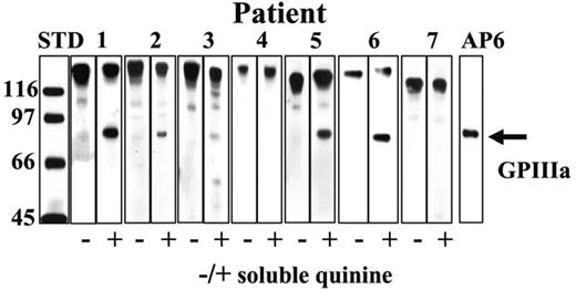
Patient samples contain drug-dependent antibodies that immunoblot GPIIIa in the presence of quinine. Patients 1, 2, 3, 5, and 6 had IgG antibodies that recognized a protein corresponding to GPIIIa in the presence but not in the absence of 0.4 mM quinine (immunoblot against whole platelet lysate). GPIIIa is marked with monoclonal AP6 specific for amino acid residues 211-221. The prominent high-molecular-weight bands correspond to naturally occurring platelet IgG.
Patient samples contain drug-dependent antibodies that immunoblot GPIIIa in the presence of quinine. Patients 1, 2, 3, 5, and 6 had IgG antibodies that recognized a protein corresponding to GPIIIa in the presence but not in the absence of 0.4 mM quinine (immunoblot against whole platelet lysate). GPIIIa is marked with monoclonal AP6 specific for amino acid residues 211-221. The prominent high-molecular-weight bands correspond to naturally occurring platelet IgG.
Five of the 6 GPIIb/IIIa-specific antibodies recognized GPIIIa in immunoblots in the presence but not in the absence of soluble quinine
Some quinine- and quinidine-dependent antibodies react with platelet GPs in immunoblotting assays performed in the presence of drug.33,34 As shown in Figure 1, serum from 5 of the 7 patients contained antibodies that recognized a band corresponding to GPIIIa in the presence but not in the absence of drug. The exceptions were patient 7, whose antibody failed to react detectably with GPIIIa in immunoblotting, and patient 4 whose antibody recognized GPIb/IX only. Although all 7 patients had drug-dependent antibodies specific for GPIb/IX, no drug-dependent reactions against either of those proteins were detected in immunoblotting.
The patients also had antibodies that recognized Qn-HSA and were inhibited by soluble quinine
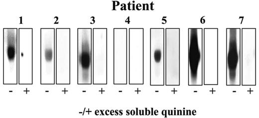
As expected, sheep antiquinine antibody reacted strongly with Qn-HSA but not unmodified HSA in immunoblots and this reaction was almost totally inhibited by preincubation of the antibody with soluble quinine but not quinidine (Figure 2). All 6 patients with GPIIb/IIIa-specific antibodies (patients 1-3 and 5-7) had antibodies that behaved similarly to the sheep antibody in that they recognized Qn-HSA and were inhibited by preincubation with soluble drug (Figure 3). Antibodies reactive with Qn-HSA were not detected in any of 17 serum samples from healthy individuals not taking quinine or in 7 samples from healthy persons who had taken quinine 325 mg daily for 10 days (data not shown).
Characterization of quinine-conjugated HSA. (Left) A quinine-specific sheep antibody reacts with Qn-HSA but not unmodified HSA in an immunoblot and is inhibited by soluble quinine 0.4 mM. (Right) Reactions of HSA and Qn-HSA with the HSA-specific monoclonal HSA-11.
Characterization of quinine-conjugated HSA. (Left) A quinine-specific sheep antibody reacts with Qn-HSA but not unmodified HSA in an immunoblot and is inhibited by soluble quinine 0.4 mM. (Right) Reactions of HSA and Qn-HSA with the HSA-specific monoclonal HSA-11.
Patient samples contain drug-specific antibodies that recognize Qn-HSA. Patients 1, 2, 3, 5, 6, and 7 had antibodies that recognized Qn-HSA in an immunoblot and were inhibited by soluble quinine 0.4 mM. None of the antibodies recognized unmodified HSA (not shown).
Patient samples contain drug-specific antibodies that recognize Qn-HSA. Patients 1, 2, 3, 5, 6, and 7 had antibodies that recognized Qn-HSA in an immunoblot and were inhibited by soluble quinine 0.4 mM. None of the antibodies recognized unmodified HSA (not shown).
“Drug-dependent” and “drug-specific” activities reside in different antibodies
Antibodies from patients 1, 2, 3, 5, and 6 were available in quantities sufficient for absorption and elution studies. These sera were absorbed with platelets in the presence of drug and washed. Platelet-bound antibody was then eluted as described in “Materials and methods.” After dialysis to remove quinine, the absorbed sera and the eluates were tested for their ability to react with intact platelets, GPIIIa, and Qn-HSA. As shown in Figure 4 (left panel), the platelet eluate prepared from serum of patient 6 reacted with intact platelets in the presence of soluble drug but the absorbed serum did not. In contrast, the absorbed serum recognized Qn-HSA but the eluate did not (right panel). Reactions of the absorbed sera and the eluates against GPIIIa in immunoblotting (not shown) were identical to those obtained with intact platelets. An identical pattern of reactions was obtained with absorbed serum and eluates from patients 1, 2, 3, and 5.
These findings demonstrate that at least 5 of the 7 patients had 2 distinct types of antibody: one that recognizes intact platelets and GPIIIa only in the presence of soluble drug and one reactive with Qn-HSA that is inhibited by soluble drug. In the “Discussion,” we will refer to the former antibodies as “platelet-reactive” and the latter as “drug-specific.”
Both types of antibody recognized quinine and desmethoxy-quinine (cinchonidine) but not other structural analogues of quinine
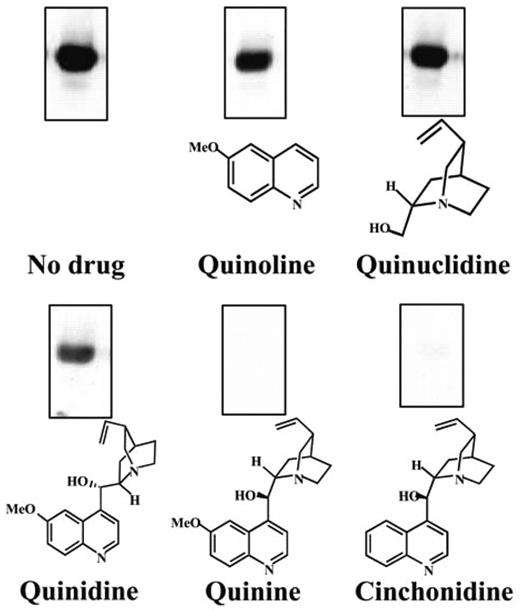
Several structural analogues of quinine were tested for their ability to promote binding of antibodies from patients 1, 2, 3, 5, and 6 to platelets and to inhibit their binding to Qn-HSA. Desmethoxyquinine (cinchonidine) was as effective as quinine in inhibiting the binding of each of the drug-specific antibodies to Qn-HSA but the other compounds, including quinuclidine and quinoline added together or separately, were completely inactive as shown for patient 6 in Figure 5. Exactly the same pattern of reactions was seen when these agents were tested for their ability to promote binding of the platelet-reactive antibodies to intact platelets (not shown). Quinine contains 2 stereoisomeric carbon atoms, C8 and C9, which comprise a bridge connecting 2 cyclic structures: quinuclidine and quinoline (Figure 5). These findings indicate that both platelet-reactive and drug-specific antibodies require the 2 stereoisomeric carbon atoms, C8 and C9, to be in a conformation specific for quinine and for the quinoline (or desmethoxy-quinoline) and quinuclidine groups to be coupled to react efficiently with their targets.
Demonstration of 2 distinct antibody populations in serum from patient 6. (Left) An eluate prepared from platelets following incubation with antibody reacted with platelets in the presence of drug. No detectable platelet-reactive antibody remained in the absorbed serum. (Right) The absorbed serum, but not the eluate, reacted with Qn-HSA. The eluted antibody failed to react with platelets in the absence of drug and the reaction of the absorbed serum with Qn-HSA was completely inhibited by soluble quinine (not shown).
Demonstration of 2 distinct antibody populations in serum from patient 6. (Left) An eluate prepared from platelets following incubation with antibody reacted with platelets in the presence of drug. No detectable platelet-reactive antibody remained in the absorbed serum. (Right) The absorbed serum, but not the eluate, reacted with Qn-HSA. The eluted antibody failed to react with platelets in the absence of drug and the reaction of the absorbed serum with Qn-HSA was completely inhibited by soluble quinine (not shown).
Inhibition of drug-specific antibody binding to Qn-HSA by quinine analogues. Reactions of serum from patient 6 with Qn-HSA were inhibited by quinine and cinchonidine (desmethoxy-quinine) but not by quinidine, quinoline, or quinuclidine.
Inhibition of drug-specific antibody binding to Qn-HSA by quinine analogues. Reactions of serum from patient 6 with Qn-HSA were inhibited by quinine and cinchonidine (desmethoxy-quinine) but not by quinidine, quinoline, or quinuclidine.
Discussion
Quinine-dependent, platelet-reactive antibodies identified in the 7 patients studied were typical of those previously described9,11 in that they reacted with the platelet GPs IIb/IIIa or Ib/IX complexes at concentrations of quinine within the pharmacologic range (10 μM) and at much higher concentrations (0.4 mM), in contrast to the behavior expected of a “hapten-specific” antibody.21,35 Because no detectable antibody binding occurred in the absence of drug and reactions were considerably weaker when soluble drug was not present at all steps of the flow cytometric assay, soluble drug appears to drive the binding of antibody to its target. It is apparent that the quinine-dependent antibodies do not behave like hapten-specific antibodies, which recognize drug-coated target cells and are inhibited by soluble drug at high concentrations.16 Moreover, quinine appears not to be metabolized to reactive intermediates that might be expected to link covalently to autologous proteins and create potential immunogens that could stimulate formation of hapten-specific antibodies.36
Although the literature contains hundreds of reports of drug-induced immune cytopenia caused by drug-dependent antibodies, antibodies that react directly with drug alone appear to be limited to those induced by penicillin and several other drugs that spontaneously form stable linkages with membrane proteins, thus becoming capable of eliciting a hapten-specific antibody response.3,22,25,37-39 Antibodies of this type have not been described in the platelet model. For these reasons, the observation that 6 of 7 patients with platelet-reactive antibodies also had quinine-specific antibodies that recognized Qn-HSA and were inhibited by soluble quinine was quite unexpected. This apparent paradox was partially resolved by showing that the “platelet-reactive” and “drug-specific” antibodies are distinct immunoglobulins that can be separated by absorption of the former with intact platelets in the presence of drug.
Despite recognizing different targets, the 2 types of antibodies were similar in their reactions with quinine and its structural analogues; that is, the platelet-reactive antibodies recognized platelets in the presence of quinine and desmethoxy-quinine (cinchonidine) but not quinidine, desmethoxy-quinidine (cinchonine) quinoline, or quininuclidine (together or separately), whereas reactions of the drug-specific antibodies were inhibited by the first 2 compounds and were unaffected by the others. Although quinine and quinidine have identical atomic compositions, stereoisomeric differences at C8 and C9 cause the quinoline and quinuclidine ring structures to orient themselves quite differently in 3 dimensions (Figure 6). Our findings indicate that both the drug-dependent (platelet-reactive) and drug-specific antibodies identified in this group of patients have recognition sites that “fit” the 3-dimensional quinine structure with or without the methoxy group, but react weakly or not at all with quinidine, quinoline, or quinuclidine, separately or in combination. Yet, one type of antibody (platelet-reactive) binds tightly to a specific epitope on a platelet GP when soluble drug is present, whereas the other (drug-specific) is inhibited by soluble drug from binding to Qn-HSA.
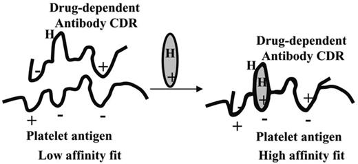
In previous studies, we showed that (1) reactions of platelet-specific antibodies induced by quinine and quinidine24 and by sulfonamide antibiotics28 can be abolished by seemingly minor structural modification of the sensitizing drug and (2) binding of a quinine-dependent antibody “traps” drug on the target as measured by resistance of tritiated quinine to removal from the platelet by repeated washes in buffer.23 These observations, together with those described here, suggest a model to explain drug-dependent binding of antibodies induced by quinine to platelet GPs (Figure 7) that builds on one of several possible models proposed earlier by Shulman and Reid.21 It is known that quinine and many other drugs bind weakly and reversibly to proteins.40,41 We propose that there are preferred sites on GPIIb/IIIa (and presumably GPIb/IX) for reversible binding of quinine and that platelet-reactive antibodies capable of causing thrombocytopenia are specific for both quinine and protein structures adjacent to a quinine-binding site. We suggest that the antibody is a partial fit for this epitope, but that the Ka value for the reaction is too low for significant binding to occur in the absence of drug. However, when drug is present it can fit into the protein-protein interface in such a way that shape and chemical complementarity between the 2 surfaces is enhanced, the coupling energy (and Ka) is increased, and the reaction is favored. This could occur even if the affinity of drug for antibody alone or for the GP alone is relatively weak because once the reaction occurs, the drug is “trapped” and its dissociation is impeded.
Structural models of quinine and quinidine. The ovals delineate the carbon bridge linking the quinoline and quinuclidine components of the molecules. Stereoisometry at the C8 and C9 positions causes the 2 molecules to assume quite different conformations in 3 dimensions.
Structural models of quinine and quinidine. The ovals delineate the carbon bridge linking the quinoline and quinuclidine components of the molecules. Stereoisometry at the C8 and C9 positions causes the 2 molecules to assume quite different conformations in 3 dimensions.
A proposed model for DDAb binding to an epitope on a platelet glycoprotein. (Left) Antibodies capable of causing drug-dependent thrombocytopenia react weakly with an epitope on a glycoprotein. The Ka for this interaction is too small to allow significant numbers of antibody molecules to bind in the absence of drug. (Right) Drug contains structural elements that are complementary to charged or hydrophobic domains (H) on the GP epitope and the complementarity-determining region (CDR) of the antibody. Drug interacts with the target protein and antibody to improve the “fit” between the 2 proteins, increasing the Ka to a value that permits binding to occur at levels of antibody, antigen, and drug achieved in the circulation after ingestion of the drug.
A proposed model for DDAb binding to an epitope on a platelet glycoprotein. (Left) Antibodies capable of causing drug-dependent thrombocytopenia react weakly with an epitope on a glycoprotein. The Ka for this interaction is too small to allow significant numbers of antibody molecules to bind in the absence of drug. (Right) Drug contains structural elements that are complementary to charged or hydrophobic domains (H) on the GP epitope and the complementarity-determining region (CDR) of the antibody. Drug interacts with the target protein and antibody to improve the “fit” between the 2 proteins, increasing the Ka to a value that permits binding to occur at levels of antibody, antigen, and drug achieved in the circulation after ingestion of the drug.
In contrast, “drug-specific” antibodies have a high affinity for free drug but lack a recognition site for a protein epitope. It is apparent that reactions of the first type of antibody with a platelet GP would be potentiated by soluble drug, whereas reactions of the second with immobilized drug (Qn-HSA) would be inhibited, in accordance with observation. Although the proposed model is speculative, it is consistent with crystallographic and functional studies of antigen-antibody complexes showing that modification of a single amino acid residue among the 30 to 40 that typically make up the antigen-antibody interface can lower the association constant by several orders of magnitude.42,43 It is not unreasonable, therefore, to suggest that a drug like quinine, by reacting favorably with amino acids on both sides of the interface, might convert an antibody that is very weakly reactive into one that binds strongly in the presence of drug.
Findings made in these studies may also have implications for the immune response leading to the production of antibodies that cause thrombocytopenia in patients with drug sensitivity. Although the drug-dependent (platelet-reactive) and drug-specific antibodies identified in our patients could be unrelated, their similar specificities for quinine and quinine analogues suggest that both arise in the course of a B-cell response to the same or a very similar antigen. Evidence suggests that maturation of marginal zone B cells is driven by weak interactions between B-cell receptors and epitopes on autologous proteins, leading to a diverse population of B cells that are weakly autoreactive.44-46 The model shown in Figure 7 proposes that drugs such as quinine can improve the fit between certain B-cell receptors and epitopes on autologous proteins such as the GPIIb/IIIa complex. Under some circumstances, for example, in a local inflammatory state, B cells might be induced to proliferate in response to drug associated with a platelet glycoprotein. After affinity maturation, some of the resulting antibodies would be expected to have the properties of immunoglobulins found in patients with drug-induced immune thrombocytopenia (ie, be capable of binding to and destroying platelets when the drug is present); others might recognize drug alone and would behave like the drug-specific antibodies identified in these investigations.
Regardless of whether the proposed model is validated by further studies, the observation that patients with quinine-induced thrombocytopenia often have drug-specific antibodies suggests that these antibodies, detected by using drug coupled to a carrier protein as a target or by another means, could provide a surrogate measure of drug sensitivity in patients with drug-induced immune thrombocytopenia. Studies to test this possibility appear warranted.
Prepublished online as Blood First Edition Paper, April 11, 2006; DOI 10.1182/blood-2006-01-009803.
Supported by grant 0235419Z from the American Heart Association–Northland Affiliate (D.W.B.) and by grant HL-13629 from the National Heart, Lung and Blood Institute (R.H.A.).
An Inside Blood analysis of this article appears at the front of this issue.
The publication costs of this article were defrayed in part by page charge payment. Therefore, and solely to indicate this fact, this article is hereby marked “advertisement” in accordance with 18 U.S.C. section 1734.


