The megakaryoblastic CHRF-288 cell line was used to investigate signal transduction pathways responsible for proplateletlike formation (PPF). The role of fibronectin (FN) and protein kinase C (PKC) activation in PPF were examined. In the presence of serum and phorbol 12-myristate 13-acetate (PMA), a PKC activator, cells exhibited full megakaryocytic differentiation, manifested by adhesion, shape change, increased cell size, polyploidy, PPF, and expression of CD41+, CD61+, and CD62P+. The same morphologic and phenotypic features were observed in serum-free cultures in the presence of FN/PMA. Only partial differentiation occurred when other integrin ligands were substituted for FN. FN alone induced minimal cell adhesion and spreading, while PMA alone induced only polyploidy without adhesion. Signal transduction changes involved the activation of the extracellular signal–regulated protein kinase 1 (ERK1)/ERK2 as well as c-Jun amino-terminal kinase 1 (JNK1)/stress-activated protein kinase (SAPK). Phosphoinositide-3 kinase and p38 were not stimulated under these conditions. Inhibitors were used to identify the causal relationship between signaling pathways and PPF. PD98059 and GF109203X, inhibitors of ERK1/ERK2 pathway and PKC, respectively, blocked PPF, while adhesion, spreading, and polyploidy were normal. These studies show that activation of ERK1/ERK2 mitogen-activated protein kinase pathway plays a critical role in PPF. The elucidation of the signal transduction pathway on megakaryocyte development and PPF is of crucial importance for understanding this unique biological process.
Introduction
Megakaryocytopoiesis involves proliferation of megakaryocyte (MK) progenitors and differentiation. Following a few mitotic cycles, an aborted mitotic process takes place in which prophase, metaphase, and initial anaphase stages are normal while late anaphase and telophase are deficient.1,2 Cytokinesis does not take place, and endoreplication cycles lead to polyploidy.3 Nuclear maturation proceeds in concert with cytoplasmic differentiation and expression of megakaryocytic markers. Cytoplasmic maturation occurs at different ploidy levels and results in platelet production (thrombopoiesis). There are different hypotheses pertaining to platelet formation and release from mature MKs. In one model, the demarcation membrane system outlines nascent platelets derived from the interior of the cytoplasm.4,5Tubular-branching demarcation membranes may be reorganized by a process of fusion-fission into flat sheets of membrane with subsequent shedding of platelets into the circulation.6 In another model, platelets are thought to be released from extruded long cytoplasmic extensions by rupture of the slender links between them.7-9 Proplateletlike formation (PPF) in vitro follows cell adhesion and polyploidization.10 11
Protein kinase C (PKC) is a family of serine/threonine protein kinases in the cytosol involved in pleiotropic processes such as cell growth, differentiation, and cytokine secretion.12,13 The role of PKC signaling in MK differentiation has been well established over the past 2 decades. In several cell lines, PKC activation by the agonist phorbol 12-myristate 13-acetate (PMA) induced MK differentiation, including cell cycle arrest, secretion of cytokines, up-regulation of MK surface antigens, polyploidization, development of proplatelet processes, and appearance of demarcation membranes.14-16 Integrins, which are heterodimeric transmembrane protein receptors, mediate cell membrane–extracellular matrix interaction and have profound effects on cell division, differentiation, and survival.17 It has become clear that integrins not only mediate the physical attachment of cells to extracellular matrix but also generate a variety of signals to the interior of the cell18-20 and regulate cell growth, survival, and gene expression.21-24 Integrin-mediated cell adhesion has been shown to strongly activate mitogen-activated protein kinase (MAPK), a key downstream effector of the Raf signaling pathway in Swiss 3T3/REF52 fibroblasts25 and NIH3T3 fibroblasts.26,27 PKC activation and integrin engagement signaling pathways play different roles in physiological processes and may generate cross-talk resulting in either up-regulation or down-regulation, according to the particular demands of different cells.
MAPKs are serine/threonine kinases that are highly conserved in eukaryotic cells from yeast to human. Several signaling cascades classified as MAPK pathways include extracellular signal–related kinase kinase (MEK)–extracellular signal–regulated protein kinase 1 (ERK1)/ERK2; MEK–c-Jun amino-terminal kinase (JNK)/stress-activated protein kinase (SAPK); and MEK-p38. These pathways have been identified as mediating cell proliferation, survival, differentiation, and apoptosis. While the ERK pathway responds mainly to mitogens and growth factors and regulates mammalian cell proliferation and differentiation, the JNK/SAPK MAPK pathway is associated with stress and apoptosis.28 The p38 MAPK pathway is activated in response to cellular stress and inflammation and is involved in many fundamental biological processes.29-31
Phosphoinositide-3 kinase (PI3K) is known to initiate several signaling pathways,32 and recently, it has also been found to stimulate the activation of the ERK1/ERK2 MAPK cascade in some signaling systems as well. Cross-talk and signal integration mechanisms among all these pathways have been described.
While primary MKs have been separated by different methods,33-36 the yield and degree of purity of the cells are relatively poor, and consistent PPF is lacking. The study of human megakaryocytopoiesis and thrombopoiesis requires the development of either long-term culture systems derived from normal MKs or permanent cell lines derived from transformed MKs. Therefore, for the purpose of clarifying the mechanism/signal transduction of PPF, we chose a human megakaryoblastic CHRF-288 cell line as a culture model that has proved to be valuable for the study of MKs and MK-associated functions.37 Our results showed that PPF in CHRF-288 cells needed 2 critical steps: fibronectin (FN) binding to the cell and activation of intracellular PKC by PMA. The activation of the MEK/ERK pathway induced by FN binding and the activation of PKC are necessary for PPF. Our results suggest substantial overlaps and cross-talk between the integrin engagement pathway, PKC activation, and the Ras/MEK/MAPK cascade.
Materials and methods
Materials
The human CHRF-288 megakaryoblastic cell line was generously provided by Dr Fern Tablin (Davis, CA) and Dr Michael Lieberman (Cincinnati, OH). PMA, extracellular matrix proteins, and BCIP (5 bromo-4-chloro-3-indolylphosphate)/nitroblue tetrazolium (NBT) liquid substrate system were from Sigma (St Louis, MO). Thrombopoietin (TPO) was from R&D (Minneapolis, MN). Proteinase inhibitors were from Boehringer Mannheim (Germany). Tris-glycine gel was from Gradipore (Frenchs Forest, New South Wales, Australia). Nitrocellulose membrane was from Amersham (Les Ulis, France). ERK1/ERK2 MAPK antibody, phosphorylated ERK1/ERK2 MAPK antibody, and goat antirabbit immunoglobulin G (IgG) alkaline phosphatase conjugate were from Promega (Madison, WI). The p38 MAPK antibody and phosphorylated p38 MAPK antibody, the JNK/SAPK antibody and phosphorylated JNK/SAPK antibody, and the PI3K/Akt antibody and phosphorylated PI3K/Akt (Ser and Thr) antibodies were from New England Biolabs (Beverly, MA). Signaling pathway inhibitors PD98059, SB203580, Wortmannin, LY294002, and GF109203X were from Calbiochem (San Diego, CA).
Cell culture
CHRF-288 cells were cultured in Fischers medium (Sigma) supplemented with 20% fetal bovine serum (FBS) (Gibco, Gaithersburg, MD), as a source of growth factors and integrin ligands, and 4 mg/L gentamicin. To identify the serum factor(s) responsible for MK differentiation, serum-free culture was carried out with X-vivo 20 culture medium (Biowhittaker, Walkersville, MD)38 in plates coated with extracellular matrix proteins, such as FN, vitronectin (VN), laminin (LN), collagen I (CI), and collagen IV (CIV). Poly-D-lysine and bovine serum albumin (BSA) were used as controls. The coating procedure involved dissolving the matrix protein in phosphate-buffered saline (PBS) at a concentration of 50 μg/mL and using it in 12-well culture plates (200 μL per well). Plates were left at 4°C overnight, washed with PBS, and incubated with a blocking agent (1% BSA) for 2 hours at 37°C before the onset of culture. PMA (10 ng/mL) was used to promote cell differentiation. The model culture system for PPF included X-vivo 20 plus FN plus PMA. When inhibitors of signaling pathways were used, they were added to the culture 1 hour prior to PMA addition.
Morphologic assay
Differentiated cells were counted by means of micrometer grids in 1-mm2 areas. Under the 10 × objective lens, the left edge of each culture well was used as the first field; the adjacent 5 fields were counted one by one. Adherent and differentiated cells were stained in situ with Wright Giemsa stain for 15 to 30 minutes, or photographed directly with an LSM510 laser microscope (Zeiss, Jenna, Germany).
Electron microscopy
Cells or plateletlike particles were fixed in 2% glutaraldehyde, washed with cacodylate buffer, postfixed with 1% osmium tetroxide and uranyl acetate, dehydrated in graded alcohol and propylene oxide, and then embedded with Epon. Ultrathin, 60-nm sections were cut, stained with uranyl acetate and lead citrate to enhance contrast, and examined in a JEOL-100CX electron microscope (Tokyo, Japan).
Flow cytometry for phenotypic analysis
Cells were counted and viability was assessed by the trypan blue exclusion method. For flow cytometry, cells were washed in PBS containing 1% BSA and then stained for 15 minutes on ice in the dark with fluorescent dye–conjugated monoclonal antibodies specific for MK markers (CD41, CD61, and CD62P) or early myeloid cell markers (CD33 and CD90) (Beckman Coulter, Brea, CA). Corresponding negative controls were fluorescein isothiocyanate–antimouse IgG, phycoerythrin-antimouse IgG, and Cy5-antimouse IgG (Beckman Coulter) used at equivalent IgG concentrations. Each sample contained 5 × 10⁵ cells. Flow cytometric analysis was performed by means of a Coulter Cytometry XL dual laser flow cytometer (Coulter, Hialeah, FL).
Ploidy analysis
Nonadherent cells were collected by centrifugation. Adherent cells were first dislodged with 0.05% trypsin in 0.33 mM EDTA, and the viable cells counted by trypan blue exclusion. Then, 5 × 10⁵ cells per sample were washed in PBS containing 1% BSA. DNA was stained with 7-amino-actinomycin D (7-AAD) (Calbiochem) following a one-step fixation-permeabilization with the ORTHO PermeaFix reagent (J & J, Raritan, NJ). The ploidy classes were then determined following flow cytometric analysis.
Protein extraction and Western blot analysis
Cells were collected at 5 minutes, 30 minutes, 1 hour, 2 hours, 24 hours, 48 hours, and 72 hours after PMA treatment in different culture conditions. Nonadherent and dislodged adherent cells were centrifuged at 400g for 5 minutes, and the cell pellets lysed in a buffer containing 30 mM Hepes, 100 mM NaCl, 10 mM benzamidine, 1 mM EDTA, 1% Triton-X-100, and 20 mM NaF, adjusted to pH 7.5. The following protein and phosphatase inhibitors were then added: 1 mM phenylmethyl sulfonyl fluoride, 10 μg/mL aprotinin, 5 μg/mL leupeptin, 2 μg/mL pepstatin, 1 mM Na3VO4. Cell lysates were cleared by centrifugation at 16 000 rpm for 5 minutes at 4°C. The protein concentrations were measured by means of protein/DC Assay (Bio-Rad, Hercules, CA) to ensure equal electrophoretic loading. Lysates were denatured by boiling for 5 minutes in Laemmli sample buffer and loaded at a concentration of 20 μg protein per lane on 10% Tris-glycine iGel. Proteins were then transferred to nitrocellulose membranes and blocked for 16 hours in Tris-buffered saline containing 0.05% Tween 20 and 1% BSA. To ensure equal protein loading, duplicate gels were stained by Ponceau S (Sigma). Antibodies against signal proteins or phosphorylated signal proteins were used to determine the activation of the kinases during cell differentiation. Each antibody was diluted in blocking buffer at the concentration recommended by the supplier and incubated with the blot at room temperature for 2 hours or at 4°C overnight, then incubated with alkaline phosphatase conjugate for 1 hour at room temperature, and color-detected by BCIP/NBT substrate liquid system.
Statistical analysis
Statistical analysis was performed by means of the 2-tailed Student t test for unpaired data in phenotypic, ploidy, and morphologic analysis.
Results
CHRF-288 cell differentiation in serum-containing medium
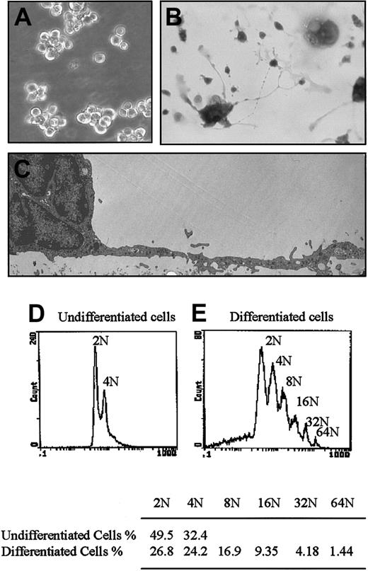
In the absence of PMA stimulation in serum-containing culture medium, CHRF-288 cells were nonadhesive and proliferated actively (Figure 1A). However, after 5 minutes of exposure to PMA (10 ng/mL), they gradually underwent full differentiation, including adhesion, spreading, size increase, polyploidization, and PPF (Figure 1B), reaching maximal differentiation after 3 days. Thereafter, cells detached from the substrate and underwent apoptosis and cell death. The cells with PPF were characterized by electron microscopy. These elongated cytoplasmic processes (Figure 1C) contained cytoskeletal structures, mitochondria, rough endoplasmic reticulum, macrovesicles, and α-granule–like organelles, but no microtubules were observed. Similar morphology was observed for the plateletlike particles, and no marginal microtubular rings, typical of platelets, were seen. The particles did not aggregate with adenosine 5′-diphosphate or thrombin. The timing of the differentiation stages was dependent on PMA concentration (1 ng/mL, 10 ng/mL, or 100 ng/mL). The higher the PMA concentration, the more rapid the differentiation pattern. Without PMA addition, cells showed few adhesion and shape change patterns (Figure 1A). TPO had no effect on the differentiation of CHRF cells.
Cell morphology in serum-containing culture medium.
Cells were cultured for 3 days. (A) Undifferentiated cells cultured in 20% FBS-containing medium. Original magnification × 200. (B) PMA-induced full differentiation in serum-containing medium, showing adherence, size increase, polyploidization, and proplatelet formation. In situ Giemsa staining. Original magnification × 200. (C) Electron micrograph showing a differentiated cell with an elongated cytoplasmic process. Original magnification × 3000. (D) Ploidy of undifferentiated cells. (E) Ploidy of differentiated cells. Nonadherent cells and adherent cells (dislodged by means of 0.05% trypsin-EDTA) were collected by centrifugation. Cells were stained with 7-AAD as indicated in “Materials and methods” and analyzed by flow cytometry. Representative of 5 independent experiments.
PMA treatment altered the cell surface expression of some membrane proteins (Table 1). CD33+, CD90+, and CD62P+ cells significantly decreased, while CD41+ and CD61+ did not change. Although cultured CHRF cells show a 2N-4N ploidy pattern in the absence of PMA (Figure 1D), the addition of PMA led to a significant increase of the DNA content and ploidy distribution, with the appearance of 4N to 64N polyploid cells (Figure 1E).
Immunocytochemical characteristics
| Antigen cluster designation, % reactivity . | Undifferentiated cells, % . | Differentiated cells, % . | P value . |
|---|---|---|---|
| CD33 | 99.5 | 81.3 | < .01 |
| CD41 | 72.5 | 81.2 | .39 |
| CD61 | 54.4 | 63.9 | .33 |
| CD62P | 49.6 | 25.3 | .01 |
| CD90 | 93.4 | 61.8 | < .01 |
| Antigen cluster designation, % reactivity . | Undifferentiated cells, % . | Differentiated cells, % . | P value . |
|---|---|---|---|
| CD33 | 99.5 | 81.3 | < .01 |
| CD41 | 72.5 | 81.2 | .39 |
| CD61 | 54.4 | 63.9 | .33 |
| CD62P | 49.6 | 25.3 | .01 |
| CD90 | 93.4 | 61.8 | < .01 |
Data are from a flow cytometric analysis of cultured CHRF-288 cells. Cells stained with specific surface antibody conjugated with fluorescent dye as indicated in “Materials and methods.” The percentages are percentages of the total number of cells. Five independent experiments were performed.
FN is the integrin ligand responsible for full differentiation
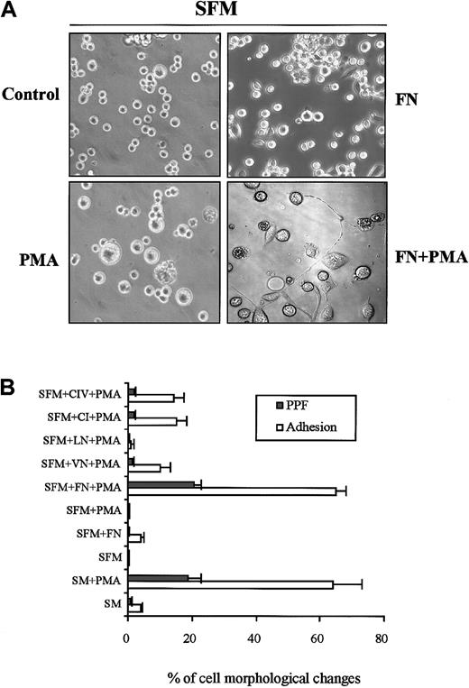
Different integrin ligands were substituted for serum in the culture medium. While FN alone induced minimal adhesion and shape change, PMA alone induced increased size and polyploidy in the absence of adhesion; FN and PMA costimulation induced full cell differentiation/PPF (Figure 2A). In this culture system, the morphologic features of PPF and functional tests of the particles were similar to the data obtained in serum-containing culture. PMA-treated cells cultured in the presence of LN increased in size without adhesion or PPF. PMA-treated cells cultured in the presence of VN-, CI- and CIV-coated wells showed minimal adhesion and PPF (Figure 2B). No adhesion or differentiation occurred with either BSA or poly-D-lysine–coated culture wells. We determined that the optimal culture model for differentiation and PPF involves seeding CHRF cells at a concentration of 2.5 to 5 × 10⁴ cells per milliliter in X-vivo 20 in the presence of PMA (10 ng/mL) in FN-coated dishes for 3 to 4 days. Using this culture model in a well-defined medium resulted in more consistent PPF than the use of serum in the culture medium.
Effect of single matrix protein on CHRF cell morphology with or without PMA treatment in serum-free medium culture.
(A) Cells cultured in serum-free medium with different combinations. Full cell differentiation, including PPF, occurred only in FN/PMA costimulation. Original magnification × 200. (B) Culture dishes were coated with a single matrix protein (50 μg/mL) overnight. Cells were cultured in serum-free medium in the presence of PMA (10 ng/mL) and a single matrix protein for 3 days. Cells were seeded at a concentration of 2.5 × 10⁴ cells per milliliter and counted in 1-mm2 areas by means of phase contrast microscopy. SM, serum-containing medium; SFM, serum-free medium. Graph depicts the means ± SE from at least 5 experiments.
Activation of ERK1/ERK2 is essential for full differentiation and PPF
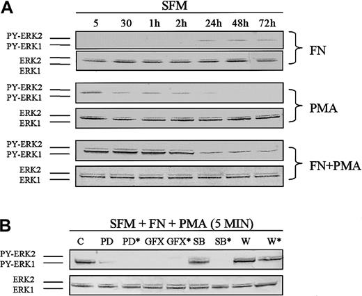
The differentiation of CHRF cells can be viewed as the result of a balance between stimulatory and inhibitory signals. Western blots were used to identify the activation of signaling molecules related to PPF. When cells were treated with FN alone, a faint phosphorylation signal of ERK1/ERK2 was detected after 1 day of culture (Figure3A, upper panels). With PMA alone, on the other hand, a weak and quick phosphorylation signal of ERK1/ERK2 was detected within 5 minutes and diminished with time (Figure 3A, middle panel). With both FN and PMA present in the culture, a strong phosphorylation of ERK1/ERK2 occurred. The activation of ERK1/ERK2 was rapid, occurring within 5 minutes of exposure to PMA. The maximum expression of phosphorylated ERK1/ERK2 occurred 30 minutes after PMA treatment, when the cells had firmly adhered to the FN substrate. PMA rapidly and persistently induced the phosphorylation of ERK1/ERK2, but the expression of the kinase tapered with time (Figure 3A, lower panel). Use of the inhibitors PD and GFX in the culture system blocked phosporylation of ERK1/ERK2, as did a high dose of SB (50 μM) (Figure 3B).
Correlation of ERK1/ERK2 MAPK activation with PPF.
Cells were cultured in FN/PMA for 3 days and analyzed by Western blot. (A) Time course of ERK1/ERK2 phosphorylation in the cultures with the FN/PMA combination or with FN or PMA alone. The upper panels show that phosphorylation of ERK1/ERK2 was faintly expressed in the presence of FN alone after 1 day of culture. The middle panel shows that phosphorylation of ERK1/ERK2 was rapid and weakly expressed in the presence of PMA alone and diminished with time. The lower panel shows that phosphorylation of ERK1/ERK2 was strongly expressed only in the presence of FN plus PMA after 5 minutes' treatment and was sustained up to 3 days, correlating with PPF. (B) Effect of signal inhibitors on phosphorylation of ERK1/ERK2. The inhibitors were added 1 hour prior to addition of PMA for 5 minutes. The upper panel shows that phosphorylation of ERK1/ERK2 was inhibited by PD, an MEK inhibitor (10 μM and 50 μM*); GFX, a PKC inhibitor (5 μM and 25 μM*); and SB, a p38 and JNK MAPK inhibitor (50 μM*). No inhibition seen with SB (10 μM) and wortmannin (W), a PI3K inhibitor (100 nM and 500 nM*). SFM indicates serum-free medium. The data are representative of 3 separate experiments.
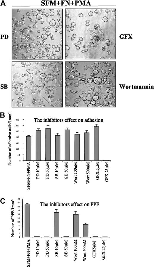
To determine a causal relationship between signaling and PPF, we supplemented our culture model system with various signaling inhibitors to identify the pathway responsible for differentiation of the CHRF-288 cells (Figure 4). The inhibitors were added to cells 1 hour before PMA addition. PD98059 (10 μM and50 μM) and GF109203X (5 μM), inhibitors of upstream of ERK1/ERK2-MAPK and PKC,39 respectively, blocked PPF but not adhesion or size increase. While a low dose of SB203580 (10 μM), an inhibitor of p38 MAPK40 and JNK/SAPK41 pathways, did not affect cell differentiation, a high dose (50 μM) of SB blocked the differentiation. Wortmannin (100 nM and 500 nM), an inhibitor of PI3K/Akt pathway,39 did not affect cell differentiation. A high dose of GF109203X (25 μM) was cytotoxic for cells as determined by the trypan blue exclusion test, and the cell body shrank.
Effect of signal inhibition on cell morphology.
Cells were cultured in serum-free medium in the presence of FN. Cells were pretreated with inhibitors for 1 hour at 37°C; then PMA (10 ng/mL) was added and culture proceeded for 3 days. (A) Morphologic observation. Cells were visualized and measured by means of phase contrast microscopy at original magnification × 200. Representative fields demonstrating the results of at least 10 separate experiments are shown. (B) Morphologic quantitation. Morphologic assay was carried out by means of a phase contrast microscope (see “Materials and methods”). The cells with PPF were defined as those bearing more than one cytoplasmic process that were at least twice the length of the cell body diameter. SFM indicates serum-free medium; PD, PD98059, an inhibitor of MEK MAPK (upstream of ERK); GFX, GF109203X, an inhibitor of PKC; SB, SB203580, an inhibitor of p38 and JNK; Wort, wortmannin, an inhibitor of PI3K. Graph depicts the means ± SE from at least 10 experiments.
Activation of JNK1/SAPK but not p38 and PI3K
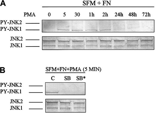
Costimulation of cells with FN/PMA induced phosphorylation of JNK1(46 kd)/SAPK within 5 minutes of PMA addition, and this activation lasted at least 2 hours (Figure 5A). Low and high doses of SB (10 and 50 μM) blocked phosphorylation of JNK1 (Figure 5B), but only a high dose of SB blocked PPF (Figure 4C). JNK1/SAPK is therefore not involved in PPF. Neither p38 MAPK nor PI3K/Akt (Ser473 and Thr308) were activated following FN/PMA stimulation (data not shown).
SAPK/JNK1 MAPK activation.
Cells were cultured in FN/PMA for 3 days and analyzed by Western blot. (A) The upper panel shows that only phosphorylation of SAPK/JNK1 (46 kd) was expressed for up to 2 hours. (B) SB203580 (10 μM and 50 μM*), an inhibitor of SAPK/JNK added 1 hour prior to PMA treatment, blocked phosphorylation of SAPK/JNK1 (46 kd). SFM indicates serum-free medium. The data are representative of 3 separate experiments.
Discussion
The identification of the signaling pathway responsible for megakaryocytic differentiation, PPF, and platelet release is still debated. In this study, we sought to characterize the MAPK-specific targets in CHRF-288 cells stimulated by FN/PMA. Rapid and prolonged activation of ERK1/ERK2 MAPK was found necessary for PPF.
We first investigated a series of integrin ligands for their effects on PPF.17 As a main component of matrix protein, FN binds to several megakaryocyte integrins, such as αIIbβ3, αvβ3, αvβ5, and α5β1. In this study, when CHRF cells were cultured in serum-free medium in FN-coated dishes, only later-term and weak phosphorylation of ERK1/ERK2 and minimal adhesion were observed. With the addition of PMA, a PKC activator used at a moderate concentration that did not down-regulate PKC,42 the cells showed a dramatic full differentiation pattern that included PPF, and concomitant activation of ERK1/ERK2. The binding to other matrix proteins, VN, CI, and CIV, with PMA treatment resulted in minimal cell adhesion and occasional PPF.
PKC activation with PMA is known to induce a series of biological processes related to differentiation correlating with the activation of MAPK.43 In our study, activation of PKC by PMA in the absence of FN resulted in weak and rapid phosphorylation of ERK1/ERK2; however, cells increased only in size and ploidy, without either adhesion or PPF. While the polyploidy pattern is similar in the presence of PMA alone and of PMA/FN, only the latter induced adhesion and PPF. Thus, while polyploidy or adhesion is essential for PPF, neither alone is sufficient to induce this morphologic change. Only FN/PMA costimulation induced a strong, rapid, and sustained phosphorylation of ERK1/ERK2 that lasted for at least 72 hours and correlated with full differentiation, including adhesion, spreading, size increase, polyploidization, and PPF. Since similar results were obtained with either serum/PMA or FN/PMA, the integrin ligand FN appears to be the component in serum responsible for the activation process leading to cell differentiation and PPF. Even though VN can also bind to αvβ3, αvβ5, and αIIbβ3, which are also receptors for FN, it could not induce full differentiation of CHRF cells with PMA treatment; neither did CI bound to αIIbβ3. The strong ERK1/ERK2 phosphorylation with FN/PMA was due to an amplification process resulting from cross-talk between the integrin- and the PKC-generated signaling pathways.
Inhibitors of signaling pathways were used in order to clarify whether there was a causal relationship between ERK1/ERK2 activation and PPF. PD98059, an MEK inhibitor upstream of ERK1/ERK2, blocked the phosphorylation of ERK1/ERK2 as well as PPF, though polyploidy and adhesion were not affected. PPF therefore required not only adhesion to an FN matrix and polyploidy, but also sustained activation of ERK1/ERK2. A causal relationship was therefore demonstrated between ERK1/ERK2 activation and PPF.
The phenotypic differences between surface markers of differentiated and undifferentiated cells are related to the maturation process as reflected by the decrease of CD90 and CD33, early myeloid surface markers. The significant decrease of CD62P in differentiated cells may be due to its translocation to the PPF. CD62P is an α-granule protein in primary MKs and platelets that is expressed on the platelet surface following platelet activation. Its expression on the surface of CHRF cells is therefore not typical for a platelet-producing cell line.
Since adhesion and polyploidy are necessary, though not sufficient, for PPF, we investigated stimulation of MAPK pathways other than ERK1/ERK2. The JNK/SAPK pathway is usually activated in response to stress/apoptosis. In this study, phosphorylation of JNK1/SAPK, also induced by FN/PMA costimulation, appeared rapidly in concert with ERK1/ERK2 activation but disappeared after 24 hours, just when cells started to develop PPF. SB203580, an inhibitor of JNK1/SAPK, prevented JNK1/SAPK phosphorylation but did not affect differentiation features of CHRF cells or PPF, when used at a concentration of 10 μM. Thus, though JNK1/SAPK activation correlates with CHRF differentiation, it is not involved in PPF. At a high concentration (50 μM), SB203580 blocked PPF, probably via inhibition of ERK1/ERK2 (Figure 3B).
The p38 MAPK is activated when apoptosis, following differentiation, is triggered.40 PI3K, on the other hand, is related to survival, growth, and differentiation.32,39 Since p38 MAPK and PI3K have been reported in some studies to be upstream of the pathway leading to ERK1/ERK2 activation,44 45 we investigated whether this was the case with CHRF cells. In this study, neither kinase was activated following treatment with the FN/PMA combination, and the inhibitors to the respective kinases did not affect cell differentiation or ERK1/ERK2 phosphorylation.
Recent reports have shown that TPO activated MK differentiation through the ERK MAPK pathway.46 TPO was ineffective in promoting differentiation features in CHRF cells (data not shown), presumably owing to the lack of the c-Mpl receptor. Only stimulation with the nonphysiological agonist PMA in the presence of FN triggers differentiation.
The ultrastructure of PPF and released particles as well as the dysfunction of particles with platelet agonists demonstrates that the CHRF-288 cell line does not produce bona fide platelets. This transformed cell line, however, provides valuable information on the mechanism of MK differentiation, ploidy, and PPF and is useful for a better understanding of molecular events associated with MK development and platelet production.
The authors are grateful to Drs Fern Tablin (University of Califonia at Davis) and Michael A. Lieberman (University of Cincinnati, OH) for the CHRF-288 megakaryoblastic cell line; Mr Phil Lefebvre for helpful discussion; and Dr Mary Dunn for assistance.
Supported in part by grant DAMD17-98-1-8327 from the US Army Breast Cancer Program and a grant from the Rehabilitation Institute of Chicago, both awarded to I.C.
The publication costs of this article were defrayed in part by page charge payment. Therefore, and solely to indicate this fact, this article is hereby marked “advertisement” in accordance with 18 U.S.C. section 1734.
References
Author notes
Fang Jiang, Northwestern University, 345 E Superior St, RIC Rm 1407, Chicago, IL 60611; e-mail:f-jiang2@northwestern.edu.

This feature is available to Subscribers Only
Sign In or Create an Account Close Modal