Using differential display polymerase chain reaction, a gene was identified in CD34+-enriched populations that had with low or absent expression in CD34− populations. The full coding sequence of this transcript was obtained, and the predicted protein has a high degree of homology to oxysterol-binding protein. This gene has been designated OSBP-related protein 3 (ORP-3). Expression of ORP-3 was found to be 3- to 4-fold higher in CD34+ cells than in CD34− cells. Additionally, expression of this gene was 2-fold higher in the more primitive subfraction of hematopoietic cells defined by the CD34+38− phenotype and was down-regulated with the proliferation and differentiation of CD34+ cells. The ORP-3 predicted protein contains an oxysterol-binding domain. Well-characterized proteins expressing this domain bind oxysterols in a dose-dependent fashion. Biologic activities of oxysterols include inhibition of cholesterol biosynthesis and cell proliferation in a variety of cell types, among them hematopoietic cells. Characterization and differential expression of ORP-3 implicates a possible role in the mediation of oxysterol effects on hematopoiesis.
Introduction
Molecular processes that maintain the stem cell pool and govern the proliferation and differentiation of hematopoietic stem and progenitor cells are widely investigated but incompletely understood. Identification of these genes will facilitate our understanding of hematopoiesis and may be used to improve clinical outcomes.
Using differential display-polymerase chain reaction (dd-PCR), we identified a differentially expressed transcript in CD34-enriched populations, with low or absent expression in CD34-depleted populations. Sequencing of this transcript revealed significant homology to oxysterol-binding protein (OSBP). On the basis of ESTs in the GenBank database, a family of up to 8 human genes that code for OSBP-related proteins is postulated to exist.1 These have been designated OSBP-related proteins (ORPs). A partial sequence for our OSBP-related gene was thus designated ORP-3.1
Study design
CD34+ purification
Umbilical cord blood (UCB) samples were obtained after uncomplicated vaginal or cesarean delivery. Adult bone marrow (ABM) cells were obtained from cell scrapings of discarded rib and femur heads after cardiothoracic or hip-replacement surgery. All samples were donated by volunteers according to approved institutional guidelines. Mononuclear cells were prepared by density-gradient separation through Ficoll-Hypaque (Amersham Pharmacia Biotech, Uppsala, Sweden). CD34+ cells were isolated using a MiniMacs bead separation kit (Miltenyi Biotec, Becton Dickinson, Sunnyvale, CA).
Flow cytometric analysis of CD34+ populations and fluorescence-activated cell sorting of CD34+subsets
CD34+ cells were labeled with anti-CD38–fluorescein isothiocyanate, anti-CD34–phycoerythrin, and anti-CD45-PECY5 (Coulter Immunotech, Fullerton, CA). CD34+ purity was determined by flow cytometric analysis (FacsCalibur, Becton Dickinson) following modified ISHAGE guidelines2 using CELLQuest software (Becton Dickinson). When required, CD34+38−/dull cells were sorted using a FacsCalibur Cell Concentrator Sorting Module.
Culture of umbilical cord blood CD34+ cells
In selected experiments, isolated CD34+ cells were cultured for 1 week at 37°C in a humidified atmosphere flushed with 5% CO2 in air, at a concentration of 0.5 × 10⁶ cells/mL in α minimum essential medium (Trace, Noble Park, Victoria, Australia) with 20% fetal bovine serum (CSL Biosciences, Parkville, Victoria, Australia), 2 mM L-glutamine, 200 U/mL penicillin–streptomycin (Sigma, St Louis, MO), 20 ng/mL recombinant human stem cell factor (Amgen, Thousand Oaks, CA), 10 ng/mL IL-1B (Endogen, Woburn, MA), 10 ng/mL IL-3, 10 ng/mL IL-6, and 10 ng/mL G-CSF (Amrad, Boronia, Victoria, Australia).
dd-PCR
RNA was extracted by RNeasy total RNA isolation kit (Qiagen, Clifton Hill, Victoria, Australia) and was DNase treated (Invitrogen, Carlsbad, CA). Reverse transcription and dd-PCR were carried out as described in the RNAimage Differential Display System (GenHunter, Nashville, TN). We used ABI Prism BigDye Terminator Cycle Sequencing Ready Reaction Kit (PE Applied Biosystems, Foster City, CA) for all sequencing reactions, and sequences were determined using an ABI PRISM 373 DNA Sequencer (PE Applied Biosystems).
mRNA extraction and rapid amplification of cDNA ends
mRNA was extracted from UCB mononuclear cells using Oligotex Direct mRNA Kit (Qiagen). 5′ and 3′ Rapid amplification of cDNA ends (RACE) was carried out using the Marathon cDNA Amplification Kit (Clontech, Palo Alto, CA). All primers used in this study are presented in Table 1.
Primer and probe sequences
| ORP-3 RACE . | |
|---|---|
| Nested reverse race primer 1 | 5′-TGA GCA GAC GGG CAG GAG GAG G-3′ |
| Nested reverse race primer 2 | 5′-TGG ACC GCC ACC TCC TGT GC-3′ |
| Nested reverse race primer 3 | 5′-AGG CAT GAC AGT GCG CCA GGT C-3′ |
| Nested reverse race primer 4 | 5′-TCT GAC GAT ACA TTC TGT GGT GGC GAA G-3′ |
| Nested reverse race primer 5 | 5′-TCA TCT CCC CCC TCA GTC CTT CCA CC-3′ |
| Nested forward race primer 1 | 5′-CCA AAG CAC CCC AAG GCT GGT ATT C-3′ |
| Nested forward race primer 2 | 5′-GTC CTG TGC TTC CTG TGA GAC TTC CTG G-3′ |
| Nested forward race primer 3 | 5′-GCT TCA AGC ACT GTC TTC CAT CTG CAT C-3′ |
| Northern hybridization | |
| ORP-3 forward | 5′-GCT CAG GAA GTT CTG TTA TCT CC-3′ |
| ORP-3 reverse | 5′-GCT AAG CTA AGC ACA AGT GAT CA-3′ |
| Real-time | |
| ORP-3 forward | 5′-CAA ACT GGA CCA TCC TGT CTT ATG-3′ |
| ORP-3 reverse | 5′-AAG CTA AGC ACA AGT GAT CAT CCT AGA-3′ |
| ORP-3 probe | fam-AAC ATT AGT GTA TTT CTC CTG TGC TTG C-tamra |
| β-actin forward | 5′-GAC AGG ATG CAG AAG GAG ATT ACT-3′ |
| β-actin reverse | 5′-TGA TCC ACA TCT GCT GGA AGG T-3′ |
| β-actin probe | fam-ATC ATT GCT CCT CCT GAG CGC AAG TAC TC-tamra |
Northern hybridization
A PCR product spanning the ORP-3 coding region was amplified from UCB mononuclear cell cDNA in a standard PCR reaction and subcloned. The ORP-3 insert was isolated and labeled using [α-32P]dATP and a Strip-Ez DNA probe synthesis kit (Ambion, Austin, TX). The labeled ORP-3 probe was hybridized to a human multiple tissue Northern blot (Clontech) with ULTRAhyb hybridization buffer (Ambion) and was visualized by autoradiography.
Taqman real-time polymerase chain reaction
Reverse transcription was carried out using the reverse transcription system (Promega, Madison, WI). Real-time PCR amplification of ORP-3 and β-actin was carried out using Taqman Universal PCR Mastermix on an ABI PRISM 7700 Sequence Detection System (PE Applied Biosystems).
Bioinformatics
Nucleic acid and protein sequences were analyzed using software available from the National Center for Biotechnology Information and Swiss Institute of Bioinformatics databases.
Results and discussion
Full ORP-3 mRNA of 6.631 kb was obtained using EST database searching and RACE technology (GenBank accession no. AY008372). The size of the complete ORP-3 cDNA has been confirmed by Northern hybridization with the presence of a transcript at approximately 7.0 kb (results not included). Our results also revealed the presence of 2 other transcripts at approximately 4.4 and 3.6 kb. These splice variants were expressed in a variety of normal human tissues and might have been produced as a result of posttranscriptional modification. Further investigation is required to determine their specific functions.
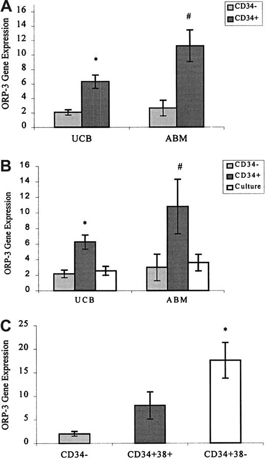
We used Taqman real-time PCR to confirm the differential expression of ORP-3. Results indicate that the expression in CD34+ cells from UCB and ABM is 3- to 4-fold higher (respectively) than corresponding CD34− populations (Figure1A). ORP-3 gene expression (normalized for the expression of β-actin24) was also significantly higher in CD34+ cells from ABM than in equivalent cell populations from UCB.
ORP-3 gene expression as measured by Taqman real-time PCR.
All gene expression results are calculated relative to β-actin and are expressed in arbitrary units (mean ± SEM).24Wilcoxon signed-rank test was used to determine the statistical significance of the differences between related groups for nonparametric data. (A) ORP-3 gene expression in CD34+ (▪) and CD34− (░) hematopoietic cells from UCB (6.3 ± 0.9, 2.0 ± 0.4, n = 21,*P ≤ .0001) and ABM (11.2 ± 2.2, 2.6 ± 1.1, n = 14, #P ≤ .001). Mean purity of the UCB and ABM CD34+-enriched populations were 57.5% ± 2.8% and 55.0% ± 4.8%, respectively. The percentage of CD34+ cells in the CD34− fractions was less than 0.01%. Average viability of the CD34+ and CD34− populations (more than 95%) did not vary significantly. ORP-3 gene expression did not correlate with sample purity (results not shown). (B) ORP-3 gene expression in CD34− (░), CD34+ (▪), and CD34+ cells after 1-week culture (■) from UCB (6.2 ± 0.9, 2.5 ± 0.6, n = 14, *P ≤ .01) and ABM (10.8 ± 3.5, 3.6 ± 1.1, n = 9, #P ≤ .01). FACS analysis of the CD34+ and CD34+culture samples showed that after 1-week culture under the conditions described, the percentage of CD34+ cells decreased by approximately 33%. Total cell number increased by approximately 10%, and viability of the 2 populations (more than 90%) did not vary significantly. (C)ORP-3 gene expression in UCB CD34−(2.0 ± 0.5), CD34+CD38+ (8.0 ± 2.9), and CD34+CD38− hematopoietic cells (17.6 ± 3.8, n = 5, P ≤ .04). CD34+38−/dullcells were sorted using sort gates constructed to include approximately 10% of the total CD34+ population. Viability (greater than 80%) of the 2 populations did not vary significantly.
The CD34+ population of cells is heterogeneous and contains multipotential and lineage-restricted cells.3,4 Therefore, we investigated ORP-3 gene expression in this population before and after 7-day culture in media containing growth factors that promote the differentiation and proliferation of hematopoietic progenitors.4,5 Our results indicate that under these culture conditions, ORP-3 gene expression in freshly isolated, uncultured CD34+ cells from UCB and ABM was 2- to 3-fold higher (respectively) than in these same cells after 7 days' culture (Figure 1B). Investigation of ORP-3 in the more primitive subset of cells defined by the CD34+38− immunophenotype6 7indicates higher expression than in the more mature CD34+38+ subset (Figure 1C). Taken together,ORP-3 expression data indicate higher expression in a less differentiated subset of hematopoietic cells. The physiological significance of this differential expression and the function of ORP-3 in these cell populations remain to be elucidated.
Translation of the ORP-3 open-reading frame yields an 887-amino acid protein that has a high degree of homology to OSBP. OSBP is a well-characterized and highly conserved protein that has been demonstrated to bind oxysterols in a dose-dependent fashion. ORP-3 and OSBP harbor the same modular domains—a C-terminal OSBP domain that is required for OS binding8 and an N-terminal pleckstrin homology domain9 that is required for translocation of the ligand protein to the Golgi apparatus.10,11 As do some OSBPs, ORP-3 contains a putative leucine zipper region.12It must be noted however, that though leucine zipper regions may be associated with DNA binding,13 this has not been demonstrated in any OSBPs.12,14 15
To date, the complete coding sequence of only one human OSBP (OSBP-Hm) has been published; however, at least 8 other human ORP partial and complete gene sequences (designated ORP 1-8) exist on the GenBank database.1 The partial sequence for the human OSBP homologue, ORP-4, was identified by Fournier et al16 using dd-PCR as a screen to identify genes associated with metastatic potential. They designated the sequence HeLa metastatic gene (HLM). Northern blot analysis of ORP-4/HLMexpression demonstrated that ORP-4/HLM expression is significantly associated with metastatic potential.
OSBPs have also been identified in lower species, and their expression has been strongly associated with cell cycle progression.17,18 Given our observations of differential expression of ORP-3 in cells with different cell cycle profiles (such as CD34+CD38+ and CD34+CD38− cells),19 we are investigating the possibility that ORP-3 may be involved in cell cycle regulation. Interestingly, it has been hypothesized that levels of OSBP-Hm vary as a function of cell cycle.20 This finding has not been confirmed, nor has its significance been reported.
Given the homology between ORP-3 and OSBP, it is likely that ORP-3 also plays a role in mediating oxysterol effects on cells. Oxysterols are hydroxylated derivatives of cholesterol that have been demonstrated to inhibit the transcription of many genes involved in cholesterol biosynthesis and cell replication and to be apoptotic to a variety of cell types.21-23 Additionally, investigations in our laboratory have demonstrated that oxysterols are potent inhibitors of HL60 and granulocyte macrophage–colony-forming unit cell growth and that they induce apoptosis in CD34+ cells (manuscript in preparation). Given the importance of these processes in hematopoiesis and the differential expression of ORP-3, we believe that characterization and investigation of the function of ORP-3 may provide useful insights into a possible regulatory role for oxysterols and their binding proteins in hematopoietic stem cell proliferation, differentiation, and self-renewal.
We thank the medical and nursing staff of The Geelong Hospital and St John of God (Geelong, Victoria, Australia) for the collection of UCB and ABM samples.
Supported in part by the Australian Red Cross Blood Service.
The publication costs of this article were defrayed in part by page charge payment. Therefore, and solely to indicate this fact, this article is hereby marked “advertisement” in accordance with 18 U.S.C. section 1734.
References
Author notes
Claudia Gregorio-King, Stem Cell Laboratory, Douglas Hocking Research Institute, Barwon Health, The Geelong Hospital, Deakin University, Geelong 3220, Victoria, Australia; e-mail:ccgk@deakin.edu.au.


This feature is available to Subscribers Only
Sign In or Create an Account Close Modal